


 Close
Close
 5th May 2025
Cardiac Causes
Nodal disease
Sinus node dysfunction (SND), also known as ‘Sick Sinus Syndrome’ or ‘Tachy-Brady Syndrome’, encompasses a spectrum of conditions that affect the specialised cardiac conduction tissue in the sinoatrial node, the atrial conduction tissue and the atrioventricular node. It commonly tends to be a disease of the older patient, with multiple postulated mechanisms whereby malfunction of the cardiac conduction system (mostly from fibrosis) occurs leading to slower sinoatrial conduction times. Electrocardiographic (ECG) features of this can be varied – from sinus bradycardia, sinus pause (>3 seconds), sinus arrest with intermittent atrial tachycardia. There is often an alternating tachycardia-bradycardia phenomenon seen in this, with intermittent sinus rhythm or sinus bradycardia. SND is usually detected on Holter monitoring but may also be picked up on serial 12-lead ECG. Definitive treatment is with pacemaker insertion, mostly for symptom management.
Heart block
Atrioventricular (AV) block, or heart block, occurs when the electrical impulse travelling from the atria to the ventricles is delayed or completely halted. There are three types of AV-block classically described, based on their ECG features – these are illustrated in Figure 1. Certain drugs (e.g. beta blockers, calcium channel blockers, tricyclic antidepressants) can cause all three types of heart block. In 1st degree AV-block, there is prolongation of the PR-interval beyond 200milliseconds with preservation of subsequent QRS complexes. No specific intervention is required for this isolated 1st degree AV-block and it is often found in asymptomatic and healthy individuals; however, in cases of symptomatic 1st degree AV-block with PR>300ms, a pacemaker may be advocated. In 2nd degree AV-block, there is loss of subsequent QRS complexes. This may be following progressive prolongation of the PR-interval, in the case of Mobitz Type 1 AV-block (also known as Wenckebach phenomenon); or, with a constant PR-interval with occasional loss of QRS complex, in the case of Mobitz Type 2 AV-block. In 3ʳᵈ degree AV-block, also known as complete heart block, there is complete disassociation of the P waves with QRS complexes. In this bradyarrhythmia, there is often a ventricular escape (junctional) rhythm characterised by a slower rate (depending on how distal to the AV-node the escape rhythm originates from) and broad QRS complexes. Due to a higher risk of asystole, pacemaker intervention is indicated in Mobitz Type 2 and 3ʳᵈ degree AV-block.
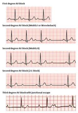
Image Source: Wikimedia Commons (2017). Atrioventricular Block, Licensed under a creative commons licence. Available for free at: https://en.wikipedia.org/wiki/Atrioventricular_block (Accessed 1 May 2025).
Ventricular disease
Abnormal conduction can also occur within the ventricles themselves, in the setting of the bundle branch block. However, this is typically distinct from any abnormal AV-conduction and is often associated with sinus rhythm.
Complete Guide to Clinical Genetics ST3 Application and Recruitment (2027)
Written by Medibuddy. Last updated: October 2026. Clinical Genetics is

Complete Guide to Clinical Radiology ST3 Application and Recruitment (2027)
Recruitment paused: NHS England is not running Clinical Radiology ST3+ recru

Complete Guide to Paediatric Surgery ST3 Application and Recruitment in United Kingdom (2026)
Written by Medibuddy. Last updated: October 2026. If you’re planning


Take your subscriptions with you
Our mobile app allows you to access your interview and exam question banks wherever you are.

