


 Close
Close
 4th May 2025
Supraventricular Tachycardia
The term Supraventricular Tachycardia (SVT) refers to any tachycardia that is not ventricular in origin. SVT is an umbrella term which includes Atrioventricular Nodal Reentry Tachycardia (AVNRT) and Atrioventricular Reentry Tachycardia (AVRT). AVNRT is used synonymously with the term SVT.
Atrioventricular Nodal Reentry Tachycardia (AVNRT)
AVNRT typically occurs in younger people. It is usually paroxysmal and may occur spontaneously. It may have a trigger such as alcohol intoxication, nicotine, stress, concurrent illness, caffeine or stimulants such as amphetamines.
Brief pathophysiology of ‘Slow-Fast AVNRT’
AVNRT results from having dual AV nodal pathways within the AV node itself. There is a fast pathway which conducts rapidly but has a long refractory period, and there is a slow pathway which conducts slowly but a short refractory period. During normal sinus rhythm, electrical impulses travel down both pathways simultaneously and cancel each other out.
However, a premature atrial contraction disrupts this as while the fast pathway is still in a refractory period, electrical impulses would then have to solely travel down the slow pathway as it is no longer in refractory period. By the time the premature impulses reach the distal end of the slow pathway, the fast pathway is no longer in a refractory period which allows impulses to travel retrogradely up the fast pathway.
Hence, a ‘re-entrant’ circuit is created as impulses are now continuously cycling around both pathways activating the bundle of His anterogradely and the atria retrogradely.
Clinical manifestations
Symptoms arise from the tachycardia generated and they include:
- Palpitations
- Chest pain
- Light-headedness or dizziness
- Syncope or presyncope
- Breathlessness
- Anxiety
ECG features
- P waves hidden within QRS complexes
- Late P waves resulting in Pseudo R wave/second R wave
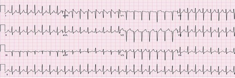
Image Source: Wikimedia Commons (2020), ECG AVNRT, Licenced under a creative commons licence. Available for free at: https://commons.wikimedia.org/wiki/File:ECG_AVNRT_181_bpm.jpg (Accessed: 1 May 2025).
Medical management
The principle in managing AVNRT is inhibiting AV nodal activity as this is the driver for the tachycardia. This can be achieved via vagal manoeuvres and medication. Vagal manoeuvres work by increasing vagal parasympathetic tone. DCCV is rarely required unless the patient is haemodynamically unstable or refractory to medical cardioversion.
1st line therapy: Vagal manoeuvres:
- Blowing into an empty 10ml syringe
- Carotid sinus massage (contraindicated in carotid disease due to the risk of stroke)
- Modified Valsalva Manoeuvre: head down, legs up
2nd line therapy: Adenosine
- This is given IV 6mg, then 12mg and then followed by 18mg
- Contraindicated in asthma due to bronchoconstriction
- Safe to give in pregnancy as does not significantly cross the placenta
- Dipyridamole enhances the effect of adenosine, whereas Theophylline is a potent adenosine receptor antagonist
3rd line therapy: Beta blockers or Calcium channel blockers
- IV Metoprolol or Verapamil
- If 3rd line treatment ineffective, DCCV should be attempted with appropriate sedation and anaesthetic support
Accessory Pathways
Background
Wolff-Parkinson-White (WPW) Syndrome is caused by an accessory conduction pathway between the atria and the ventricles leading to an atrioventricular reentry tachycardia (AVRT). This accessory pathway is also called the bundle of Kent which allows electrical impulses generated by the Sino-atrial node to bypass the AV node. This accessory pathway allows electrical impulses to be conducted quicker than the AV node, resulting in a shorter PR interval which brings about the characteristic Delta wave seen in ECGS. WPW has an incidence of 0.1-0.3 per 1000.
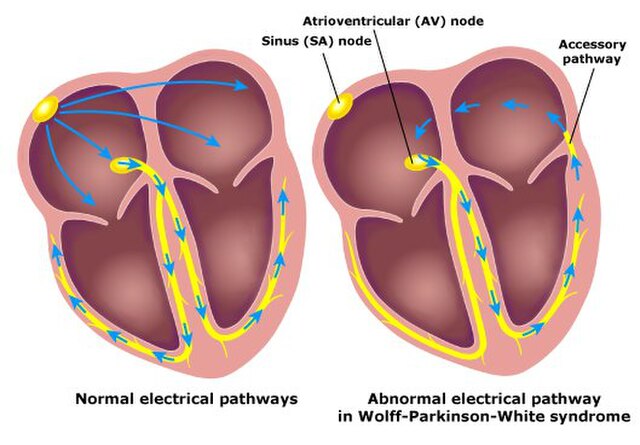
Image Source: Wikimedia Commons (2011). Wolf, Parkinson and White. Licenced under a creative commons licence. Available for free at: https://commons.wikimedia.org/w/index.php?search=wolf+parkinson+white&title=Special:MediaSearch&go=Go&type=image (Accessed: 1 May 2025).
Associations with WPW syndrome
- Hypertrophic Cardiomyopathy (HCM)
- Mitral valve prolapse
- Ebstein’s Anomaly
Clinical manifestations
Most patients who present with an arrhythmia will present with one or more of the following symptoms:
- Palpitations
- Chest pain
- Light-headedness or dizziness
- Syncope or presyncope
- Sudden cardiac death
ECG features in WPW
- Short PR interval <120ms
- Slurred upstroke of a wide QRS complex – ‘delta wave’
- Prolonged QRS >110ms
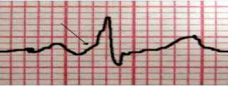
Image Source: Wikimedia Commons (2020). Delta Wave, Licensed under a creative commons licence. Available for free at: https://upload.wikimedia.org/wikipedia/commons/thumb/e/e9/DeltaWave09.JPG/640px- DeltaWave09.JPG (Accessed: 1 May 2025).
Arrhythmias associated with WPW
Atrial Fibrillation is seen in about 10-30% of patients with WPW syndrome. The presence of an accessory pathway in WPW allows the rapid conduction of atria signals to the ventricles as it bypasses the AV node. This results in rapid ventricular rates that may result in VT or VF.
Medical management
The general principle in managing patients with WPW is to prevent electrical impulses from travelling through the accessory pathway. Hence any AV nodal blocking drugs such as Adenosine, Beta blockers and Calcium channel blockers are contraindicated as blocking the AV node will allow uncontrolled electrical impulses down the accessory pathway potentially leading to VT/VF and cardiac arrest.
Drugs used to treat WPW include
- Flecainide (In the absence of structural heart disease)
- Amiodarone
- Procainamide
Surgical management
Definitive treatment options include Catheter ablation or Surgical ablation. Efficacy of catheter- based ablation is around 85%to 95% but this is mainly dependent on the location of the accessory pathway.
Complete Guide to Clinical Genetics ST3 Application and Recruitment (2027)
Written by Medibuddy. Last updated: October 2026. Clinical Genetics is

Complete Guide to Clinical Radiology ST3 Application and Recruitment (2027)
Recruitment paused: NHS England is not running Clinical Radiology ST3+ recru

Complete Guide to Paediatric Surgery ST3 Application and Recruitment in United Kingdom (2026)
Written by Medibuddy. Last updated: October 2026. If you’re planning


Take your subscriptions with you
Our mobile app allows you to access your interview and exam question banks wherever you are.

