


 Close
Close
 7th May 2025
Patent Ductus Arteriosus
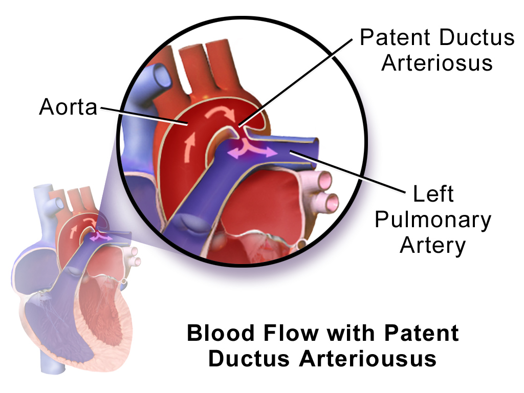
Image Source: Wikimedia Commons (2014). Blausen 0707 Patent Ductus Arteriousus, Licensed under a creative commons licence. Available for free at:
https://upload.wikimedia.org/wikipedia/commons/a/a0/Blausen_0707_PatentDuctusArteriosus.png?20140528155004 (Accessed 2 May 2025).
Patent Ductus Arteriosus (PDA) refers to the persistence of the ductus arteriosus after the first 48 hours of life. The ductus arteriosus is a foetal vascular structure that allows blood passage from the pulmonary artery to the aorta in utero, thus bypassing the foetal lungs. Therefore, PDA describes the CHD where the ductus arteriosus fails to close postnatally, allowing blood to flow from the aorta to the pulmonary artery. This is down a pressure gradient. The details of PDA are summarised in Table Five.
| Feature | Summary |
|---|---|
| Epidemiology | Incidence of 3-8 per 10,000 live births
2:1 female-to-male ratio May be present with other congenital heart defects It can present in some genetic syndromes, e.g. Noonan, Downs, DiGeorge |
| Aetiology | In utero, patency is maintained by hypoxaemia and circulating prostaglandins (PGE2 and PG12).
In the post-natal period, increased oxygen saturations and reduced circulating prostaglandin secondary to placental loss result in the closure of the ductus. The pathology of PDA is due to the left-to-right shunting of blood from the aorta to the pulmonary artery. |
| Clinical History | This varies depending on the age of the initial presentation and the size of the PDA.
An older child or adult can present with shortness of breath and exercise intolerance. |
| Clinical Examination | Small PDA
Moderate PDA
Large PDA
|
| Diagnostic Investigations | Echocardiogram – visualisation of the defect and shunt.
Chest X-ray – normal if PDA is small, slight cardiomegaly if moderate, left heart enlargement if large. Ecg – normal if PDA is small, biventricular hypertrophy and left atrial anomaly if large. |
| Management | In adults, closure is often warranted if there is sizeable left-to-right shunting and/or pulmonary arterial hypertension. Often percutaneous closure is utilised with prophylactic antibiotics for six months. However, closure is contraindicated if the pulmonary vascular resistance is too high. In some cases, surgical ligation is used in larger PDAs. |
| Follow Up | Patient monitoring is required until the definitive closure of the PDA is attained. There are several complications which include congestive cardiac failure, pulmonary hypertension, and endarteritis. |
Table 5: This table details the main features of patent ductus arteriosus. Sourced and adapted from Johnson et al. Patent Ductus Arteriosus: BMJ Best Practice 2022 and Doyle et al. Clinical manifestations and diagnosis of patent ductus arteriosus (PDA) in term infants, children, and adults.: UpToDate; 2022.
Complete Guide to Clinical Genetics ST3 Application and Recruitment (2027)
Written by Medibuddy. Last updated: October 2026. Clinical Genetics is

Complete Guide to Clinical Radiology ST3 Application and Recruitment (2027)
Recruitment paused: NHS England is not running Clinical Radiology ST3+ recru

Complete Guide to Paediatric Surgery ST3 Application and Recruitment in United Kingdom (2026)
Written by Medibuddy. Last updated: October 2026. If you’re planning


Take your subscriptions with you
Our mobile app allows you to access your interview and exam question banks wherever you are.

