


 Close
Close
 7th May 2025
Right-to-Left Shunt
Tetralogy of Fallot
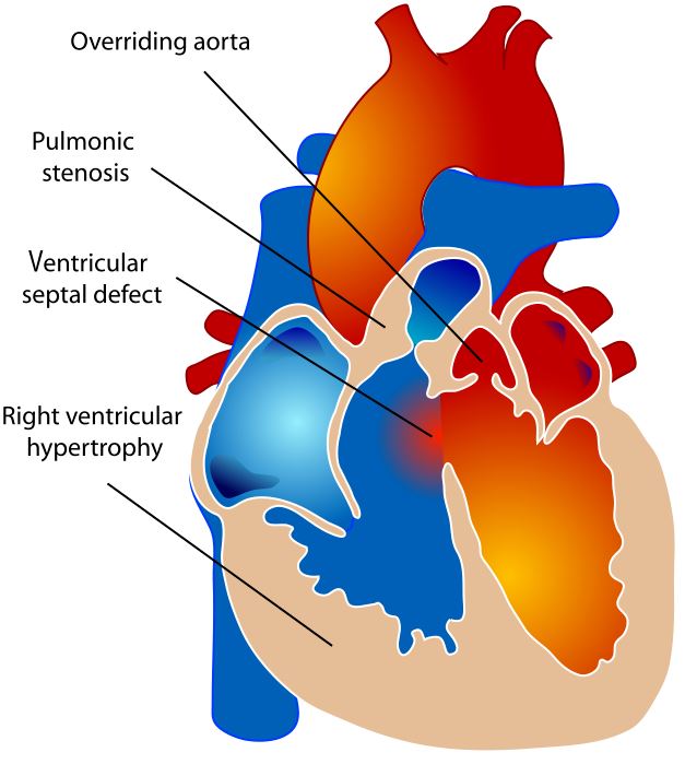
Image Source: Wikimedia Commons (2016). Tetralogy of Fallot, Licensed under a creative commons licence. Available for free at:
https://upload.wikimedia.org/wikipedia/commons/thumb/e/e6/Tetralogy_of_Fallot.svg/1228px-Tetralogy_of_Fallot.svg.png?20160125111000 (Accessed 2 May 2025).
Tetralogy of Fallot (TOF) refers to a cyanotic type of CHD in which four structural abnormalities coexist and most often present within the neonate. These are pulmonary stenosis (narrowing of the right ventricular outlet), right ventricular hypertrophy (due to increased pulmonary resistance), ventricular septal defect (shunt between both ventricles) and an overriding aorta (expansion of the aorta due to increased blood flow from both ventricles via the VSD). The main features are summarised in Table Seven.
| Feature | Summary |
|---|---|
| Epidemiology | 7-10 % of all CHD patients and is the most common cyanotic CHD. |
| Aetiology | Epigenetics
Genetic syndromes, e.g. Trisomy 21, 18 and 13, DiGeorge Maternal risk factors, e.g. diabetes, retinoic acid or trimethadione ingestion |
| Clinical History | Often in the newborn with cyanosis and a murmur noted on auscultation.
If present later, then increasing cyanosis, murmur and hypercyanotic episodes (crying with deep rapid breathing and increased cyanosis). |
| Clinical Examination | Variable, depending on the level of right ventricular outlet obstruction. But very often, a harsh systolic ejection murmur loudest at the left sternal edge, tachypnoea and variable degree of cyanosis. |
| Diagnostic Investigations | Pulse oximetry (pre and post-ductal readings show low oxygen saturations).
Echocardiogram (visualisation of pulmonary stenosis, ventricular septal defect, right ventricular hypertrophy and an overriding aorta). ECG (may show signs of right ventricular hypertrophy). Chest X-ray (“boot” shaped heart) Hyper-oxygenation Testing (no significant increase noted in PaO2) |
| Diagnostic Criteria | Evidence of the following on echocardiogram:
|
| Management | Acute Setting Hypercyanotic Episodes
Non-remitting severe cyanosis (due to severe pulmonary obstruction)
Severe cyanosis and limited pulmonary blood flow
Ongoing Complete Surgical Repair
Monitoring and potential pulmonary valve replacement
Antimicrobial Prophylaxis
|
| Follow Up | Lifelong Follow-Up, including those with surgical repair for complication surveillance.
Pre-conception counselling, including monitoring during pregnancy. Complications include progressive right ventricular failure and pulmonary regurgitation, arrhythmias (ventricular and atrial), sudden cardiac death and heart failure. After surgical repair, the prognosis is individual, based on the patient’s anatomy and the occurrence of any complications. Overall, long-term survival rates are improving. |
Table 7: This table details the main features of Tetralogy of Fallot. Sourced and adapted from Doyle T, Kavanaugh-McHugh A. Management and outcome of tetralogy of Fallot: UpToDate; 2023 and Gossett J, Kamp A. Tetralogy of Fallot: BMJ Best Practice; 2022.
Complete Guide to Clinical Genetics ST3 Application and Recruitment (2027)
Written by Medibuddy. Last updated: October 2026. Clinical Genetics is

Complete Guide to Clinical Radiology ST3 Application and Recruitment (2027)
Recruitment paused: NHS England is not running Clinical Radiology ST3+ recru

Complete Guide to Paediatric Surgery ST3 Application and Recruitment in United Kingdom (2026)
Written by Medibuddy. Last updated: October 2026. If you’re planning


Take your subscriptions with you
Our mobile app allows you to access your interview and exam question banks wherever you are.

