


 Close
Close
 5th June 2025
Connective Tissue Diseases and the Skin
Scleroderma (Systemic Sclerosis)
Classification
Scleroderma is a term that translates to hardened skin. However, the general term ‘scleroderma’ is often used for both systemic sclerosis (systemic scleroderma) and morphoea (localised scleroderma). Though the skin changes in the two conditions are virtually indistinguishable, on both a clinical and histopathological level, morphoea does not result in damage and fibrosis of the lungs, heart, kidneys and/or gastrointestinal tract.
Systemic sclerosis (SSc) is an autoimmune inflammatory condition that causes fibrosis, inflammation and vasculopathy and involves multiple systems, such as skin, lungs, heart, gastrointestinal tract and kidneys. There are two subtypes of systemic sclerosis according to distribution of skin involvement – limited cutaneous SSc (lcSSc) and diffuse cutaneous SSc (dcSSc).
Epidemiology
Different world populations have different prevalence, with higher rates in the US compared to Europe or Asia. The prevalence is 88 per million seen in England compared to 250 per million adults in the US. Women are more often affected, with a ratio of 5:1 female to male. SSc is rare in childhood, more common in young adults and has a peak incidence in patients’ 50s.
Aetiology
Aetiology is unknown but genetic, immunological and environmental factors have been suggested:
- Genetic: <5% of monozygotic and dizygotic twins are clinically concordant for systemic sclerosis; however, most monozygotic twins are concordant for antinuclear antibodies
- Immunology: 90% of patients have positive ANA on indirect immunofluorescence; this group of patients shows subsets of mutually exclusive auto-antibodies that are associated with distinct clinical phenotypes
- Environmental: exposure to silica dust and multiple solvents have been proposed, but no clear link has been identified
Pathophysiology
The precise mechanism is unknown, but many models have been proposed. At the start of the disease, the immune system, endothelial cells and fibroblasts are all activated. A sub-population of hyper-proliferative fibroblasts is selected for, which are confirmed to be over-represented in scleroderma. These cells produce high levels of collagen. Activated T-cells contribute to disease by producing pro-fibrotic cytokines and downregulating cytokines that inhibit growth or synthesis.
Diagnosis
LcSSc and dcSSc both show the same signs and symptoms, but the skin distribution and course of the disease is different. Two thirds of patients with scleroderma have dcSSc, which has widespread skin involvement including proximal limbs. This is often rapidly progressive, has significant internal organ involvement and has a worse prognosis. One third of patients with scleroderma have lcSSc, where the sclerosis is limited to the digits, face and distal limbs (does not spread further than elbows or knees). It has a slower progression and less organ involvement, but carries a risk of pulmonary artery hypertension. Up to 20% of patients have an overlap syndrome with another connective tissue disease.
Systemic features
- Gastrointestinal: dysmotility, gastroesophageal reflux (GORD), dysphagia, bloating, nausea and vomiting, alternating diarrhoea/constipation
- Cardiopulmonary: interstitial lung disease (ILD), pulmonary artery hypertension, cardiomyopathy
- Renal dysfunction: patients may present with abrupt onset of moderate/marked hypertension, which is a sign of scleroderma renal crisis
- Musculoskeletal: friction rubs over joints and tendons, synovitis, weakness and limited movement causing contractures
- Ophthalmological: tight eyelids, reduced tear secretion, retinopathy
- Fatigue, arthralgia, myalgia
- Sicca symptoms and Sjogren syndrome
Cutaneous features
- Skin sclerosis, commonly sclerodactyly (hardening of the skin causing the fingers to curl inward) or microstomia
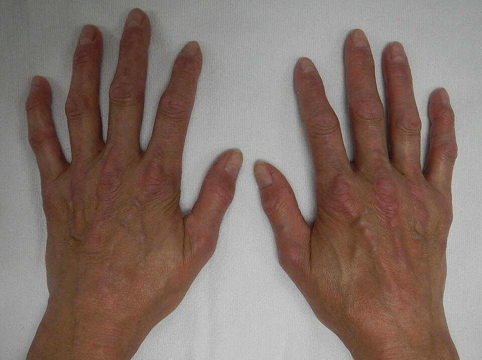 Image Source: Wikimedia Commons (2015). Crest 1, Licensed under a creative commons licence. Available for free at: https://commons.wikimedia.org/wiki/File:CREST1.JPG (Accessed 19 May 2025).
 - Puffy hands, often one of the first symptoms
- Raynaud’s phenomenon
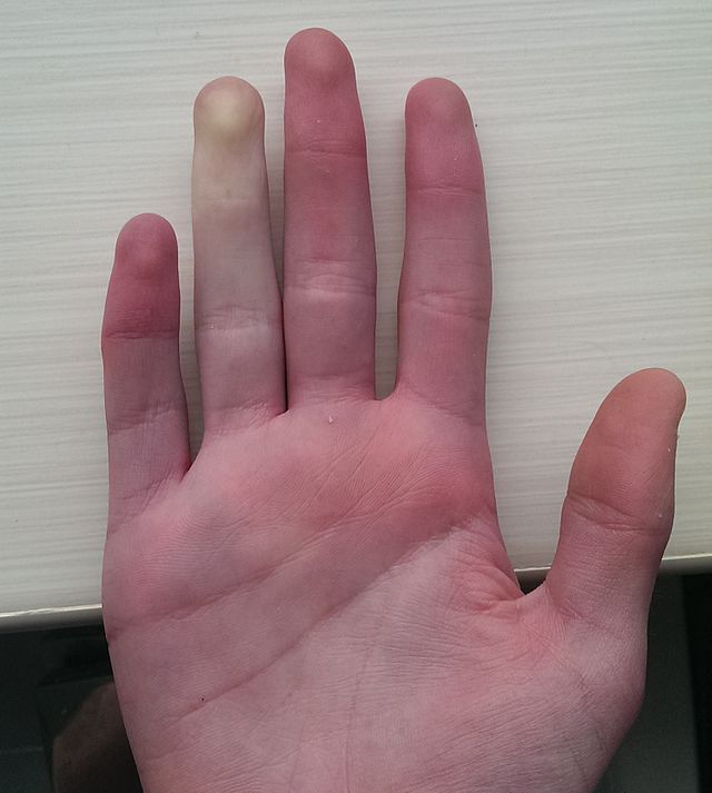 Image Source: Wikimedia Commons (2014). Raynaud Phenomenon, Licensed under a creative commons licence. Available for free at: https://commons.wikimedia.org/w/index.php?search=Raynaud%E2%80%99s+phenomenon&title=Special:MediaSearch&go=Go&type=image (Accessed 19 May 2025).
 - Digital pits and/or ulceration
- Abnormal nail fold capillaries
- Telangiectases – often located on the face, hands and mucous membranes
- Subcutaneous calcinosis – small, hard masses on pressure points (eg. Fingers, forearms)
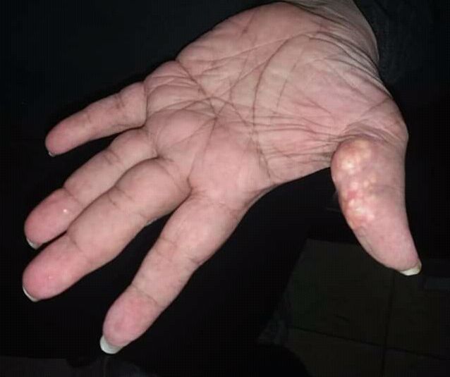 Image Source: Wikimedia Commons (2018). Calcinosis Cutis, Licensed under a creative commons licence. Available for free at: https://commons.wikimedia.org/wiki/File:Calcinosis_cutis_new_image.jpg (Accessed 19 May 2025).
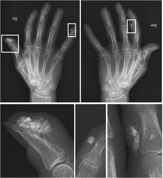 Image Source: Wikimedia Commons (2018). Calcinosis of Crest Syndrome, Licensed under a creative commons licence. Available for free at: https://commons.wikimedia.org/wiki/File:Calcinosis_of_CREST_syndrome.jpg (Accessed 19 May 2025).
 - Salt and pepper dyspigmentation – hyperpigmentation with vitiligo-like depigmentation
Though the previous name, CREST syndrome, of lcSSc is no longer used, it remains a useful mnemonic to remember the key features:
- Calcinosis
- Raynaud’s phenomenon
- (O)esophageal dysmotility
- Sclerodactyly
- Telangiectasia
Investigations
Initial investigations look at auto-antibodies, with over 90% of patients having a positive ANA. Within this group there are subsets of mutually exclusive antibodies, which are associated with distinct clinical pictures:
- Anti-topoisomerase I (anti-Scl70)
- Seen in 20% of patients
- Associated with an increased risk of ILD and diffuse skin involvement
- Anti-RNA polymerase III antibody
- Seen in 20% of patients
- Associated with renal crisis
- Anti-centromere antibodies
- Seen in 20-25% of patients
- Associated with limited skin involvement and overall better prognosis
- In the remaining 40% no specific auto-antibody has been identified yet
Further investigations to assess for systemic involvement are required. Pulmonary involvement can be investigated with pulmonary function tests and high-resolution CT of the lungs. Cardiac involvement requires ECG and echocardiogram (ECHO). If the ECHO finds elevated right ventricular systolic pressure, the patient should be referred for a right-heart catheterisation and full evaluation, as ECHO findings may not be indicative of the true pulmonary artery pressures. For gastrointestinal symptoms such as dysmotility and reflux, a barium swallow can be a useful test. Anaemia or melaena can indicate chronic GI blood loss from gastric antral vascular ectasia syndrome (GAVE) and required upper GI endoscopy. If inflammatory myositis develops, patients will require blood tests for serum muscle enzymes, possibly followed by a nerve conduction study and a muscle biopsy.
Complications
The greatest complications of systemic sclerosis are interstitial lung disease (ILD) and renal crisis. Development of ILD will need specialist referral and can ultimately require lung transplantation.
Severe and life-threatening renal disease occurs in 10-15% of patients and was the largest mortality cause before ACE inhibitors (ACEi) were introduced. Early dcSSc patients are at highest risk of developing a renal crisis. The signs of a renal crisis are onset of acute renal failure, abrupt onset of moderate or marked hypertension, urinalysis indicating proteinuria with few cells or casts and micro-angiopathic haemolytic anaemia. ACEi must be titrated to the maximal tolerated dose and if required, further anti-hypertensives can be added (usually calcium channel blockers).
Small pericardial effusions can occur when cardiac involvement is noted, which can progress to cardiac tamponade. Oral prednisolone is given for pericardial effusions, but strict monitoring is required, as corticosteroids can precipitate a renal crisis. Tamponade is managed with a pericardial window and systemic corticosteroids.
Patients should be monitored with regular ECHOs to look for the development of pulmonary hypertension, with referral for right heart catheterisation if needed.
Raynaud’s phenomenon can lead to digital ulceration and skin sclerosis can cause fixed contractures. Patients may develop gastric antral vascular ectasia (GAVE). Heartburn can lead to strictures and Barrett’s metaplasia and needs early intervention.
Management
Treatment options are according to the organ system affected. As patients with systemic sclerosis often have multiple systems affected, multi-modal therapies may be required. Involvement of multiple specialists may also be required to manage the complications, such as respiratory, renal and gastroenterology input.
Prognosis
DcSSc has a poorer prognosis compared to lcSSc, though both can develop significant visceral involvement as detailed above. Mean survival is approximately 12 years after diagnosis, but with a wide variation in disease severity. At the milder end of the disease, survival is close to the general population.
Dermatomyositis
Dermatomyositis is an idiopathic inflammatory autoimmune disorder with characteristic myopathy and cutaneous signs.
Epidemiology
Dermatomyositis is a rare disease that can occur at any age. Two peaks of incidence have been associated – between 5-15 years old and between 40-60 years old.
Data on incidence and prevalence is available from a US-population study. The estimated annual incidence of all subtypes is 9.63 per million people, with 13.98 per million in females and 4.68 per million in males. The overall prevalence in all subtypes was estimated at 21.42 in 100,000 people, with 33.09 in 100,000 females and 8.35 in 100,000 in males.
Juvenile-onset dermatomyositis has an annual incidence of 2-3 cases per million. Females are more commonly affected with the mean age of onset being 10 years old. Adult-onset dermatomyositis affects females more than males, with a ratio of 2.5:1. It is more commonly seen in the black population compared to the white population. Malignancy-associated dermatomyositis affects males and females equally but is more common in over 50s, with risk increasing with age.
Aetiology and Pathophysiology
The aetiology of dermatomyositis is unknown – the current opinion is that it is due to an interaction between environmental factors and genetic susceptibility.
- Genetic: a genetic predisposition can be inferred from the familial associations investigated
- Immunological: certain HLA subtypes appear to be connected to an increased risk of developing dermatomyositis
- Environmental: overall only weak associations found but the strongest evidence is available for ultraviolet radiation intensity
- Infection: some reports are available of patients with antibodies to Borrelia burgdorferi and Toxoplasma gondii developing myositis; other reports suggest this with viral antibodies, particles, nucleic acids
- Drug-induced: D-penicillamine, hydroxyurea, statins, phenylbutazone, terbinafine
The pathogenesis is unclear, but it is widely accepted to be an autoimmune condition.
Diagnosis
Clinical signs and symptoms
Muscle and skin signs develop simultaneously in approximately 60% of patients. In 30%, skin lesions precede muscle weakness, though the time in between can be up to several months. Between 10-20% of patients develop skin signs, but never develop any apparent muscle disease (known as amyopathic dermatomyositis). As it is considered an autoimmune disorder, dermatomyositis may also present with systemic manifestations.
Myopathy
This often manifests as a symmetrical weakness of the proximal muscles, though the severity varies between patients. Oftentimes, this manifests as difficulty carrying out certain activities – climbing stairs, rising from a sitting or crouching position, lifting objects, washing or combing hair, difficulty swallowing. Myalgia is present in less than 30% of patients. On examination, muscle tenderness is seen in 50% of patients. Muscle atrophy can be seen in long-standing disease, but is not common and so an alternative diagnosis should be considered in these cases.
Cutaneous signs
- Heliotrope rash – bilateral violaceous to erythematous discolouration of eyelids with swelling of the eyelids and surrounding skin; pathognomonic sign of dermatomyositis
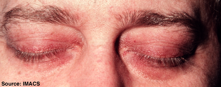 Image Source: Wikimedia Commons (2010). Dermatomyositis, Licensed under a creative commons licence. Available for free at: https://commons.wikimedia.org/wiki/File:Dermatomyositis10.jpg (Accessed 19 May 2025)
 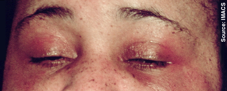 Image Source: Wikimedia Commons (2010). Dermatomyositis, Licensed under a creative commons licence. Available for free at: https://commons.wikimedia.org/wiki/File:Dermatomyositis11.jpg (Accessed 19 May 2025).
 - Shawl sign – fixed redness of the back, shoulders, chest and neck
- V-sign – fixed redness of the face, neck and upper chest
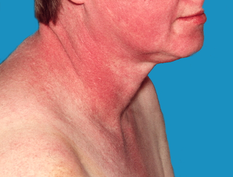 Image Source: Wikimedia Commons (2011). Dermatomyositis 18, Licensed under a creative commons licence. Available for free at: https://commons.wikimedia.org/wiki/File:Dermatomyositis18.jpg (Accessed 19 May 2025).
 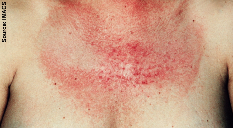 Image Source: Wikimedia Commons (2010). Dermatomyositis 17, Licensed under a creative commons licence. Available for free at: https://commons.wikimedia.org/wiki/File:Dermatomyositis17.jpg (Accessed 19 May 2025).
 - Photosensitivity
- Gottron’s papules – pathognomonic of dermatomyositis; violaceous papules and plaques on dorsal knuckles, and more rarely on wrists, elbows, knees, malleoli
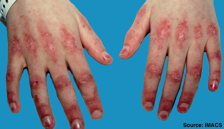 Image Source: Wikimedia Commons (2010). Stomach blood supply, Licensed under a creative commons licence. Available for free at: https://commons.wikimedia.org/wiki/File:Dermatomyositis2.jpg (Accessed 19 May 2025).
 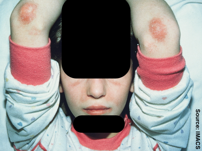 Image Source: Wikimedia Commons (2010). Dermatomyositis 4, Licensed under a creative commons licence. Available for free at: Https://commons.wikimedia.org/wiki/File:Dermatomyositis4.jpg (Accessed 19 May 2025).
 - Itchy scalp with inflammation and diffuse, non-scarring hair thinning
- Nailfold changes – periungal erythema, cuticular overgrowth (ragged cuticles), dilated capillaries of the nailfold
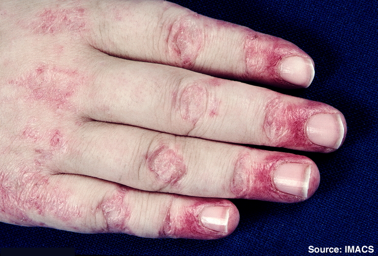 Image Source: Wikimedia Commons (2010). Dermatomyositis 7, Licensed under a creative commons licence. Available for free at: https://commons.wikimedia.org/wiki/File:Dermatomyositis7.jpg (Accessed 19 May 2025).
 - Mechanic’s hands – hyperkeratotic changes in hands seen in antisynthetase syndrome
- Holster sign – erythema over buttocks and lateral thighs
- Calcinosis cutis (calcium deposits)
Systemic manifestations
- Inflammatory polyarthritis – when present, has a similar distribution to rheumatoid arthritis
- Interstitial lung disease
- Cardiac involvement
- Raynaud’s phenomenon
Investigations
The following investigations should be considered in dermatomyositis:
- Serum creatinine kinase (CK) – the most sensitive muscle enzyme test
- Serum aldolase – glycolytic pathway enzyme found mainly in skeletal muscle, brain and liver; not as specific or sensitive as CK, but can be elevated in dermatomyositis when CK is normal
- Muscle biopsy – should be performed in all suspected dermatomyositis cases
- Electromyography (EMG) – recommended if raised CK and typical muscle biopsy findings are present
- Skin biopsy – required in patients that only present with skin manifestations to differentiate from other connective tissue diseases, biopsy appearance is similar to cutaneous lupus
- Antinuclear antibody (ANA) – positive in 80% of patients
- Anti-double-stranded DNA, anti-Ro, anti-La, anti-Sm, anti-ribonucleoprotein (RNP) – useful for differentiation from other connective tissue diseases
- Myositis-specific antibodies (MSAs) and myositis-associated antibodies (MAAs)
MSAs and MAAs are found in most dermatomyositis patients and are associated with different clinical subsets. They are not widely available, but can offer information regarding clinical courses and therapeutic responses.
- Anti-Mi-2 – directed against a helicase involved in transcriptional activation
- Strongly associated with the classical presentation of dermatomyositis, but found in only 10% of patients
- Anti-U1 ribonucleoprotein (RNP) and anti-Ku found in overlap syndromes that present similarly to anti-Mi-2 positive dermatomyositis
- Anti-transcription intermediary factor 1 gamma (TIF1-gamma)
- Most commonly isolated antibody
- Strongly associated with malignancy
- Anti-signal recognition particle (SRP)
- Found almost exclusively in polymyositis and only occasionally in dermatomyositis
- When present, associated with acute severe, treatment-resistant necrotising myositis
- Anti-Jo-1 and other anti-amioacyl-tRNA synthetase antibodies (including anti-PL-7, anti-PL-12, anti-EJ, anti-OJ, anti-KS) -> anti-synthetase syndrome
- Patients positive for the above present with a distinct clinical picture called anti-synthetase syndrome
- Main clinical features include interstitial lung disease, fever, myositis, polyarthritis, mechanic’s hands, Raynaud’s phenomenon
- Anti-Jo-1 associated with a higher frequency of myositis
- The other are associated with a higher frequency of interstitial lung disease
- Anti-melanoma differentiation-associated gene 5 (MDA5, also known as CADM-140) – specific to clinically amyopathic dermatomyositis
- Strongly associated with a rapidly progressive interstitial lung disease with poor prognosis
- May present with tender palm papules and diffuse alopecia
- Anti-NPX2
- Strongly associated with juvenile-onset dermatomyositis; a high proportion of these patients suffer from cutaneous calcinosis
- Anti-PM-Scl
- Found in patients with overlapping myositis and scleroderma
Management
Treatment depends on the presentation of dermatomyositis – though there can be some overlap in management, muscle and skin manifestations are treated with different agents. Dermatomyositis is considered to be the most responsive idiopathic inflammatory myopathy, but even with good control of myopathy, skin lesions may remain treatment-resistant.
In severe disease or an acute flare, intravenous corticosteroids are used in the short-term until better control is achieved and the patient can be switched to oral agents. Patients with anti-synthetase syndrome may be corticoid-resistant and may require concomitant immunosuppressives. If anti-SRP positive, patients often require immunosuppressives, IVIG or rituximab early in the disease due to the association with acute severe recalcitrant necrotising myositis. If the patient is MDA5 positive, which is associated with rapidly progressive ILD, they often also require high-dose systemic corticosteroids from an early stage.
Management of myopathy
- First-line: high doses of oral corticosteroids
- Second-line:
- Methotrexate – often preferred over azathioprine as it acts quicker; a low dose is useful for both muscle and skin involvement; effective corticosteroid-sparing agent
- Azathioprine – appears to take longer to have an effect (6 months or more)
- Third-line – can be used as a monotherapy or combined with corticosteroids or other immunosuppressants
- Ciclosporin – corticosteroid-sparing agent in some patients
- Tacrolimus and mycophenolate
- Cyclophosphamide – useful in refractory disease associated with vasculitis, ILD or respiratory muscle involvement
- Chlorambucil – successful in a few studies of refractory disease
- In some cases, the disease remains active regardless of any immunosuppressive therapy; Adjunctive IVIG in patients with significant muscle weakness has demonstrated some benefit in small studies and case reports
Management of skin manifestations
- First-line: topical therapy
- Patient education regarding adequate sun protection due to photosensitive nature of skin lesions
- Topical corticosteroids – beneficial even in patient who are already on systemic corticosteroids; help control erythema and pruritus
- Topical antipruritic treatments as adjuncts to topical corticosteroids – emollients with added menthol, phenol or camphor or topical antihistamines (e.g., Doxepin)
- If severe pruritus unresponsive, trial of oral antihistamine
- Second-line: antimalarials
- Hydroxychloroquine – only effective for skin lesions and scalp pruritus with no effect on muscle symptoms
- Chloroquine – risk of retinopathy greater than with hydroxychloroquine
- Topical tacrolimus – only anecdotal evidence exists for this agent
- Third-line: immunosuppressives
- Methotrexate – can be used in combination with topical treatment and antimalarials; useful for both muscle and skin manifestations
- Azathioprine – only anecdotal evidence exists of benefit in methotrexate-resistant dermatomyositis
- Mycophenolate – shown to be beneficial in recalcitrant skin disease
- Ciclosporin – beneficial in resistant skin disease and early ILD; contra-indicated in renal impairment and hypertension
- Anti-inflammatory agents such as thalidomide and dapsone are alternatives to immunosuppressives – case reports and anecdotal evidence exist
- Adjunctive IVIG can be used in refractory skin involvement.
Prognosis
Classic dermatomyositis has a 5-year survival of 95% and 10-year survival of 84%. Despite improved survival rates with newer treatments, one third of patients will suffer from permanent disability. Classic dermatomyositis has a relapsing-remitting course and patients will require long-term treatment.
Amyopathic dermatomyositis have a good response to treatment of myopathy, but cutaneous disease is often more resistant to treatment. Follow-up is required to check for any myopathy, but prognosis is overall good.
Poor prognostic factors include
- Delay in treatment of more than 6 months after symptom onset
- Presenting with greater muscle weakness
- Respiratory muscle involvement
- ILD
- Dysphagia
- Malignancy
- Cardiac involvement
Lupus
Lupus erythematosus (LE) encompasses a group of auto-inflammatory conditions. Systemic lupus erythematosus (SLE) is a generalised disorder that can affect any body system. Cutaneous lupus, or skin involvement, is seen in 80% of patients with SLE.
Epidemiology
Incidence and prevalence have been difficult to assess worldwide – studies have mainly focused on white populations in small geographical areas. The incidence in the UK is estimated to be 4.91 per 100,000 person-years, with the highest reported incidence seen in North America (23.2 per 100,000 person-years). Incidence is higher in women compared to men, with reported ratios ranging from 2:1 to 15:1. Peak age of onset is 30-70 for women and 50-70 for men. Prevalence of SLE in the US is estimated to be 72.8 per 100,000 person-years. This prevalence estimate is 9 times higher among women compared to men. There is also a racial and ethnic component, with the highest prevalence seen in African-Americans (230.9 per 100,000 in women, 26.7 per 100,000 in men), followed by Hispanics (120.7 per 100,000 in women, 18 per 100,000 in men), and lowest in white populations (84.7 per 100,000 in women, 8.9 per 100,000 in men). Global data appears to confirm these differences noted in the US data.
Aetiology
The aetiology of SLE is not understood, but it is generally agreed that an environmental factor affecting a genetically susceptible individual is fundamental in its pathogenesis. The high female preponderance also suggests hormonal factors may play a role.
The importance of genetic factors is confirmed by familial aggregation and higher-than-expected rates of concordance in twin studies, with the heritability of SLE estimated at 43.9%. Genome-wide association studies have identified more than 60 genetic risk loci, with the one most consistently associated with SLE shown below:
- IRF5 (interferon regulatory factor 5) codes a transcription factor involved in the regulation of pro-inflammatory cytokines
- STAT4 (signal transducer and activator of transcription 4) acts as a transcription activator and helps mediate responses to interleukin-12 in lymphocytes and regulate T helper cell differentiation
- BANK1 (B-cell scaffold protein with ankyrin repeats 1) codes for a B-cell-specific scaffold protein and may contribute to SLE by altering B-cell signalling
- ITGAM (integrin alpha M) – polymorphism of this gene has a significant association with SLE in multiple ethnic populations
- PTPN22 (protein tyrosine phosphatase, non-receptor type 22) inhibits T-cell receptor signalling and selectively promotes type I interferon responses; a missense single-nucleotide polymorphism of this is associated with an increased risk for SLE
Related environmental factors may be non-infectious or infectious.
- Infectious agents include Epstein-Barr virus (EBV), parvovirus B19, cytomegalovirus (CMV) and human immunodeficiency virus 1 (HIV1).
- Non-infectious agents can include exposure to sunlight, drugs, hormones, toxins such as cigarette smoke or stress and emotional upset.
- Drugs can either be an environmental factor triggering a flare or unmasking of latent LE, or can specifically cause drug-induced lupus erythematosus, which is a lupus-like syndrome different from idiopathic LE.
Drug-induced LE has a time relationship with the causative drug, with symptoms beginning after starting treatment with the causative drug. However, the latent time between starting the medication and the symptoms beginning can range between 1 month and 10 years, which can make it very difficult to establish a causative relationship. Unlike idiopathic LE, drug-induced LE resolves once the causative agent is stopped. Some of the drugs commonly associated with LE are procainamide, minocycline, terbinafine, sulfasalazine, isoniazid, phenytoin and carbamazepine.
Pathophysiology
SLE is an auto-immune disease where high-affinity immunoglobulin G antibodies target double-stranded DNA as well as nuclear proteins. When there is rapid clearance of cells via apoptosis, the nuclear antigens do not have time to be exposed to the immune system and recognised as native. Failure of this process, along with other processes that ensure immune tolerance for nuclear antigens, results in antinuclear antibodies being produced. The complement system is activated, causing inflammation and tissue injury.
Diagnosis
Systemic signs
SLE is a systemic disease so presenting signs and symptoms may involve a range of organ systems.
- Constitutional: fatigue, fever and weight loss are common symptoms; unexplained fever is common and characteristic of SLE
- Lymphadenopathy: peripheral lymphadenopathy more often regional than generalised; nodes are usually non-tender, vary in size from a few mm to 3-4cm, in cervical and axillary regions; histology of a lymph node frequently shows reactive hyperplasia
- Mucocutaneous signs and symptoms are covered in more detail below, commonly seen
- Musculoskeletal: joint pain symptoms are common; correctable ulnar deviation and joint subluxations in the hands in the absence of radiological damage are characteristic; may also develop myositis leading to muscle weakness and pain
- Raynaud’s phenomenon: can develop secondary to SLE; recent onset of triphasic colour change (white – ischaemia, blue – deoxygenation and red – reperfusion) in hands and feet in a young woman due to exposure to cold or emotional stress should prompt further investigation into SLE.
- Renal: involvement of this system is seen in 50-70% of patients, though more common in male patients; this can manifest as lupus nephritis (more common in Hispanic and black patients), glomerulonephritis (more common in patients positive for anti-dsDNA antibodies), hypertension, nephrotic syndrome, renal failure; most patients are asymptomatic
- Central nervous system (CNS): major CNS involvement is uncommon; when it is seen, it usually manifests as seizures, cranial nerve abnormalities (visual field defects, blindness, papilloedema, nystagmus, ptosis, facial palsy) and psychiatric illnesses (psychosis, depressive disorders, organic brain syndromes); cerebral infarcts may occur secondary to co-existing positive antiphospholipid antibodies
- Cardiac: pericarditis, myocarditis, endocarditis, arterial and venous thrombosis, premature atherosclerotic coronary artery disease; the risk of cardiovascular events such as MI and stroke is 2-3x higher in SLE patients
- Pulmonary: pleuritis, pleural effusions (usually unilateral and exudative), diffuse interstitial lung disease, pulmonary hypertension, rarely pulmonary haemorrhage; pleural embolisms may occur particularly if antiphospholipid antibodies are positive; shrinking lung syndrome is a rare respiratory manifestation characterised by dyspnoea, chest pain, a raised hemidiaphragm and a restrictive pattern on pulmonary function tests
- Haematological: anaemia, leukopenia and thrombocytopenia are commonly seen; anaemia is often due to chronic disease and improves as disease activity becomes controlled; haemolytic anaemia can rarely occur but it is severe when it presents
- Gastrointestinal tract: oral ulcers are common; oesophageal hypomobility can less commonly cause dysphagia; lupus peritonitis or mesenteric artery occlusion can cause abdominal pain, vomiting and diarrhoea; pancreatitis may occur due to SLE; chronic active hepatitis can occur
- Serositis: pleuritis and pericarditis is more common than peritonitis
Mucocutaneous signs
Skin involvement is the first sign of SLE in 25% of patients but up to 80% will develop cutaneous LE. Skin involvement can present as LE-specific or LE-non-specific manifestations. LE-specific lesions are aggravated by UV exposure and are localised in sun-exposed sites such as the face, neck, upper chest and upper back. Cutaneous lupus (CLE) has specific acute, subacute, intermittent and chronic manifestations. Typically, SLE will present with acute CLE whereas only 5% of patients with chronic CLE have SLE, due to only the skin being involved.
Acute CLE: typically, SLE patients present with acute CLE manifestations
- Malar or ‘butterfly’ face rash – violaceous erythema extending from the cheeks over the bridge of the nose and sparing the nasolabial folds; usually lasts for a few days and resolves without scarring; often recurs after sun exposure; seen in 30-40% of patients and may be more common in females
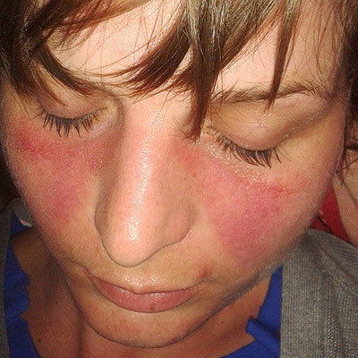 Image Source: Wikimedia Commons (2013). Lupus, Licensed under a creative commons licence. Available for free at: https://commons.wikimedia.org/wiki/File:Lupusfoto.jpg (Accessed 19 May 2025).
 - Bullous systemic lupus erythematosus – presenting sign in one third of SLE patients;
- Tense vesicles, bullae and erosions on a background of normal or erythematous skin usually in a sun-exposed area (often the face is the area affected);
- Blisters heal without scarring and the pruritus is absent or mild;
- Often associated with a flare of lupus nephritis
- Three types which can only be distinguished by immunohistochemistry; type I is associated with auto-antibodies to type VII collagen
- Treatment of choice is dapsone, which leads to a rapid clinical improvement and this also differentiates bullous SLE from SLE with a blistering rash
- Maculopapular rash resembling a morbilliform drug eruption – morbilliform rash can be seen in SLE
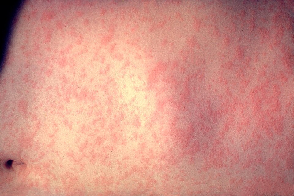 Image Source: Wikimedia Commons (2008). Morbillivirus Measles Infection, Licensed under a creative commons licence. Available for free at: https://commons.wikimedia.org/wiki/File:Morbillivirus_measles_infection.jpg (Accessed 19 May 2025).
 - Mucosal erosions and ulcerations (mouth, genitals, lips, nose) – mouth ulcers, and less commonly nose ulcers, occur in up to 45% of patients; usually large and painless
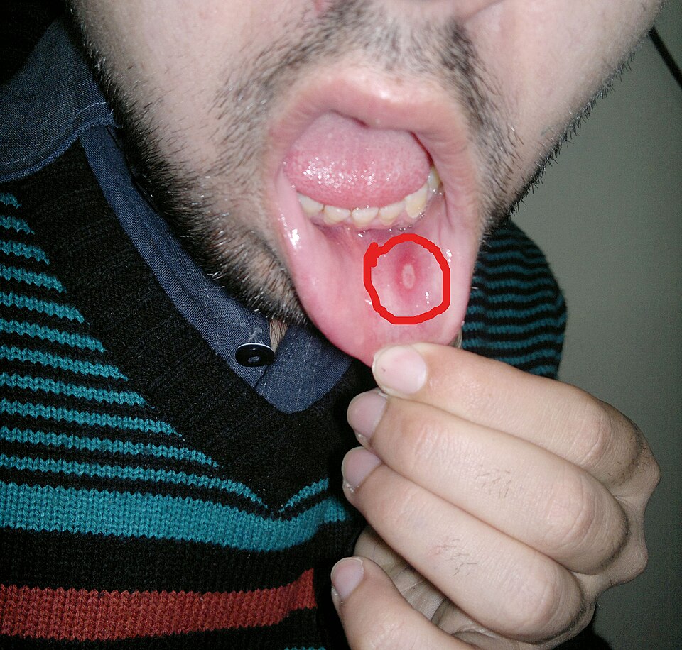 Image Source: Wikimedia Commons (2014). Aphthous Stomatitis on the Labial Mucosa, Licensed under a creative commons licence. Available for free at: https://commons.wikimedia.org/wiki/File:Aphthous_stomatitis_on_the_labial_mucosa.jpg (Accessed 19 May 2025).
 - Photosensitivity, which can be mild or severe
- Diffuse hair loss (nonscarring alopecia) with brittle hair shafts
Subacute CLE: less commonly associated with SLE (50% have a mild form of SLE); 20-40% are thought to have drug-induced subacute CLE; comprises 10-15% of CLE presentations
- Psoriasiform papulosquamous rash on upper back, chest and upper arms
- Annular, polycyclic plaques with central clearing
- Lesions seen on trunk and arms resolve to leave dyspigmentation and telangiectases, but no scarring
- Signs precipitated or aggravated by sun exposure
Intermittent (lupus tumidus)
- Affects sun-exposed areas – chest, back, upper arms, face
- Erythematous patches and plaques with a smooth surface, with round or annular shapes
- Signs resolve with no scarring left in the winter
Chronic CLE: affects 25% of SLE patients
- Classic discoid lupus is the most common manifestation – scaly plaques with follicular prominence (carpet tack sign) that cause post-inflammatory dyspigmentation and/or scarring
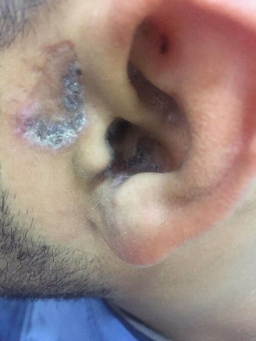 Image Source: Wikimedia Commons (2018). Stomach blood supply, Licensed under a creative commons licence. Available for free at: https://commons.wikimedia.org/wiki/File:Shuster_sign_of_discoid_lupus_ertherymatouses.jpg (Accessed 19 May 2025).
 - Hypertrophic (warty) lupus – variant of discoid LE with thickened warty plaques resembling viral warts
- Mucosal lupus – plaques, ulcers and scaling of the lips, nose, mouth and sometimes genitals
- Lupus profundus/panniculitis – inflammation of the subcutaneous without vasculitis; presents as firm, deep, tender nodules which leave dents in the skin
- Chilblain lupus – rare variant involving mainly the fingers and toes of smokers; triggered by exposure to a moist cold environment
Non-specific cutaneous SLE – features related to underlying illness rather than an autoimmune attack; can occur in other connective tissue or autoimmune diseases
- Nail fold capillary telangiectasia
- Raynaud phenomenon
- Diffuse hair thinning
- Cutaneous vasculitis – palpable purpura or urticarial vasculitis
- Livedo reticularis – in 20-30% of SLE patients
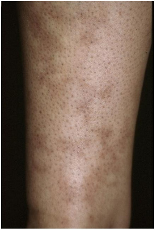 Image Source: Wikimedia Commons (2012). Livedo Reticularis of an Arm, Licensed under a creative commons licence. Available for free at: https://commons.wikimedia.org/wiki/File:Livedo_reticularis_of_an_arm.png (Accessed 19 May 2025).
 - Papular mucinosis (mucin deposits)
- Calinosis cutis (calcium deposits)
Investigations
Initial investigations should include:
- Antinuclear antibody (ANA) – a positive ANA does not diagnose SLE, but a diagnosis of SLE requires a positive ANA
- Anti-Ro antibody – rarely, ANA can be negative in anti-Ro-antibody positive lupus
- Double-stranded DNA, Smith antigen – highly specific for SLE and often confirmatory of diagnosis if present
- Full blood count – often leukopenia caused by neutropenia, anaemia
- Clotting screen – activated partial thromboplastin time may be prolonged if antiphospholipid antibodies present
- Kidney function and urinalysis – deranged if renal system affected
- ESR and CRP – elevated due to systemic inflammation
Subsequent investigations which can be considered:
- Coombs’ test to assess for haemolysis
- Complement levels – often raised in SLE
- Antiphospholipid antibodies
- Skin or renal biopsy
Management
Treatment is divided into preventative measures, topical therapy and systemic therapy.
Preventative measures to minimise flares and organ damage include:
- Diligent minimisation of sun exposure with clothes, accessories, sunscreen
- Smoking cessation
- Psychological interventions
Topical therapy includes potent topical corticosteroids and calcineurin inhibitors (tacrolimus ointment and pimecrolimus cream).
Systemic therapy choice depends on which organ systems are most affected. The medications generally used are as follows:
- NSAIDs – first line for musculoskeletal stiffness or pain
- Hydroxychloroquine – recommended in all SLE patients; response rates are about 80% in CLE; risk of retinal toxicity
- Corticosteroids – mainstay of treatment in the ill SLE patient; can give intravenous methylprednisolone for immediate therapeutic effect before switching to oral
- Immunosuppressive agents include methotrexate, azathioprine, mycophenolate
- Biological agents – B-cell targeting agents such as belimumab and rituximab
Prognosis
SLE often presents as flares with periods of remission. In some patients, all signs of active disease resolve. The 5-year survival is 95% and 10-year survival is 92%, with the most common causes of mortality being cardiovascular disease, infection and severe disease activity.
Rheumatoid arthritis
Rheumatoid arthritis (RA) is a chronic autoimmune systemic inflammatory disorder that most often symmetrically affects the small joints of the hands and feet.
Epidemiology
RA is a common condition, affecting about 1% of the UK population. It is 2-4 times more common in women compared to men and the peak age of onset is 30-50 years old with incidence increasing with age. The peak age of incidence in the UK for both men and women is around 70 years of age. Extra-articular involvement is seen in 40% of cases and skin changes are seen in 30%.
Aetiology
The aetiology of RA is multifactorial with no clear cause found. RA is an autoimmune condition with antibodies seen against rheumatoid factor or anti-citrullinated protein antibodies. It is also associated with genetic factors, most notably HLA-DR4 which may be involved in the pathogenesis. Some genetic variants of PTPN22 and other genes have been identified as risk factors. It has also been proposed that an infection may be the trigger RA in a susceptible individual, but no specific causative factor has been identified.
Pathophysiology
Central to the pathogenesis is the formation of inflamed synovium in joints. Increased inflammation in the synovium causes the synovial lining to become hyperplastic with immune cell infiltration in the sublining. Increased cytokines are seen, with tumour necrosis factor (TNF), interleukin-1 (IL-1) and interleukin-6 (IL-6) most abundantly seen. Angiogenesis serves the hypertrophic synovium. The inflammatory setting, along with increased levels of metalloproteinase activity is thought to drive the joint destruction.
Diagnosis
Clinical features
Patients often present with symmetrical synovitis of the small joints of the hands and feet, though any joint can be affected. This consists of pain often worse at rest, swelling of the joint, heat and early morning stiffness lasting over 1 hour. Extra-articular changes are often skin-related and can be numerous:
- Atrophic, dry, friable skin
- Raynaud phenomenon
- Nail changes e.g., onycholysis, nail ridging and splitting, clubbing
- Rheumatoid nodules – only seen in RA
- solitary or multiple skin-coloured nodules on sites of recurrent pressure or trauma, generally asymptomatic
- Rheumatoid vasculitis – only seen in RA
- An unusual complication of long-standing severe rheumatoid arthritis involving small to medium-sized vessels
- Can present as palpable purpura, nailfold infarcts, digital necrosis, ulcers, and urticarial vasculitis
- A specific manifestation of this vasculitis is a Bywaters lesion – painless purpuric papules of <1mm diameter found on the fingertip or around the nail
- Rheumatoid neutrophilic dermatitis – only seen in RA
- Rare severe skin manifestation of severe long-standing RA
- Symmetrical asymptomatic eruption of erythematous papules, nodules, and plaques which may show an annular distribution at the extremities
- Associated with a disease flare and resolves with treatment
There are three classical deformities seen in RA, though they are no longer commonly seen in presenting patients due to the advent of disease-modifying anti-rheumatic drugs (DMARDs).
- Swan neck deformity – DIP hyperflexion, PIP hyperextension
- Boutonniere deformity – PIP flexion, DIP hyperextension
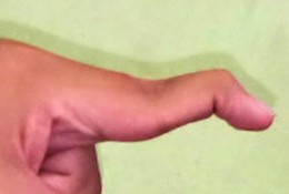 Image Source: Wikimedia Commons (2021). Swan Neck Deformation Finger, Licensed under a creative commons licence. Available for free at: https://commons.wikimedia.org/wiki/File:Swan_neck_deformation_finger.jpg (Accessed 19 May 2025).
 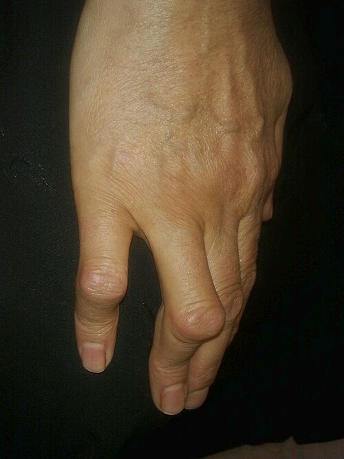 Image Source: Wikimedia Commons (2013). Boutonniare Deformity, Licensed under a creative commons licence. Available for free at: https://commons.wikimedia.org/wiki/File:Boutonni%C3%A8re_deformity.jpg (Accesed 19th May 2025)
 - Ulnar deviation – inflammation of MCP joints causing finger joint dislocation and deviation to the ulnar side
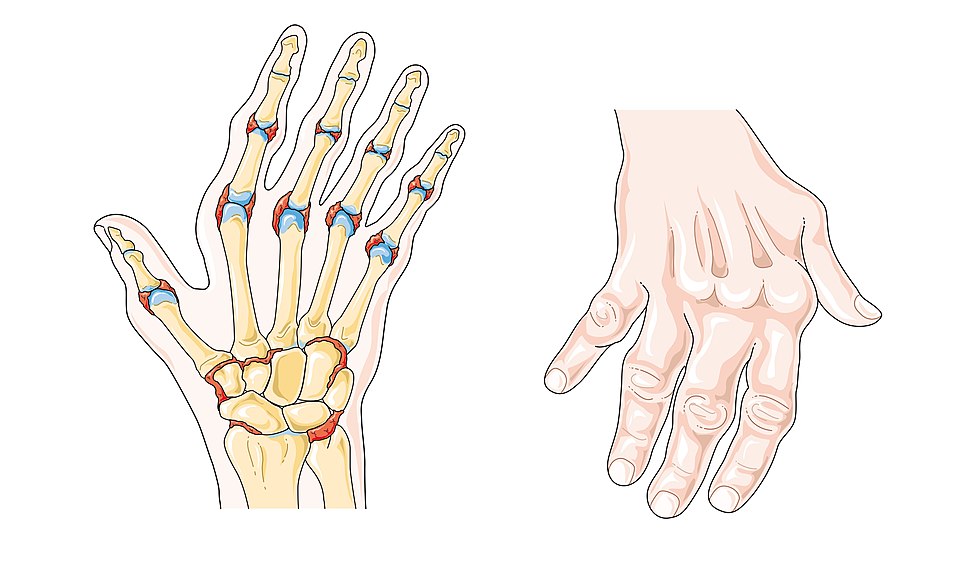 Image Source: Wikimedia Commons (2019). Rheumatoid Arthritis – Smart Servier, Licensed under a creative commons licence. Available for free at: https://commons.wikimedia.org/wiki/File:Rheumatoid_arthritis_–_Smart-Servier_(cropped).jpg (Accessed 19 May 2025).
Investigations
Diagnosis is clinical however investigations can speed up the process. The first line investigations include:
- Rheumatoid factor (RF) – positive in 60-70% of RA patients, found in 90% of patients with rheumatoid nodules
- Anti-cyclic citrullinated peptide (anti-CCP) antibodies – found in 80% of RA patients
- X-rays of hands and feet – can aid diagnosis but often helpful to monitor progress of disease
- Ultrasonography – may detect synovitis of the wrist and fingers at the initial presentation
All people suspected to have RA should have a rheumatology appointment within 3 weeks to confirm this. If the small joints of the hands and feet are affected, more than one joint is affected or there has been a delay of 3 months or more between onset of symptoms and patient presenting, a referral should be made within 3 working days.
Complications
- Joint destruction causing work disability and requiring joint replacement surgeries
- Shortened life span – 8-10 years on average
- Increased risk for coronary artery disease
- Interstitial lung disease (ILD)
- Felty syndrome – defines as the presence of RA, splenomegaly, and an abnormally low white blood cell count; seen in less than 1% of patients
- Carpal tunnel syndrome – seen in 10-20%
- Secondary to immunosuppressive effects of treatment – infection, malignancy, methotrexate-related liver toxicity
Management
Management recommendations depend on the disease severity at presentation.
For mild disease at initial presentation:
- 1st line: DMARD monotherapy
- 1st option: Hydroxychloroquine
- 2nd options: Sulfasalazine, Methotrexate or Leflunomide
- Consider corticosteroids and NSAIDs in acute flares
For moderate-to-severe disease at initial presentation.
- 1st line: Methotrexate monotherapy
- 2nd options: Sulfasalazine, Hydroxychloroquine, Leflunomide
- 2nd line: Add biological agent or targeted DMARD
- Biological agent options: etanercept, infliximab, adalimumab, certolizumab pegol, golimumab, abatacept, rituximab, tocilizumab, sarilumab
- DMARD options: tofacitinib, baricitinib, upadacitinib, filgotinib
- Consider corticosteroids and NSAIDs in acute flares
For pregnant or planning pregnancy patients:
- 1st line: Corticosteroids
- 2nd line: Hydroxychloroquine or Sulfasalazine
Ehlers-Danlos syndrome
Epidemiology
The Ehlers-Danlos syndromes (EDS) are a group of 13 inherited connective tissue disorders. They are estimated to affect between 1 in 5,000 and 1 in 100,000, but this is believed to be an underestimate. The commonest subtype is hypermobile EDS (hEDS), followed by classical EDS (cEDS), with the other 11 subtypes being very rare.
Aetiology
EDS is caused by genetic abnormalities in connective tissue proteins and/or their metabolism, such as collagen, fibrillin and/or other matrix proteins. The specific gene and protein affected depends on the EDS subtype and can be seen in the table below.
Classification
The 2017 international classification recognises 13 subtypes:
| EDS Subtype | Autosomal recessive or dominant | Gene affected | Resulting protein affected |
|---|---|---|---|
| 1. Classical (cEDS) | Autosomal dominant | COL5A1, COL5A2,
COL1A |
Collagen Type V Collagen Type I |
| 2. Classical-like (clEDS) | Autosomal recessive | TNXB | Tenascin XB |
| 3. Cardiac-valvular (cvEDS) | Autosomal recessive | COL1A2 | Collagen Type I |
| 4. Vascular (vEDS) | Autosomal dominant | COL3A
COL1A1 |
Collagen Type III
Collagen Type I |
| 5. Hypermobile (hEDS) | Autosomal dominant | unknown | |
| 6. Arthrochalasia (aEDS) | Autosomal dominant | COL1A1, COL1A2 | Collagen Type 1 |
| 7. Dermatosparaxis (dEDS) | Autosomal recessive | ADAMTS2 | ADAMTS-2 |
| 8. Kyphoscoliotic (kEDS) | Autosomal recessive | PLOD1
FKBP14 |
LH1
FKBP22 |
| 9. Brittle cornea syndrome (BCS) | Autosomal recessive | PRDM5
ZNF469 |
PRDM5
ZNF469 |
| 10. Spondylodysplastic (spEDS) | Autosomal recessive | B4GALT7
B3GALT6 SLC39A13 |
beta4GalT7
beta3GalT6 ZIP13 |
| 11. Musculocontractural (mcEDS) | Autosomal recessive | CHST14
DSE |
D4ST1
DSE |
| 12. Myopathic (mEDS) | Autosomal dominant/recessive | COL12A1 | Collagen Type XII |
| 13. Periodontal (pEDS) | Autosomal dominant | C1R
C1S |
C1r
C1s |
Table 1: Summary of ICD Classification of Ehlers-Danlos Syndrome subtypes
Clinical features & Pathophysiology
The specific signs and symptoms and their severity depends on the EDS subtype and also on the individual – some individuals are asymptomatic or only develop minor symptoms, whereas others can experience significant complications. The features generally seen in all subtypes are related to the connective tissue abnormality – ligament laxity leads to hypermobility, resulting fragility of tissues leads to easy bruising and impaired healing can be seen with atrophic scars.
Other clinical features include:
- Translucent skin with underlying vessels and/or tendons easily visible
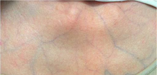 Image Source: Wikimedia Commons (2013). Stomach blood supply, Licensed under a creative commons licence. Available for free at: https://commons.wikimedia.org/wiki/File:PMC3567970_1752-1947-7-35-1_(cropped).png (Accessed 19 May 2025).
 - Soft skin with a velvet feel
- Hyperelastic skin
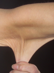 Image Source: Wikimedia Commons (2012). Hyperelastic Skin in a case of Ehlers-Danlos Syndrome, Licensed under a creative commons licence. Available for free at: https://commons.wikimedia.org/wiki/File:Hyperelastic_skin_in_a_case_of_Ehlers-Danlos_syndrome_(cropped).png (Accessed 19 May 2025).
 - Atrophic scars
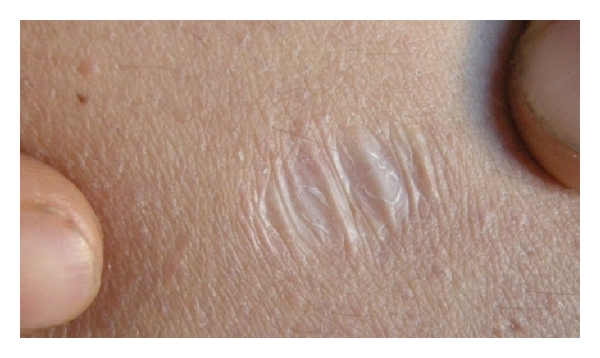 Image Source: Wikimedia Commons (2012). Atrophic Scar of Ehlers-Danlos Syndrome, Licensed under a creative commons licence. Available for free at: https://commons.wikimedia.org/wiki/File:Atrophic_scar_of_Ehlers_Danlos_syndrome.jpg (Accessed 19 May 2025).
 - Molluscoid pseudotumours, fleshy calcified lesions that form at pressure points
- Subcutaneous spheroids, former fat cells that lose their blood supply and become calcified, seen on forearms and shins
- Easy bruising
- Generalised hypermobility
- Recurrent joint dislocation
- Hypotonia
- Congenital or early-onset kyphoscoliosis
- Congenital talipes equinovarus (clubfoot), feet point downwards and inwards
- Arachnodactyly
- Thin cornea
- Autonomic dysfunction resulting in palpitations, postural hypotension, dizziness, chest pain, shortness of breath
- Motor delay in infancy – once walking initiated, motor delay resolves
- Chronic pain
- Fatigue
- History of delayed onset to local anaesthetic – seen in two thirds of patients
- Presence of hernias
- Non-specific GI signs
- Pneumothorax
- Heart murmur
Diagnosis
EDS is initially diagnosed clinically – musculoskeletal and skin manifestations are the primary features, with only joint hypermobility seen across all subtypes. A full skin examination may reveal some of the above-mentioned features. Hypermobility is assessed using the Beighton 9-point scale (see Table () below) – a score of 5 or more suggests generalised hypermobility. There are major and minor criteria defined for all subtypes in order to achieve a clinical diagnosis. This can then be followed by genetic testing for 11 out of the 12 subtypes that have known related genes. Hypermobile EDS, despite being the most common, has an unknown aetiology.
| Joint | Finding | Points |
| Fifth/little finger (left) | With the forearm and palm of the hand resting on a flat surface, dorsiflexion MCP >90 degrees | 1 |
| Dorsiflexion MCP </= 90 degrees | 0 | |
| Fifth/little finger (right) | With the forearm and palm of the hand resting on a flat surface, dorsiflexion of MCP >90 degrees | 1 |
| Dorsiflexion MCP </= 90 degrees | 0 | |
| Left thumb | With the arm straightened, hands pronated, wrist flexed, the thumb can dorsiflexed to touch the forearm | 1 |
| Thumb cannot be dorsiflexed to tough the forearm | 0 | |
| Right thumb | With the arm straightened, hands pronated, wrist flexed, the thumb can dorsiflexed to touch the forearm | 1 |
| Thumb cannot be dorsiflexed to tough the forearm | 0 | |
| Left elbow | With the arm straightened and hand supinated, elbow hyperextends more than 10 degrees | 1 |
| Elbow does not hyperextend beyond 10 degrees | 0 | |
| Right elbow | With the arm straightened and hand supinated, elbow hyperextends more than 10 degrees | 1 |
| Elbow does not hyperextend beyond 10 degrees | 0 | |
| Left knee | While standing with knees locked, the knee hyperextends more than 10 degrees | 1 |
| The knee does not hyperextend beyond 10 degrees | 0 | |
| Right knee | While standing with knees locked, the knee hyperextends more than 10 degrees | 1 |
| The knee does not hyperextend beyond 10 degrees | 0 | |
| Spine | Patient can bend forwards and touch the ground with palms lying flat without bending knees | 1 |
| Patient cannot touch floor with palms without bending knees | 0 | |
Table 2: Summary of Beighton Score 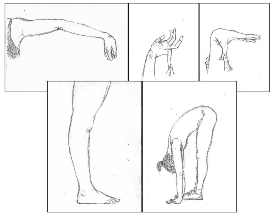 Image Source: Wikimedia Commons (2017). Ehlers-Danlos Skala Beighton, Licensed under a creative commons licence. Available for free at: https://commons.wikimedia.org/wiki/File:Ehlers-Danlos_skala_Beighton%27a.png (Accessed 19 May 2025).
Complications
Complications can occur with surgeries due to difficulty with suturing in friable skin and due to easy rupture of blood vessels or organ tissue. It is advised that they are avoided as much as possible. Pregnancy complications such as increase in pain, premature delivery, difficulties with tear and episiotomy healing, issues with anaesthetic resistance, uterine prolapse can occur. In addition, vascular EDS is associated with a shortened lifespan with a mean survival age of 51. This subtype carries a high risk of arterial dissection, organ rupture and obstetric complications.
Management
EDS management depends on the subtype and the individual. Asymptomatic patients do not require any specific treatment. Generally, advice is centred on preventative management – EDS patients are advised to avoid contact sports or physically demanding jobs, and to engage with regular exercise to minimise risk of injury. All patients should have genetic counselling and be educated with regards to the inheritance of the specific subtype.
In terms of specific symptoms, all systems can be affected in EDS. Pain management can be tricky and can sometimes require an MDT approach. Pain and joint instability can be aided by physiotherapy and occupational therapy. Splints or orthotics may be required for non-responding joints. Autonomic dysfunction and weakness of supporting structures in cardiology, neurology, ophthalmology, gastroenterology, surgery and/or gynaecology will need specialist referral.
References
BMJ Best Practice: https://bestpractice.bmj.com/topics/en-gb/570
DermNet: https://dermnetnz.org/topics/ehlers-danlos-syndrome
BMJ Best Practice Systemic sclerosis (scleroderma): https://bestpractice.bmj.com/topics/en-gb/295?q=Systemic%20sclerosis%20(scleroderma)&c=suggested
DermNet Systemic sclerosis: https://dermnetnz.org/topics/systemic-sclerosis
DermNet Scleroderma: https://dermnetnz.org/topics/scleroderma
BMJ Best Practice: https://bestpractice.bmj.com/topics/en-gb/105?q=Rheumatoid%20arthritis&c=suggested
NICE CKS: https://cks.nice.org.uk/topics/rheumatoid-arthritis/
DermNet: https://dermnetnz.org/topics/rheumatoid-arthritis-and-the-skin
DermNet Systemic lupus erythematosus: https://dermnetnz.org/topics/systemic-lupus-erythematosus
DermNet Cutaneous lupus erythematosus: https://dermnetnz.org/topics/cutaneous-lupus-erythematosus
DermNet Drug-induced lupus erythematosus: https://dermnetnz.org/topics/drug-induced-lupus-erythematosus
DermNet Bullous systemic lupus erythematosus: https://dermnetnz.org/topics/bullous-systemic-lupus-erythematosus
BMJ Best Practice Systemic lupus erythematosus: https://bestpractice.bmj.com/topics/en-gb/103/prognosis
Dermatology Made Easy by Amanda Oakley
BMJ Best Practice: Dermatomyositis: https://bestpractice.bmj.com/topics/en-gb/595?q=Dermatomyositis&c=suggested
DermNet Adult-onset dermatomyositis: https://dermnetnz.org/topics/adult-onset-dermatomyositis
Complete Guide to Clinical Genetics ST3 Application and Recruitment (2027)
Written by Medibuddy. Last updated: October 2026. Clinical Genetics is

Complete Guide to Clinical Radiology ST3 Application and Recruitment (2027)
Recruitment paused: NHS England is not running Clinical Radiology ST3+ recru

Complete Guide to Paediatric Surgery ST3 Application and Recruitment in United Kingdom (2026)
Written by Medibuddy. Last updated: October 2026. If you’re planning


Take your subscriptions with you
Our mobile app allows you to access your interview and exam question banks wherever you are.

