


 Close
Close
 5th June 2025
Diagnosis
Clinical signs and symptoms
Muscle and skin signs develop simultaneously in approximately 60% of patients. In 30%, skin lesions precede muscle weakness, though the time in between can be up to several months. Between 10-20% of patients develop skin signs, but never develop any apparent muscle disease (known as amyopathic dermatomyositis). As it is considered an autoimmune disorder, dermatomyositis may also present with systemic manifestations.
Myopathy
This often manifests as a symmetrical weakness of the proximal muscles, though the severity varies between patients. Oftentimes, this manifests as difficulty carrying out certain activities – climbing stairs, rising from a sitting or crouching position, lifting objects, washing or combing hair, difficulty swallowing. Myalgia is present in less than 30% of patients. On examination, muscle tenderness is seen in 50% of patients. Muscle atrophy can be seen in long-standing disease, but is not common and so an alternative diagnosis should be considered in these cases.
Cutaneous signs
- Heliotrope rash – bilateral violaceous to erythematous discolouration of eyelids with swelling of the eyelids and surrounding skin; pathognomonic sign of dermatomyositis
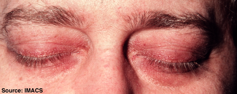 Image Source: Wikimedia Commons (2010). Dermatomyositis, Licensed under a creative commons licence. Available for free at: https://commons.wikimedia.org/wiki/File:Dermatomyositis10.jpg (Accessed 19 May 2025)
 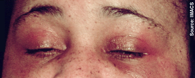 Image Source: Wikimedia Commons (2010). Dermatomyositis, Licensed under a creative commons licence. Available for free at: https://commons.wikimedia.org/wiki/File:Dermatomyositis11.jpg (Accessed 19 May 2025).
 - Shawl sign – fixed redness of the back, shoulders, chest and neck
- V-sign – fixed redness of the face, neck and upper chest
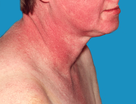 Image Source: Wikimedia Commons (2011). Dermatomyositis 18, Licensed under a creative commons licence. Available for free at: https://commons.wikimedia.org/wiki/File:Dermatomyositis18.jpg (Accessed 19 May 2025).
 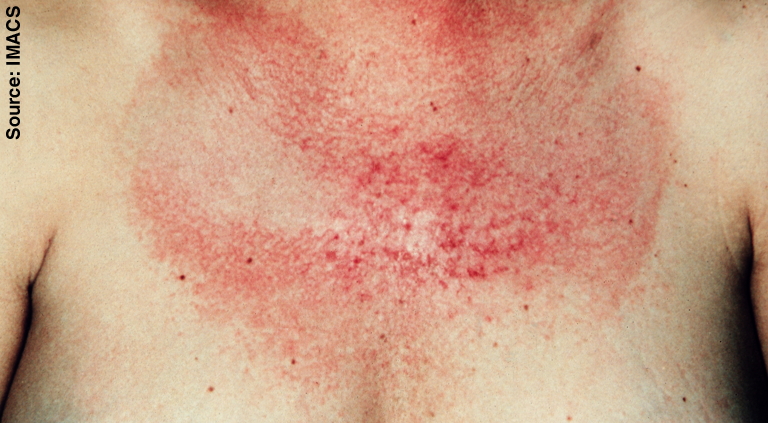 Image Source: Wikimedia Commons (2010). Dermatomyositis 17, Licensed under a creative commons licence. Available for free at: https://commons.wikimedia.org/wiki/File:Dermatomyositis17.jpg (Accessed 19 May 2025).
 - Photosensitivity
- Gottron’s papules – pathognomonic of dermatomyositis; violaceous papules and plaques on dorsal knuckles, and more rarely on wrists, elbows, knees, malleoli
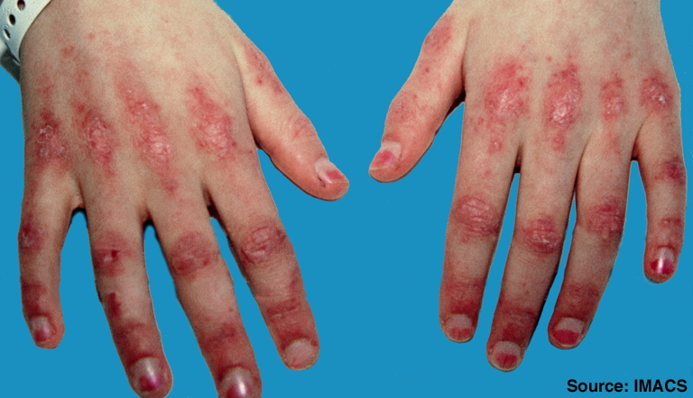 Image Source: Wikimedia Commons (2010). Stomach blood supply, Licensed under a creative commons licence. Available for free at: https://commons.wikimedia.org/wiki/File:Dermatomyositis2.jpg (Accessed 19 May 2025).
 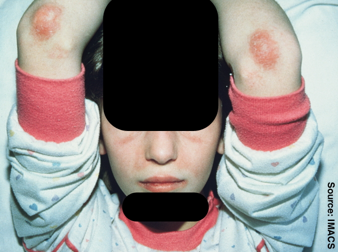 Image Source: Wikimedia Commons (2010). Dermatomyositis 4, Licensed under a creative commons licence. Available for free at: Https://commons.wikimedia.org/wiki/File:Dermatomyositis4.jpg (Accessed 19 May 2025).
 - Itchy scalp with inflammation and diffuse, non-scarring hair thinning
- Nailfold changes – periungal erythema, cuticular overgrowth (ragged cuticles), dilated capillaries of the nailfold
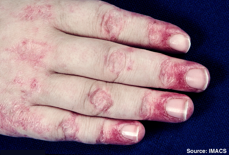 Image Source: Wikimedia Commons (2010). Dermatomyositis 7, Licensed under a creative commons licence. Available for free at: https://commons.wikimedia.org/wiki/File:Dermatomyositis7.jpg (Accessed 19 May 2025).
 - Mechanic’s hands – hyperkeratotic changes in hands seen in antisynthetase syndrome
- Holster sign – erythema over buttocks and lateral thighs
- Calcinosis cutis (calcium deposits)
Systemic manifestations
- Inflammatory polyarthritis – when present, has a similar distribution to rheumatoid arthritis
- Interstitial lung disease
- Cardiac involvement
- Raynaud’s phenomenon
Complete Guide to Clinical Genetics ST3 Application and Recruitment (2027)
Written by Medibuddy. Last updated: October 2026. Clinical Genetics is

Complete Guide to Clinical Radiology ST3 Application and Recruitment (2027)
Recruitment paused: NHS England is not running Clinical Radiology ST3+ recru

Complete Guide to Paediatric Surgery ST3 Application and Recruitment in United Kingdom (2026)
Written by Medibuddy. Last updated: October 2026. If you’re planning


Take your subscriptions with you
Our mobile app allows you to access your interview and exam question banks wherever you are.

