


 Close
Close
 5th June 2025
Ehlers-Danlos syndrome
Epidemiology
The Ehlers-Danlos syndromes (EDS) are a group of 13 inherited connective tissue disorders. They are estimated to affect between 1 in 5,000 and 1 in 100,000, but this is believed to be an underestimate. The commonest subtype is hypermobile EDS (hEDS), followed by classical EDS (cEDS), with the other 11 subtypes being very rare.
Aetiology
EDS is caused by genetic abnormalities in connective tissue proteins and/or their metabolism, such as collagen, fibrillin and/or other matrix proteins. The specific gene and protein affected depends on the EDS subtype and can be seen in the table below.
Classification
The 2017 international classification recognises 13 subtypes:
| EDS Subtype | Autosomal recessive or dominant | Gene affected | Resulting protein affected |
|---|---|---|---|
| 1. Classical (cEDS) | Autosomal dominant | COL5A1, COL5A2,
COL1A |
Collagen Type V Collagen Type I |
| 2. Classical-like (clEDS) | Autosomal recessive | TNXB | Tenascin XB |
| 3. Cardiac-valvular (cvEDS) | Autosomal recessive | COL1A2 | Collagen Type I |
| 4. Vascular (vEDS) | Autosomal dominant | COL3A
COL1A1 |
Collagen Type III
Collagen Type I |
| 5. Hypermobile (hEDS) | Autosomal dominant | unknown | |
| 6. Arthrochalasia (aEDS) | Autosomal dominant | COL1A1, COL1A2 | Collagen Type 1 |
| 7. Dermatosparaxis (dEDS) | Autosomal recessive | ADAMTS2 | ADAMTS-2 |
| 8. Kyphoscoliotic (kEDS) | Autosomal recessive | PLOD1
FKBP14 |
LH1
FKBP22 |
| 9. Brittle cornea syndrome (BCS) | Autosomal recessive | PRDM5
ZNF469 |
PRDM5
ZNF469 |
| 10. Spondylodysplastic (spEDS) | Autosomal recessive | B4GALT7
B3GALT6 SLC39A13 |
beta4GalT7
beta3GalT6 ZIP13 |
| 11. Musculocontractural (mcEDS) | Autosomal recessive | CHST14
DSE |
D4ST1
DSE |
| 12. Myopathic (mEDS) | Autosomal dominant/recessive | COL12A1 | Collagen Type XII |
| 13. Periodontal (pEDS) | Autosomal dominant | C1R
C1S |
C1r
C1s |
Table 1: Summary of ICD Classification of Ehlers-Danlos Syndrome subtypes
Clinical features & Pathophysiology
The specific signs and symptoms and their severity depends on the EDS subtype and also on the individual – some individuals are asymptomatic or only develop minor symptoms, whereas others can experience significant complications. The features generally seen in all subtypes are related to the connective tissue abnormality – ligament laxity leads to hypermobility, resulting fragility of tissues leads to easy bruising and impaired healing can be seen with atrophic scars.
Other clinical features include:
- Translucent skin with underlying vessels and/or tendons easily visible
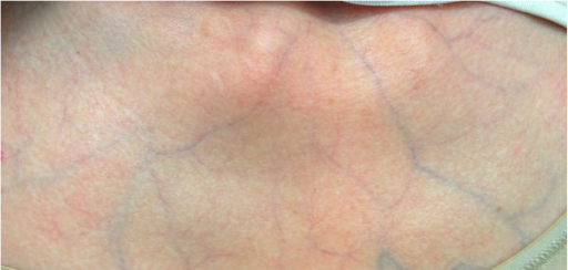 Image Source: Wikimedia Commons (2013). Stomach blood supply, Licensed under a creative commons licence. Available for free at: https://commons.wikimedia.org/wiki/File:PMC3567970_1752-1947-7-35-1_(cropped).png (Accessed 19 May 2025).
 - Soft skin with a velvet feel
- Hyperelastic skin
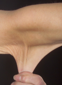 Image Source: Wikimedia Commons (2012). Hyperelastic Skin in a case of Ehlers-Danlos Syndrome, Licensed under a creative commons licence. Available for free at: https://commons.wikimedia.org/wiki/File:Hyperelastic_skin_in_a_case_of_Ehlers-Danlos_syndrome_(cropped).png (Accessed 19 May 2025).
 - Atrophic scars
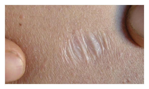 Image Source: Wikimedia Commons (2012). Atrophic Scar of Ehlers-Danlos Syndrome, Licensed under a creative commons licence. Available for free at: https://commons.wikimedia.org/wiki/File:Atrophic_scar_of_Ehlers_Danlos_syndrome.jpg (Accessed 19 May 2025).
 - Molluscoid pseudotumours, fleshy calcified lesions that form at pressure points
- Subcutaneous spheroids, former fat cells that lose their blood supply and become calcified, seen on forearms and shins
- Easy bruising
- Generalised hypermobility
- Recurrent joint dislocation
- Hypotonia
- Congenital or early-onset kyphoscoliosis
- Congenital talipes equinovarus (clubfoot), feet point downwards and inwards
- Arachnodactyly
- Thin cornea
- Autonomic dysfunction resulting in palpitations, postural hypotension, dizziness, chest pain, shortness of breath
- Motor delay in infancy – once walking initiated, motor delay resolves
- Chronic pain
- Fatigue
- History of delayed onset to local anaesthetic – seen in two thirds of patients
- Presence of hernias
- Non-specific GI signs
- Pneumothorax
- Heart murmur
Diagnosis
EDS is initially diagnosed clinically – musculoskeletal and skin manifestations are the primary features, with only joint hypermobility seen across all subtypes. A full skin examination may reveal some of the above-mentioned features. Hypermobility is assessed using the Beighton 9-point scale (see Table () below) – a score of 5 or more suggests generalised hypermobility. There are major and minor criteria defined for all subtypes in order to achieve a clinical diagnosis. This can then be followed by genetic testing for 11 out of the 12 subtypes that have known related genes. Hypermobile EDS, despite being the most common, has an unknown aetiology.
| Joint | Finding | Points |
| Fifth/little finger (left) | With the forearm and palm of the hand resting on a flat surface, dorsiflexion MCP >90 degrees | 1 |
| Dorsiflexion MCP </= 90 degrees | 0 | |
| Fifth/little finger (right) | With the forearm and palm of the hand resting on a flat surface, dorsiflexion of MCP >90 degrees | 1 |
| Dorsiflexion MCP </= 90 degrees | 0 | |
| Left thumb | With the arm straightened, hands pronated, wrist flexed, the thumb can dorsiflexed to touch the forearm | 1 |
| Thumb cannot be dorsiflexed to tough the forearm | 0 | |
| Right thumb | With the arm straightened, hands pronated, wrist flexed, the thumb can dorsiflexed to touch the forearm | 1 |
| Thumb cannot be dorsiflexed to tough the forearm | 0 | |
| Left elbow | With the arm straightened and hand supinated, elbow hyperextends more than 10 degrees | 1 |
| Elbow does not hyperextend beyond 10 degrees | 0 | |
| Right elbow | With the arm straightened and hand supinated, elbow hyperextends more than 10 degrees | 1 |
| Elbow does not hyperextend beyond 10 degrees | 0 | |
| Left knee | While standing with knees locked, the knee hyperextends more than 10 degrees | 1 |
| The knee does not hyperextend beyond 10 degrees | 0 | |
| Right knee | While standing with knees locked, the knee hyperextends more than 10 degrees | 1 |
| The knee does not hyperextend beyond 10 degrees | 0 | |
| Spine | Patient can bend forwards and touch the ground with palms lying flat without bending knees | 1 |
| Patient cannot touch floor with palms without bending knees | 0 | |
Table 2: Summary of Beighton Score
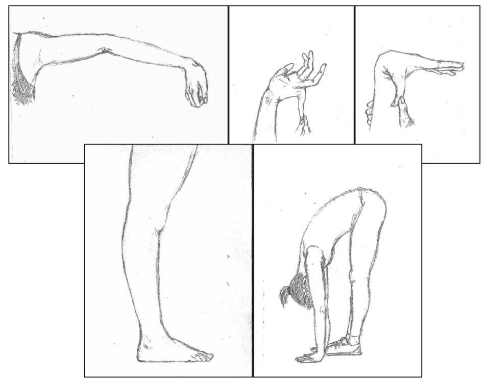 Image Source: Wikimedia Commons (2017). Ehlers-Danlos Skala Beighton, Licensed under a creative commons licence. Available for free at: https://commons.wikimedia.org/wiki/File:Ehlers-Danlos_skala_Beighton%27a.png (Accessed 19 May 2025).
Complications
Complications can occur with surgeries due to difficulty with suturing in friable skin and due to easy rupture of blood vessels or organ tissue. It is advised that they are avoided as much as possible. Pregnancy complications such as increase in pain, premature delivery, difficulties with tear and episiotomy healing, issues with anaesthetic resistance, uterine prolapse can occur. In addition, vascular EDS is associated with a shortened lifespan with a mean survival age of 51. This subtype carries a high risk of arterial dissection, organ rupture and obstetric complications.
Management
EDS management depends on the subtype and the individual. Asymptomatic patients do not require any specific treatment. Generally, advice is centred on preventative management – EDS patients are advised to avoid contact sports or physically demanding jobs, and to engage with regular exercise to minimise risk of injury. All patients should have genetic counselling and be educated with regards to the inheritance of the specific subtype.
In terms of specific symptoms, all systems can be affected in EDS. Pain management can be tricky and can sometimes require an MDT approach. Pain and joint instability can be aided by physiotherapy and occupational therapy. Splints or orthotics may be required for non-responding joints. Autonomic dysfunction and weakness of supporting structures in cardiology, neurology, ophthalmology, gastroenterology, surgery and/or gynaecology will need specialist referral.
Complete Guide to Clinical Genetics ST3 Application and Recruitment (2027)
Written by Medibuddy. Last updated: October 2026. Clinical Genetics is

Complete Guide to Clinical Radiology ST3 Application and Recruitment (2027)
Recruitment paused: NHS England is not running Clinical Radiology ST3+ recru

Complete Guide to Paediatric Surgery ST3 Application and Recruitment in United Kingdom (2026)
Written by Medibuddy. Last updated: October 2026. If you’re planning


Take your subscriptions with you
Our mobile app allows you to access your interview and exam question banks wherever you are.

