


 Close
Close
 5th June 2025
Diagnosis
Systemic signs
SLE is a systemic disease so presenting signs and symptoms may involve a range of organ systems.
- Constitutional: fatigue, fever and weight loss are common symptoms; unexplained fever is common and characteristic of SLE
- Lymphadenopathy: peripheral lymphadenopathy more often regional than generalised; nodes are usually non-tender, vary in size from a few mm to 3-4cm, in cervical and axillary regions; histology of a lymph node frequently shows reactive hyperplasia
- Mucocutaneous signs and symptoms are covered in more detail below, commonly seen
- Musculoskeletal: joint pain symptoms are common; correctable ulnar deviation and joint subluxations in the hands in the absence of radiological damage are characteristic; may also develop myositis leading to muscle weakness and pain
- Raynaud’s phenomenon: can develop secondary to SLE; recent onset of triphasic colour change (white – ischaemia, blue – deoxygenation and red – reperfusion) in hands and feet in a young woman due to exposure to cold or emotional stress should prompt further investigation into SLE.
- Renal: involvement of this system is seen in 50-70% of patients, though more common in male patients; this can manifest as lupus nephritis (more common in Hispanic and black patients), glomerulonephritis (more common in patients positive for anti-dsDNA antibodies), hypertension, nephrotic syndrome, renal failure; most patients are asymptomatic
- Central nervous system (CNS): major CNS involvement is uncommon; when it is seen, it usually manifests as seizures, cranial nerve abnormalities (visual field defects, blindness, papilloedema, nystagmus, ptosis, facial palsy) and psychiatric illnesses (psychosis, depressive disorders, organic brain syndromes); cerebral infarcts may occur secondary to co-existing positive antiphospholipid antibodies
- Cardiac: pericarditis, myocarditis, endocarditis, arterial and venous thrombosis, premature atherosclerotic coronary artery disease; the risk of cardiovascular events such as MI and stroke is 2-3x higher in SLE patients
- Pulmonary: pleuritis, pleural effusions (usually unilateral and exudative), diffuse interstitial lung disease, pulmonary hypertension, rarely pulmonary haemorrhage; pleural embolisms may occur particularly if antiphospholipid antibodies are positive; shrinking lung syndrome is a rare respiratory manifestation characterised by dyspnoea, chest pain, a raised hemidiaphragm and a restrictive pattern on pulmonary function tests
- Haematological: anaemia, leukopenia and thrombocytopenia are commonly seen; anaemia is often due to chronic disease and improves as disease activity becomes controlled; haemolytic anaemia can rarely occur but it is severe when it presents
- Gastrointestinal tract: oral ulcers are common; oesophageal hypomobility can less commonly cause dysphagia; lupus peritonitis or mesenteric artery occlusion can cause abdominal pain, vomiting and diarrhoea; pancreatitis may occur due to SLE; chronic active hepatitis can occur
- Serositis: pleuritis and pericarditis is more common than peritonitis
Mucocutaneous signs
Skin involvement is the first sign of SLE in 25% of patients but up to 80% will develop cutaneous LE. Skin involvement can present as LE-specific or LE-non-specific manifestations. LE-specific lesions are aggravated by UV exposure and are localised in sun-exposed sites such as the face, neck, upper chest and upper back. Cutaneous lupus (CLE) has specific acute, subacute, intermittent and chronic manifestations. Typically, SLE will present with acute CLE whereas only 5% of patients with chronic CLE have SLE, due to only the skin being involved.
Acute CLE: typically, SLE patients present with acute CLE manifestations
- Malar or ‘butterfly’ face rash – violaceous erythema extending from the cheeks over the bridge of the nose and sparing the nasolabial folds; usually lasts for a few days and resolves without scarring; often recurs after sun exposure; seen in 30-40% of patients and may be more common in females
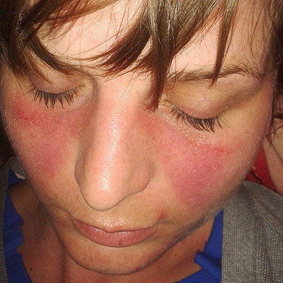 Image Source: Wikimedia Commons (2013). Lupus, Licensed under a creative commons licence. Available for free at: https://commons.wikimedia.org/wiki/File:Lupusfoto.jpg (Accessed 19 May 2025).
 - Bullous systemic lupus erythematosus – presenting sign in one third of SLE patients;
- Tense vesicles, bullae and erosions on a background of normal or erythematous skin usually in a sun-exposed area (often the face is the area affected);
- Blisters heal without scarring and the pruritus is absent or mild;
- Often associated with a flare of lupus nephritis
- Three types which can only be distinguished by immunohistochemistry; type I is associated with auto-antibodies to type VII collagen
- Treatment of choice is dapsone, which leads to a rapid clinical improvement and this also differentiates bullous SLE from SLE with a blistering rash
- Maculopapular rash resembling a morbilliform drug eruption – morbilliform rash can be seen in SLE
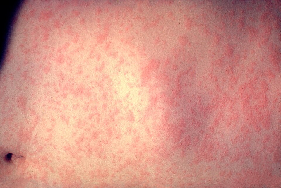 Image Source: Wikimedia Commons (2008). Morbillivirus Measles Infection, Licensed under a creative commons licence. Available for free at: https://commons.wikimedia.org/wiki/File:Morbillivirus_measles_infection.jpg (Accessed 19 May 2025).
 - Mucosal erosions and ulcerations (mouth, genitals, lips, nose) – mouth ulcers, and less commonly nose ulcers, occur in up to 45% of patients; usually large and painless
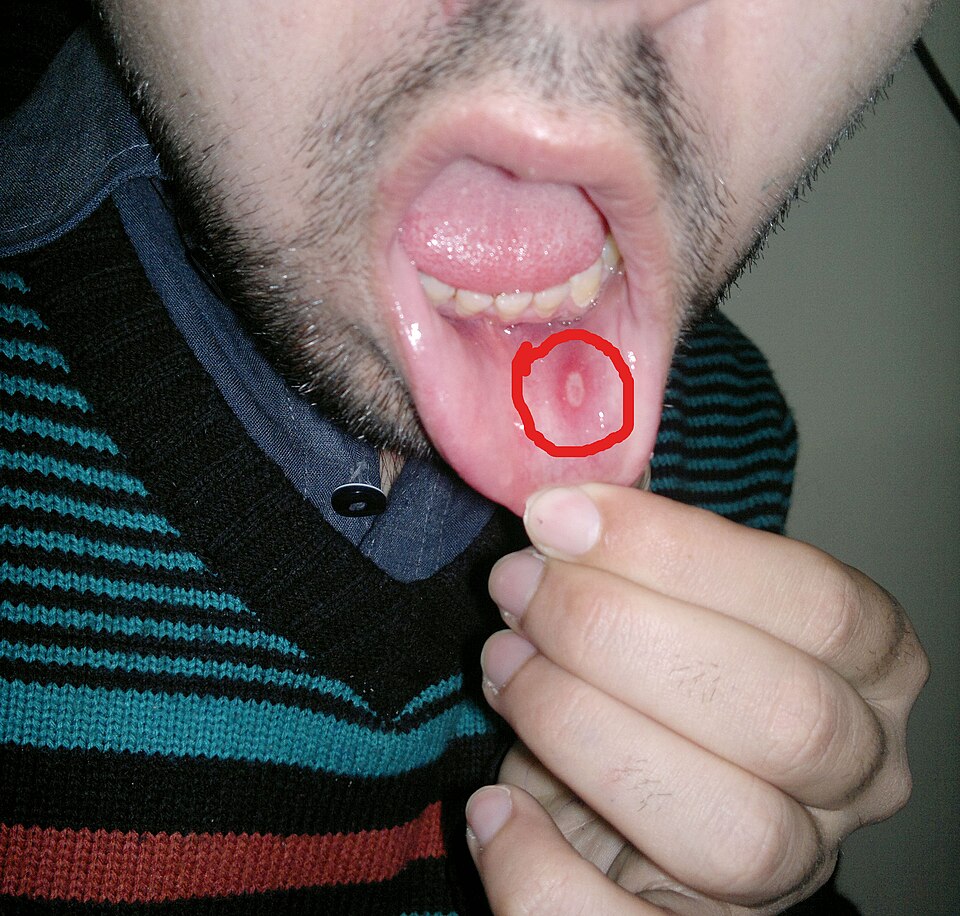 Image Source: Wikimedia Commons (2014). Aphthous Stomatitis on the Labial Mucosa, Licensed under a creative commons licence. Available for free at: https://commons.wikimedia.org/wiki/File:Aphthous_stomatitis_on_the_labial_mucosa.jpg (Accessed 19 May 2025).
 - Photosensitivity, which can be mild or severe
- Diffuse hair loss (nonscarring alopecia) with brittle hair shafts
Subacute CLE: less commonly associated with SLE (50% have a mild form of SLE); 20-40% are thought to have drug-induced subacute CLE; comprises 10-15% of CLE presentations
- Psoriasiform papulosquamous rash on upper back, chest and upper arms
- Annular, polycyclic plaques with central clearing
- Lesions seen on trunk and arms resolve to leave dyspigmentation and telangiectases, but no scarring
- Signs precipitated or aggravated by sun exposure
Intermittent (lupus tumidus)
- Affects sun-exposed areas – chest, back, upper arms, face
- Erythematous patches and plaques with a smooth surface, with round or annular shapes
- Signs resolve with no scarring left in the winter
Chronic CLE: affects 25% of SLE patients
- Classic discoid lupus is the most common manifestation – scaly plaques with follicular prominence (carpet tack sign) that cause post-inflammatory dyspigmentation and/or scarring
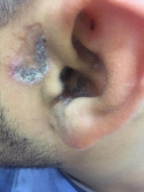 Image Source: Wikimedia Commons (2018). Stomach blood supply, Licensed under a creative commons licence. Available for free at: https://commons.wikimedia.org/wiki/File:Shuster_sign_of_discoid_lupus_ertherymatouses.jpg (Accessed 19 May 2025).
- Hypertrophic (warty) lupus – variant of discoid LE with thickened warty plaques resembling viral warts
- Mucosal lupus – plaques, ulcers and scaling of the lips, nose, mouth and sometimes genitals
- Lupus profundus/panniculitis – inflammation of the subcutaneous without vasculitis; presents as firm, deep, tender nodules which leave dents in the skin
- Chilblain lupus – rare variant involving mainly the fingers and toes of smokers; triggered by exposure to a moist cold environment
Non-specific cutaneous SLE – features related to underlying illness rather than an autoimmune attack; can occur in other connective tissue or autoimmune diseases
- Nail fold capillary telangiectasia
- Raynaud phenomenon
- Diffuse hair thinning
- Cutaneous vasculitis – palpable purpura or urticarial vasculitis
- Livedo reticularis – in 20-30% of SLE patients
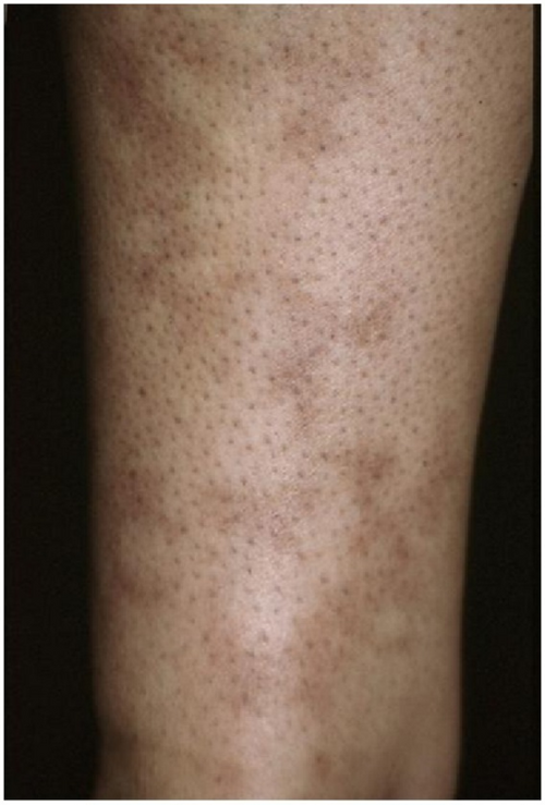 Image Source: Wikimedia Commons (2012). Livedo Reticularis of an Arm, Licensed under a creative commons licence. Available for free at: https://commons.wikimedia.org/wiki/File:Livedo_reticularis_of_an_arm.png (Accessed 19 May 2025).
- Papular mucinosis (mucin deposits)
- Calinosis cutis (calcium deposits)
Complete Guide to Clinical Genetics ST3 Application and Recruitment (2027)
Written by Medibuddy. Last updated: October 2026. Clinical Genetics is

Complete Guide to Clinical Radiology ST3 Application and Recruitment (2027)
Recruitment paused: NHS England is not running Clinical Radiology ST3+ recru

Complete Guide to Paediatric Surgery ST3 Application and Recruitment in United Kingdom (2026)
Written by Medibuddy. Last updated: October 2026. If you’re planning


Take your subscriptions with you
Our mobile app allows you to access your interview and exam question banks wherever you are.

