


 Close
Close
 30th July 2026
Stevens-Johnsons syndrome/Toxic Epidermal Necrolysis
Background
Steven-Johnson’s Syndrome (SJS) and Toxic Epidermal Necrolysis (TEN) are severe skin reactions affecting both cutaneous and mucosal tissue. The two conditions are a continuum of the same disease process differentiated by the total body surface area (TBSA) affected. Although rare, SJS/TEN are associated with a high mortality rate, 10% and 30% respectively. The main causative factor is medication however infection and malignancy can also trigger the reaction.
Epidemiology
Both SJS and TEN are relatively rare with an estimated incidence of between 4 to 7 cases per million per year. The conditions have a female predominance, affect all ages and are twice as common in Asian and black populations. Several HLA allotypes, in particular, HLA-B*1502 in Han Chinese, Thai, Malaysian and South Indian patients taking anticonvulsants and Europeans with HLA-B*5701 allotype taking abacavir or HLA-A*3101 taking carbamazepine have been associated with an increased risk of SJS/TEN.
Aetiology
Medications remain the most common cause of SJS/TEN. The commonest causes are summarised in the table below:
| Medications | Other causes |
|---|---|
| Anti-convulsants e.g. lamotrigine, carbamazepine, phenytoin, sodium valproate) | Epstein Barr Virus |
| Antibiotics e.g. minocycline, penicillin, cephalosporin quinolones | Herpes Simplex Virus |
| Aciclovir | Cytomegalovirus |
| Retinoids | Mycoplasma pneumonia |
| Anti-cancer drugs | Cancer |
| Biologic drugs | Vaccinations |
| Corticosteroids | |
| Allopurinol | |
| Contrast media | |
| Sulfasalazine | |
| Azathioprine | |
| Non-steroidal anti-inflammatory drugs (NSAIDS) | |
| Anti-HIV medications in particular nevirapine | |
| Antifungals | |
| Antimalarials |
Table 3: Summary of causes of SJS/TEN
The exact mechanism of SJS/TEN is unknown but it is theorised to be an immunological reaction mediated by cytotoxic T-cells (CD8). The condition leads to the separation of the epidermis and papillary dermis at the epidermal-dermal junction with keratinocyte apoptosis.
Diagnosis
Definition
- SJS: <10% TBSA (total body surface area) affected
- SJS/TEN overlap: 10%-30% TBSA (total body surface area) affected
- TEN: >30% body surface area TBSA (total body surface area) affected
Classification may change as the presentation evolves in the earlier phases.
Routine drug testing is not recommended but may be considered in circumstances where the trigger is unknown, or accidental exposure to the drug is possible.
Clinical manifestations
Diagnosis is largely clinical and with presentation evolving in the initial phases of the disease.
Non-specific symptoms include sore throat, fever, headache, arthralgia, vomiting/diarrhoea, and lethargy. These are followed by mucocutaneous disease which peaks at 4 days.
Skin lesions include erythema, blistering which coalesces to form large areas of skin detachment, and macules or purpuric spots.
Mucosal lesions may cause conjunctivitis, oral ulceration and crusted lips, genital ulceration and urinary symptoms, dysphagia and diarrhoea (due to GI tract ulceration), and respiratory symptoms.
Investigations
- Full blood count: anaemia, leucopenia
- Urea and electrolytes
- Liver function tests: mildly raised liver enzymes
- Coagulation profile
- Skin biopsy: histopathology shows epidermal necrosis, keratinocyte necrosis, minimal lymphocytic infiltrate in the dermis.
- Serum granulysin: elevated in initial phase
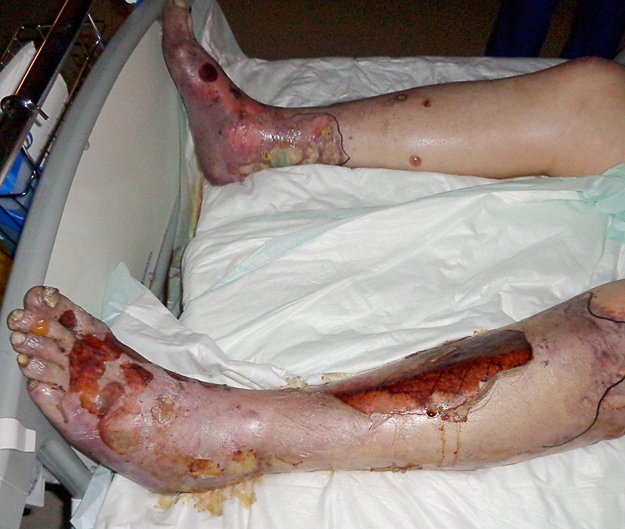
Skin detachment in the lower limbs secondary to TEN.
Image Source: Wikimedia Commons (2017). Necrolysis Epidermalix Toxica, Licensed under a creative commons licence. Available for free at: https://commons.wikimedia.org/wiki/File:Necrolysis_epidemalis_toxica_02.JPG (Accessed 19 May 2025).
Prognostic criteria
Two prognostic models exist, the SCORTON (SCORe of Toxic Epidermal Necrolysis) score and the ABCD 10 score. Both scores predict in-hospital risk of mortality.
SCORTON score
A score of 1 point is given for each of the following criteria:
- Age > 40 years
- Malignancy
- Heart rate > 140 beats per minute
- Body surface area detachment is > 10% at presentation
- Serum urea level > 10mmol/L
- Serum glucose > 14mmol/L
- Serum bicarbonate < 20mmol/L
| Score | Percentage risk of mortality (%) |
|---|---|
| 0 – 1 | 3.2 |
| 2 | 12.1 |
| 3 | 35.3 |
| 4 | 58.3 |
| 5 | > 90 |
Table 4: SCORTON scoring and associated mortality
ABCD 10 score
Points are allocated based on the following criteria:
- A: age > 50 years (1 point)
- B: bicarbonate > 20mmol/L
- C: cancer present and active (2 points)
- D: dialysis prior to admission (3 points)
- 10: epidermal detachment > 10% of BSA (1 point)
| Score | Percentage risk (%) |
|---|---|
| 0 | 2.3 |
| 1 | 5.4 |
| 2 | 12.3 |
| 3 | 25.5 |
| 4 | 45.7 |
| 5 | 67.4 |
| 6 | 83.6 |
Table 5: ABCD10 score and associated risk mortality
Differential diagnosis
- Drug related eosinophilia and systemic symptoms (DRESS) syndrome
- Other severe cutaneous adverse reactions (SCARs)
- Erythema multiforme major
- Staphylococcal scalded skin syndrome
- Pemphigus vulgaris or paraneoplastic pemphigus
Medical management
The mainstay of treatment is aimed at adequate wound management to maintain the skin barrier and reduce fluid loss.
Other steps in management include:
- Stop suspected causative agent
- VTE prophylaxis
- Adequate analgesia using validated pain scores/tools
- Wound management
- Strict barrier nursing to reduce infection risk
- Wound swabs from three areas regularly throughout the acute phase of infection
- Wound cleansing with saline +/- antimicrobial such as chlorhexidine
- Emollient use over all epidermis
- Topical anti-microbial to sloughy areas
- Decompression of blisters either by piercing or aspiration of fluid
- Transfer to burns unit if >30% of BSA affected and:
- Clinical deterioration
- Extension of epidermal detachment
- Sub-epidermal pus
- Local sepsis
- Wound conversion
- Delayed healing
- Surgical management
- Removal of necrotic tissue and cleaning under general anaesthetic
- Surgical debridement of wounds
- Closure of wounds using allograft/xenograft of biobrane
- Fluid management
- Fluid input/output
- Replacement as appropriate
- Nutrition
- Continuous enteral nutrition of 20-25kcal/kg/day in the acute phase and 25-30kcal/kg/day in the recovery phase
- Ocular involvement
- Ophthalmologic review
- Ocular hydration drops +/- topical corticosteroid drops
- Prophylactic topical antibiotic
- Urogenital involvement
- Daily urogenital review
- Wound management with white soft paraffin ointment
- Urinary catheter
- Mouth involvement
- Wound management with white soft paraffin ointment
- Anti-inflammatory +/- steroid mouthwash
- Daily saline mouthwash
- Airway involvement
- Transfer to ICU/burn unit
- Fibre-optic bronchoscopy if there is respiratory involvement
Complete Guide to Clinical Genetics ST3 Application and Recruitment (2027)
Written by Medibuddy. Last updated: October 2026. Clinical Genetics is

Complete Guide to Clinical Radiology ST3 Application and Recruitment (2027)
Recruitment paused: NHS England is not running Clinical Radiology ST3+ recru

Complete Guide to Paediatric Surgery ST3 Application and Recruitment in United Kingdom (2026)
Written by Medibuddy. Last updated: October 2026. If you’re planning


Take your subscriptions with you
Our mobile app allows you to access your interview and exam question banks wherever you are.

