


 Close
Close
 14th July 2026
Dermatological Cancers
Squamous Cell Carcinoma
Background
Squamous cell carcinoma (SCC) occurs in keratinocytes, located in the epidermis. SCC is the second most common skin cancer, secondary to basal cell carcinoma. Risk factors include UV exposure and immunosuppression. Treatment includes both non-surgical options (cryotherapy, topical and systemic chemotherapy) and surgical options (surgical excision, Mohs surgery).
Epidemiology
The second most common non-melanoma skin cancer, after basal cell carcinoma. Frequently seen in those >40 years of age, women, and in those with Fitzpatrick skin type I and II.
Aetiology
Solar UV exposure, ionising radiation, arsenic exposure, burns, previous psoralen, UV-A light therapy, HPV and immunocompromised states are known to increase risk of SCC. SCC occurs in keratinocytes, which have proliferated uncontrollably due to mutations and malignant transformation of the cells.
Risk factors can be separated into low risk, high risk and very-high risk as per the British Association of Dermatologists guidelines:
- Low risk:
- Patient factors: no lymphovascular or perineural invasion, Breslow thickness ≤ 4mm, tumour diameter ≤ 20mm
- Tumour factors: immune-competent
- High risk:
- Patient factors: immunosuppression or biological therapies, HIV therapy stabilised on HAART
- Tumour factors: lymphovascular invasion, poorly differentiated histology, tumour site, Breslow thickness of >4-6mm
- Very high risk:
- Patient factors: history of chronic lymphocytic leukaemia or myelofibrosis, solid organ transplant recipients
- Tumour factors: bone invasion, Breslow thickness >6mm, diameter >40mm
Diagnosis
SCC can be classified into four types, namely actinic keratosis (precursor lesion), SCC in situ (otherwise known as Bowen’s disease), invasive and metastatic.
SCC variants:
- Keratoacanthoma: rapidly growing dome-shaped nodule with keratin-filled central crater, grows over weeks and then shrinks after months.
- Verrucous carcinoma: fungating nodules or plaques, locally destructive, rarely metastasises
- Marjolin ulcer: aggressive, ulcerating, high rate of metastasis
History and examination
- Typically present with multiple actinic keratoses (erythematous, poorly defined, and irregularly shaped macules or plaques on sun-exposed areas of the skin).
- Most commonly seen on head and neck, and on upper extremities.
- Lesions are friable and can bleed easily.
- Lesions often have a scale or haemorrhagic crust.
SCC in situ (Bowen’s disease) is non-melanocytic and is characterised by a slow-growing and longstanding erythematous thin scaly patch/plaque with irregular and well-demarcated borders. (Figure 1)
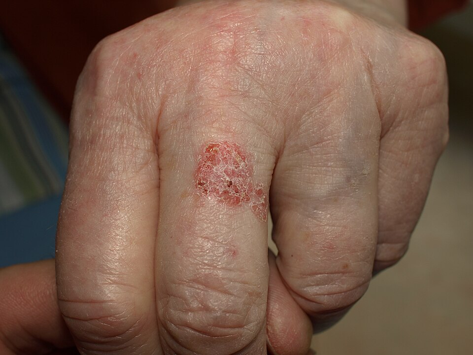
Bowen’s disease (cutaneous SCC in situ)
Image Source: Wikimedia Commons (2020), ECG AVNRT, Licenced under a creative commons licence. Available for free at: https://en.wikipedia.org/wiki/Cutaneous_squamous-cell_carcinoma#/media/File:Bowen11.jpg (Accessed: 25 May 2026).
SCCs typically are painful and fast-growing.
Invasive SCC presents as exophytic (outward-growing) and ulcerated tumours.
Regional lymph nodes are involved in 85% of metastatic SCCs.
Clinical history and examination are key to a diagnosis, although a skin biopsy is needed to confirm diagnosis.
Investigations
Dermoscopy:
- Actinic keratosis: erythematous pseudonetwork, surface scale, linear waxy vessels
- Bowen’s disease: erythema, keratosis, and glomerular vessels.
- Invasive: ulceration, keratinisation, irregular groups of white follicular cells
Histology typically shows nests of squamous cells with keratinisation, keratin pearls, large vesicular nuclei, and scant cytoplasm (Figure 2):
- Bowen’s disease: full-thickness keratinocyte atypia, confined to epidermis with an intact basement membrane
- Invasive SCC: extend beyond basement membrane
Imaging may be warranted for high-risk SCC: CT/PET/MRI
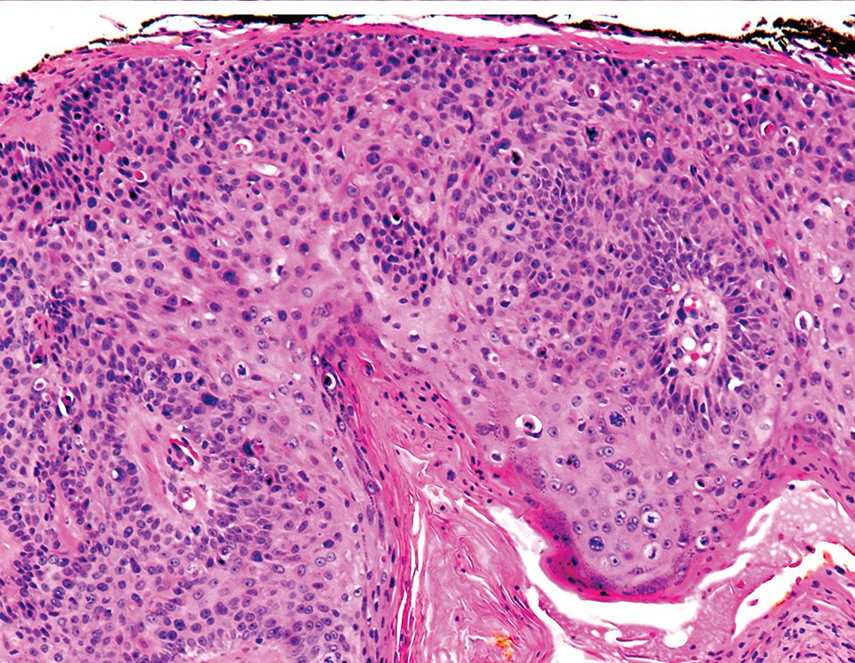
Histology of SCC
Image Source: Wikimedia Commons (2020), ECG AVNRT, Licenced under a creative commons licence. Available for free at: https://commons.wikimedia.org/wiki/File:Micrograph_of_squamous_cell_carcinoma_in_situ_-_100x.jpg (Accessed: 20 May 2026).
Management
Management differs depending on subtype of SCC:
- Bowen’s disease:
- Cryotherapy: liquid nitrogen leads to the delay in formation of vesicles or bullae.
- Topical chemotherapy: fluorouracil 5% (twice daily for 3-6 weeks) or imiquimod 5% (one daily, 2-3 times a week for 3-6 weeks).
- Second line:
- Surgical excision or Mohs: for tumours that do not respond
- Radiotherapy: for unresectable lesions or poor surgical candidates
- Invasive
- Conventional surgical excision (non-cosmetically sensitive), <2cm in diameter, and
- Excision with a ≥4mm if low risk, ≥6mm margin if high risk or ≥10mm margin if very high risk
- Mohs surgery (if cosmetically sensitive, i.e., face), >2cm in diameter
- Radiotherapy: may be indicated in aggressive subtypes
- Conventional surgical excision (non-cosmetically sensitive), <2cm in diameter, and
- Metastatic
- Surgical excision (non-cosmetically sensitive) PLUS radiotherapy
- Mohs surgery (cosmetically sensitive), >2cm PLUS radiotherapy
Complications
High likelihood: scarring
Medium likelihood: deformities due to local destruction,
References
- https://bestpractice.bmj.com/topics/en-gb/270/pdf/270/Squamous%20cell%20carcinoma%20of%20the%20skin.pdf
- https://www.uptodate.com/contents/cutaneous-squamous-cell-carcinoma-cscc-clinical-features-and-diagnosis
- https://patient.info/doctor/squamous-cell-carcinoma-of-skin
Basal Cell Carcinoma
Background
Basal cell carcinoma (BCC) is the most common carcinoma worldwide. Typical presentation is a pearly white/pink papulo-nodule or plaque. Usually locally aggressive, with metastasis being rare. Management is usually with surgical excision, with low recurrence rates.
Epidemiology
The most common malignancy of the skin with increasing incidence. Incidence in the United Kingdom is thought to be around 110 per 100,000 people.
Aetiology
Frequent and repetitive exposure to the sun is a well-known risk factor, as UV radiation leads to DNA damage of keratinocytes. Other risk factors include exposure to arsenic. BCC arises from pluripotent cells in the epidermis and follicular epithelium. A background of xeroderma pigmentosum, Gorlin-Goltz syndrome (nevoid basal cell carcinoma due to mutations in the PTCH1 tumour suppressor gene) or solid organ transplantation also increases risk for BCC. BCCs arise from pluripotent cells in the epidermis and follicular epithelium. It is thought that both p53 gene mutations and aberrant activation of the Sonic Hedgehog signalling cascade contribute to the development of BCCs.
Diagnosis
A biopsy is needed in order to differentiate subtype, which will impact treatment.
Classification:
- Nodular: most common form of BCC, a pearly white nodule with telangiectasia on surface
- Superficial: poorly defined margins. Dermoscopy reveals short fine telangiectasia, maple leaf areas, without grey/blue ovoid nests
- Micronodular: typically appears as multiple small nests in the dermis, with an increased risk of recurrence
- Metatypical: squamous differentiation
- Morpheaform/infiltrating/sclerosing: similar to plaques of morphea (scleroderma), poorly defined margins, perineural invasion common
- Infundibulocystic: stimulates trichoepithelioma, composed of mitotically active hyperchromatic blue cells
- Mixed: mixture of two or more of the above types of BCC
- Periocular: slow growing, painless and non-resolving, presents as a small white nodule on the eyelid
Typically presents as pearly papules +/- plaques, non-healing scars, nodules/plaques with rolled borders and papules with telangiectasias.
Investigations
Dermoscopy findings include arborising telangiectasia, focal ulceration, shiny white/red structureless areas, short white streaks/lines under polarised light, blue-grey ovoid nests, and leaf-like areas (Figure 3).
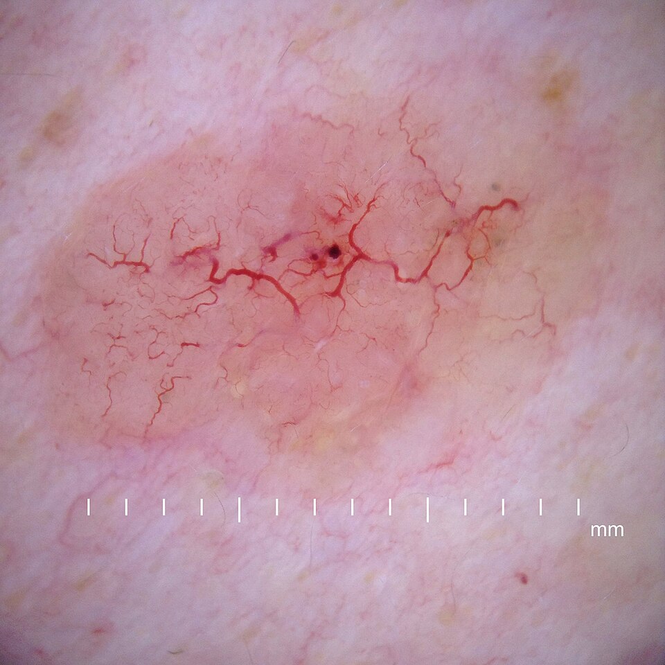
Dermoscopy of BCC
Image Source: Wikimedia Commons (2020), ECG AVNRT, Licenced under a creative commons licence. Available for free at: https://en.wikipedia.org/wiki/Basal-cell_carcinoma#/media/File:Derm oscopy_nodular_basal_cell_carcinoma.jpg (Accessed: 20 May 2026).
Either a shave or punch biopsy should be performed to confirm diagnosis. Punch biopsy is typically used in non-challenging cosmetical areas, as closure with stitches is usually required.
Histopathology shows proliferation of basal cells, well-defined linear and branching serpentine vessels, basophilic (blue) hyperchromatic ovoid nests, blue-grey globules and spoke wheel areas (Figure 4).
Histology of BCC
Image Source: Wikimedia Commons (2020), ECG AVNRT, Licenced under a creative commons licence. Available for free at: https://en.wikipedia.org/wiki/Basal-cell_carcinoma#/media/File:High-magnification_micrograph_of_basal-cell_carcinoma.jpg (Accessed: 20 May 2026).
Medical management
Low risk:
- Cryosurgery: effective for superficial variants
- Topical 5% imiquimod, a toll-like receptor-7 agonist chemotherapy agent, can be used in patients with superficial basal cell carcinoma. This is thought to result in an inflammatory response to BCC via induction of interferon alpha. The treatment course is five times a week for six weeks.
Sites not amenable to surgery: radiotherapy
Metastatic disease:
- Hedgehog pathway inhibitors: vismodegib and sonidegib
Surgical management
Surgery is the mainstay of treatment to ensure complete removal and prevent recurrence.
Low risk:
- Non-cosmetically challenging: excision or curettage +/- electrodesiccation and biopsy
- Cosmetically challenging (e.g., head and neck): Mohs surgery
High risk:
- Mohs surgery
Complications
Low likelihood: lymph node spread, distant metastasis
Medium likelihood: local spread of BCC
References
- https://bestpractice.bmj.com/topics/en-gb/269/pdf/269/Basal%20cell%20carcinoma.pdf
- https://www.ncbi.nlm.nih.gov/books/NBK1133/
- https://www.ncbi.nlm.nih.gov/books/NBK430921/
- https://www.uptodate.com/contents/treatment-and-prognosis-of-basal-cell-carcinoma-at-low-risk-of-recurrence
- https://patient.info/doctor/basal-cell-carcinoma
Malignant Melanoma
Background
Malignancy arises from melanocytes and is one of the most common forms of cancer in young adults. Typical presentation includes a pigmented skin lesion in sun-exposed regions. Most commonly seen in those with Fitzpatrick type I skin. Diagnosis is confirmed with biopsy. Management options include surgical excision and, in those cases where surgery is not indicated, topical chemotherapy or radiotherapy.
Epidemiology
Melanoma is amongst the most common forms of cancer and causes of cancer-related deaths. Men and those with Fitzpatrick type 1 skin are more likely to develop melanoma.
Aetiology
Exposure to UV radiation, history of sunburn during childhood, family history of melanoma, Fitzpatrick type I and II skin (Table 1), genetic syndromes (xeroderma pigmentosum), the proportion of congenital and acquired melanocytic nevi and genetic susceptibility. Melanoma arises from melanocytes. The CDKN2A gene is associated with familial melanoma. The MAPK pathway activates the BRADF and NRAS oncogenes and the CDKN2/RB1 pathway. BRAF mutations are present in around 40 to 50% of melanomas.
| Type | Characteristics |
|---|---|
| I | Always burns, pale white skin |
| II | Burns easily, tans minimally, white skin |
| III | Burns moderately, tans uniformly, light brown skin |
| IV | Burns minimally, always tans well, moderate brown skin |
| V | Rarely burns, tans profusely, dark brown skin |
| VI | Never burns, deeply pigmented dark brown to black skin |
Table 1. Fitzpatrick skin types
Risk factors for metastasis in those with stage I or II:
- > 55 years of age
- Head and neck tumour
- Breslow’s thickness > 4mm
- BRAF mutations
- Vascular invasion
Diagnosis
Subtypes of melanoma:
- Superficial spreading (Figure 5): most common, usually found on the torso, incidence increases with age
- Nodular: second most common, typically associated with a late diagnosis and subsequent poor prognosis
- Lentigo maligna melanoma: more common in older population and on sun-damaged skin, typically slow growing. Dermoscopy findings include annular granular pigmentation, atypical globules, atypical network, rhomboidal structures, and blue-white veil.
- Acral lentiginous: ~1-3% of melanomas, most commonly seen in those with Fitzpatrick type V or VI.
- Rate variants: amelanotic (non-pigmented), spitzoid, desmoplastic
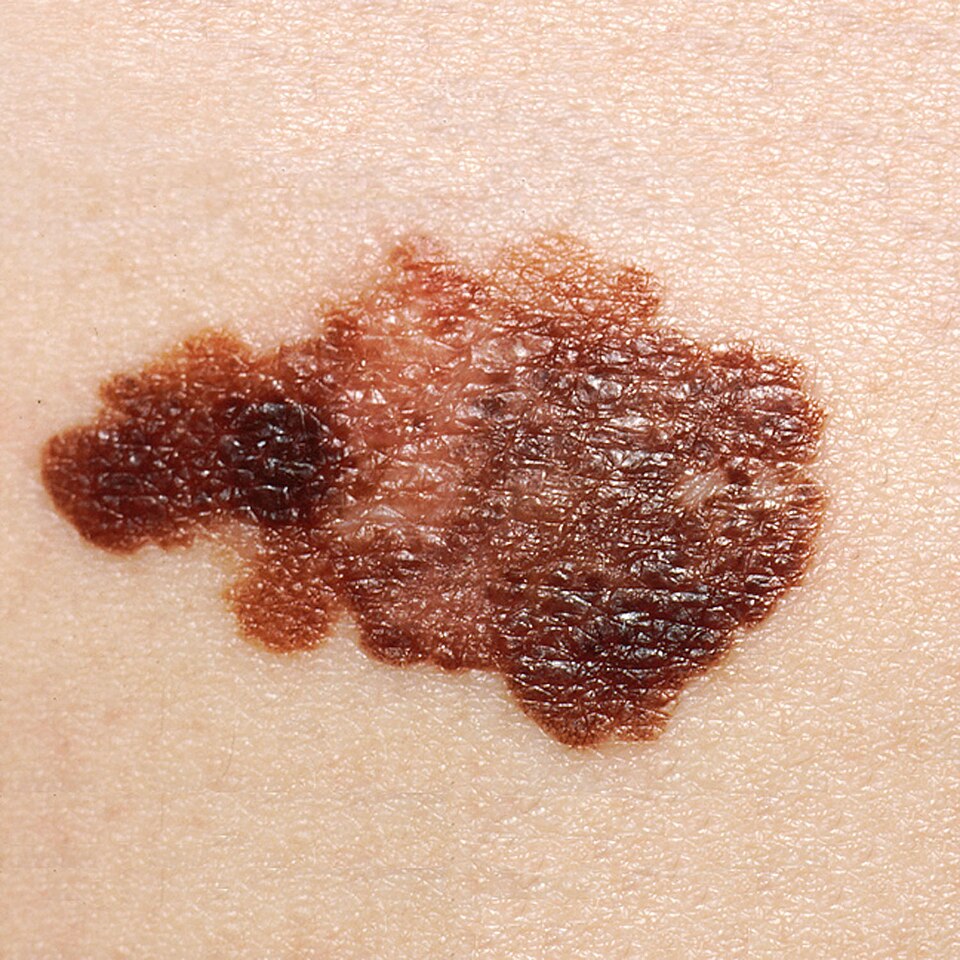
Superficial spreading melanoma
Image Source: Wikimedia Commons (2020), ECG AVNRT, Licenced under a creative commons licence. Available for free at: https://commons.wikimedia.org/wiki/File:Melanoma_(2).jpg (Accessed: 20 May 2026).
History and examination
History includes questions around size, shape, colouration, or ulceration of the lesion. Bleeding or pruritic lesions should have a high clinical suspicion for melanoma.
ABCDE is a useful way to assess lesions:
- A: asymmetry
- B: border irregularity
- C: colour
- D: diameter > 6mm
- E: evolution
Investigations
A vitamin D level should be checked at diagnosis in all patients with melanoma
Dermoscopy: atypical globules and dots, atypical network, blue-white veil
Biopsy and histopathology:
- Biopsy: full-thickness excision of lesion with 1-3mm margin
- Prognostic indicators: Breslow’s thickness (depth from granular cell layer to deepest tumour penetration in dermis), ulceration, mitotic count, lymphovascular invasion, microscopic satellites, serum LDH
- Abnormal and severe melanocytic proliferation in epidermis +/- dermis, asymmetrical growth pattern
Immunohistochemistry: markers used in analysis include Melan-A/MART-1, S-100, HMB-45, SOX10, MiTF and tyrosinase.
BRAF mutation testing should be performed in those with stage IIIB onwards.
Staging of cutaneous melanoma:
- Localised disease (stage I-III)
- Regional disease (stage III)
- Distant metastatic disease (stage IV)
Clark’s level
- Level 1: in-situ melanoma
- Level 2: invasion within papillary dermis
- Level 3: invasion filling papillary dermis
- Level 4: invasion within reticular dermis
Management
Dependent on subtype of melanoma and stage of cancer.
In patients where surgery is not indicated (i.e., multi-comorbid patients or difficult tumour location), topical chemotherapy (imiquimod), or radiotherapy may be used.
In those with melanoma in situ, topical imiquimod should be considered if removal of 5mm margins would cause disfigurement.
Unresectable advanced stage (stage III or IV):
- Systemic therapy (nivolumab and ipilimumab, nivolumab or pembrolizumab)
- Where surgery cannot be performed for metastasis, talimogene laherparepvec or electrochemotherapy may be indicated in some patients.
Surgical management
Early stage (melanoma in situ, stage I and stage II):
- Wide local excision with 5-10 mm margins (5mm for lentigo maligna).
- Mohs’ surgery can be considered in those with minimally invasive tumours on sites such as the ear or face.
- Sentinel lymph node biopsy (SNLB) may be used in those with stage I or stage II, and is an important prognostic factor in this patient group.
Resectable advanced stage (stage III or IV):
- Wide local excision PLUS adjuvant systemic therapy (nivolumab, pembrolizumab or dabrafenib and trametinib).
Monitoring
Early stage (I/IIA): follow-up recommended (up to four times in first year after treatment completion)
Those with stage IB-IIB should be followed up every 3 months for the first three years after treatment completion, followed by every six months for the next two years, with the aim of discharge after five years.
Advanced disease (stage III or IV) and receiving systemic therapy: regular imaging every 3 months to assess progression
Complications
High likelihood: adverse effects of treatment, SNLB complications (post-operative lymphodema), sequelae of metastatic disease
Low likelihood: local surgical complications (bleeding/infection), SNLB complications, local recurrence,
References
- https://bestpractice.bmj.com/topics/en-gb/268/pdf/268/Melanoma.pdf
- https://www.nice.org.uk/guidance/ng14/resources/melanoma-assessment-and-management-pdf-1837271430853
- https://dermnetnz.org/topics/superficial-spreading-melanoma
- https://www.nccn.org/guidelines/category_1
Merkel cell carcinoma
Background
Merkel cell carcinoma (MCC) is an aggressive primary cutaneous neuroendocrine tumour. Risk factors include UV exposure, increasing age and Merkel cell polyomavirus. Early aggressive treatment is needed.
Epidemiology
MCC is a rare skin tumour with rising incidence rates.
Aetiology
Commonly seen in elderly Caucasian individuals and appears in the sixth or seventh decade of life. Risk factors include increasing age, sun exposure, immunosuppression, and Merkel cell polyomavirus.
Diagnosis
History and examination
Rapidly enlarging, shiny, smooth-surfaced solitary red nodule. Commonly seen on the head and neck. Ulceration is rare and typically only occurs in advanced stages. Metastasis and lymph node spread are common.
AEIOU can be used to aid diagnosis:
- A: asymptomatic/non-tender
- E: expanding rapidly (≤ 3 months)
- I: immunosuppressed
- O: older than 50 years
- U: UV-exposed sites
Dermoscopy:
- Linear, irregular, and polymorphous vessels, poorly focused vessels, milky pink/white areas, and architectural disorder.
Immunohistochemistry:
- Positive immunostaining with low molecular weight cytokeratins, e.g., cytokeratin 20 (CK20), and neuroendocrine markers such as neuron-specific enolase, Chromogranin A and synaptophysin are typically positive.
- Thyroid transcription factor (TTF1) is usually negative.
Histopathology:
- Uniformly small, round blue cells with scanty cytoplasm and vesicular nuclei
Prognosis
- Favourable prognostic factors include female gender, age <50, tumour size ≤ 2cm, local disease, presence of Merkel cell polyomavirus or increased viral abundance.
- Poor prognostic factors include those who are immunosuppressed and those with nodal involvement. Histology features that would suggest poor prognosis include tumour size ≥ 5mm, diffuse growth pattern and high mitotic rate.
Management
Dependent on the stage of the tumour, a multidisciplinary approach is needed.
Early aggressive treatment is needed.
Early stage MCC: surgical excision with wide resection margins followed by sentinel lymph node biopsy and postoperative radiotherapy
Localised MCC: surgical excision followed by radiotherapy
Advanced/metastatic disease: radiotherapy, chemotherapy, immunotherapy +/- molecular-targeted therapy.
References
- https://bestpractice.bmj.com/topics/en-gb/269/differentials#collapseDiff-47fe7fd8-09aa-404f-806a-5a4954c1a525
- https://dermnetnz.org/topics/merkel-cell-carcinoma
Benign Skin Tumours
Background
Benign skin tumours encompass cherry angiomas, actinic keratosis, sebaceous hyperplasia, seborrheic keratosis, keratoacanthomas, lipoma, sebaceous cysts, and pyogenic granuloma.
Cherry angiomas, or Campbell de Morgan spots, are common benign non-blanching lesions of the skin. Management involves reassurance and, in a few cases, removal for cosmetic reasons.
Actinic keratosis, otherwise known as solar keratosis, is typically seen in fair-skinned people after long-term sun exposure caused by UV rays. Most lesions do not progress to malignancy. Dermoscopy reveals erythema, keratin plugs and scale.
Sebaceous hyperplasia is a common benign condition associated with enlarged sebaceous glands, which appear as small yellow bumps.
Seborrheic keratosis is associated with hyperpigmented plaques with a ‘stuck-on’ appearance. Dermoscopy reveals pseudo comedones with milia-like cysts and hairpin vessels.
Keratoacanthomas are benign, rapidly growing lesions which typically spontaneously resolve over four to six months.
Lipomas are slow-growing benign tumours. Most lipomas do not cause any symptoms and usually do not require treatment.
Sebaceous cysts typically present as a painless lump and, on occasion, are filled with discharge. If uncomplicated, usually left alone; however, if inflamed or infected, sebaceous cysts may warrant further management.
Pyogenic granuloma are rapidly growing benign lesion. Treatment options include 5% imiquimod cream, steroid injections, or excision.
Epidemiology
Cherry angiomas are the most common cutaneous vascular proliferation, which increase in frequency and size with age. Actinic keratosis is commonly seen in individuals with fair skin and is associated with increasing age and cumulative UV exposure. Seborrheic keratoses are one of the most common skin tumours, typically seen in adults over the age of 30. The incidence of keratoacanthomas is around 1 in 1000 in the UK. The peak incidence is in those over the age of 60. Lipomas can be seen in any age group, but are more common in those between 40-60 years of age. Incidence is similar to keratoacanthomas at 1 in 1000 people. The mean age for pyogenic granulomas is 5–8-year-olds; from then on, the incidence decreases with increasing age. A slight increase in incidence is seen in women.
Aetiology
Cherry angiomas are the most common cutaneous vascular proliferation, which increase in frequency with advancing age. Actinic keratosis is most commonly seen in fair-skinned people and those with increased sun exposure. In some cases, actinic keratosis may progress to SCC. Sebaceous hyperplasia is commonly seen in elderly and immunosuppressed patients. There is a link between UV-induced skin ageing and seborrheic keratosis. Keratoacanthomas are most commonly seen in those with increased sun exposure, workers exposed to tar, and those immunocompromised.
Malignancy in those with lipoma is rare but can be seen in lesions with the clinical appearance of a lipoma, liposarcoma. Characteristics in keeping with liposarcoma include:
- > 5cm diameter
- Lesion in the extremities, retroperitoneally or abdominal wall
- Rapid growth or bone invasion
Diagnosis
Cherry angiomas are usually found on the trunk or on upper extremities and are typically asymptomatic.
Actinic keratosis
- Presents as a small, scaly plaque
- Risk of malignant change
- Clinical diagnosis and can be classified as follows:
- Grade 1: slightly palpable
- Grade 2: moderately thick
- Grade 3: very thick, hyperkeratotic
- 2-week wait referral warranted if any suspicious features of SCC:
- Recent growth, bleeding/ulceration, nodular lesion
Sebaceous hyperplasia is mainly found on the face and presents as white/yellow papules. On dermoscopy, sebaceous hyperplasia is typically associated with group white globules with crown vessels.
Seborrheic keratosis presents as flat lesions that appear ‘stuck’ on the skin. They are usually pigmented and have a well-defined border. Typically, lesions are asymptomatic; however, they may easily become inflamed or itchy. Dermoscopy reveals thickened epidermis with multiple milia-like cysts and comedo-like openings, fingerprint-like structures and gyri and sulci (Figure 6). On immunohistochemistry, seborrheic keratosis typically presents as uniform hairpin (looped) vessels.
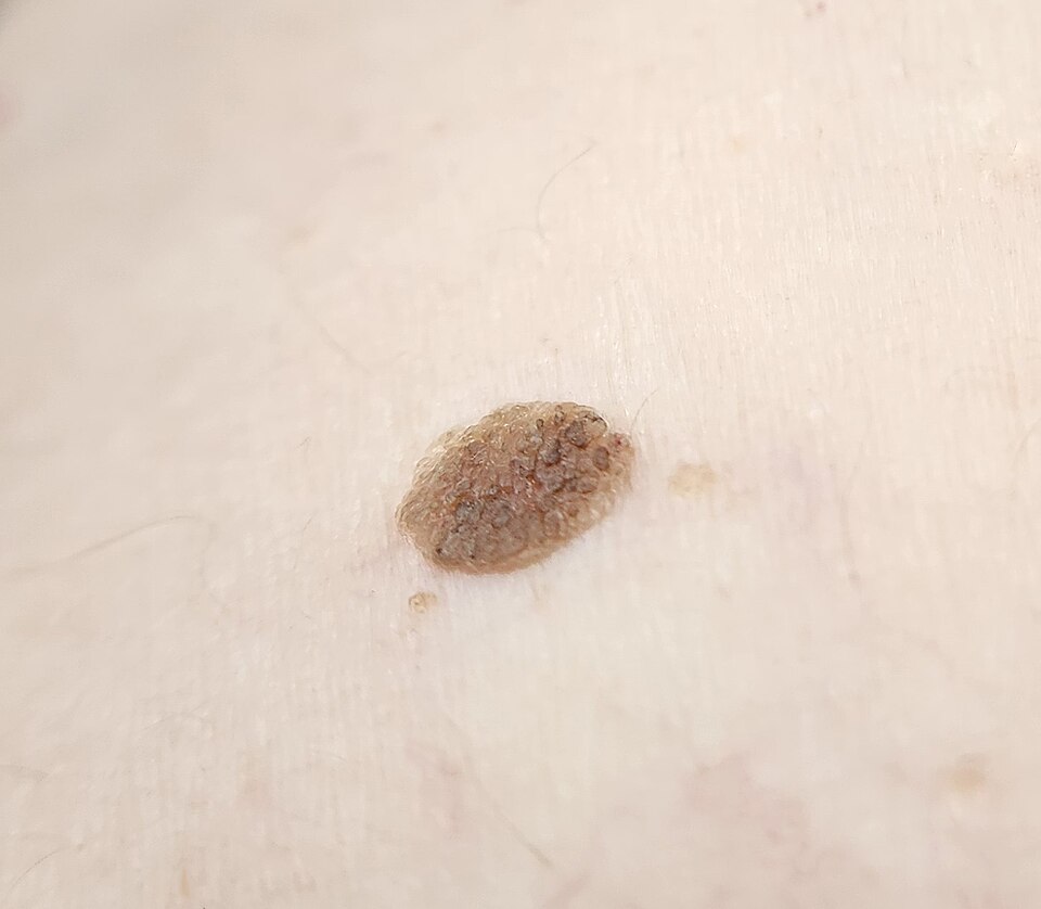
Seborrheic keratosis
Image Source: Wikimedia Commons (2020), ECG AVNRT, Licenced under a creative commons licence. Available for free at: https://commons.wikimedia.org/wiki/File:Seborrheic_keratosis_closup.jpg (Accessed: 20 May 2026).
Keratoacanthomas
- Presents as rapidly growing lesions over a few weeks to months, on sun-exposed areas, usually solitary and round papules that may progress to dome-shaped nodules with a shiny surface.
- 2-week wait referral as difficult to distinguish from SCC
- Excisional or deep biopsy required
A lipoma is a soft/firm smooth nodule, usually less than 5cm in diameter. They are typically situated in the deep fatty layer, are mobile and non-painful. Diagnosis is based on clinical appearance.
Pyogenic granulomas are solitary red or purple papules/nodules which vary in size. They have a polypoid appearance and typically develop a ‘stalk’ at their base. Typically seen to bleed, crust or ulcerate. They are fast-growing and most commonly occur on the head, neck or extremities.
Management
Cherry angiomas are benign lesions and typically require no treatment. If removal is required, usually for cosmetic reasons, treatment options include curettage, electrocautery, or excision.
Actinic keratosis:
- Prevention and education: limit sun exposure, vigilant for any suspicious features
- Topical:
- 5-fluorouracil: individual lesions
- Imiquimod 5%: multiple lesions
- Diclofenac gel: thin areas
- Tirbanibulin 1%: used focally or in broad areas
- Cryotherapy: single or multiple discrete lesions
- Photodynamic therapy: more effective than cryotherapy and associated with better cosmetic results
- Curettage or excisional surgery: discrete hyperkeratotic lesions
Sebaceous hyperplasia is benign and does not require treatment unless for cosmetic reasons. If so, curettage or cryotherapy may be used.
Seborrheic keratosis:
- Irritated/itchy: corticosteroids
- Raised: curettage or cautery
- Flat: cryotherapy
Keratoacanthomas
- Complete excision, if not amenable to surgery, topical treatments such as retinoids and intralesional methotrexate may be used
Lipomas
- Usually not removed
- Complete excision is sometimes performed in those with fast-growing lipomas or for cosmetic reasons.
Pyogenic granulomas are typically treated due to bleeding. Options include:
- Topical:
- Imiquimod 5%
- Timolol gel 0.5%
- Topical beta-blockers
- Intralesional steroid injection
- Curettage and cautery
- Excision with primary closure
References
- https://dermnetnz.org/cme/dermoscopy-course/dermoscopy-of-seborrhoeic-keratosis
- https://patient.info/doctor/seborrhoeic-wart
- https://patient.info/doctor/epidermoid-and-pilar-cysts-sebaceous-cysts-pro
- https://patient.info/doctor/keratoacanthoma-pro
- https://bestpractice.bmj.com/topics/en-gb/617/pdf/617/Seborrhoeic%20keratosis.pdf
Cutaneous T-cell Lymphoma
Background
Cutaneous T-cell lymphoma (CTCL) is a group of uncommon disorders characterised by the accumulation of T lymphocytes in the skin. Mycosis fungoides (MF) and its leukaemic variant Sézary syndrome (SS) are subtypes of CTCL; MF behaves similarly to an indolent lymphoma, whilst SS is a much more aggressive form of the disease.
Epidemiology
MF is a primary cutaneous non-Hodgkin’s lymphoma of T-cell origin and is the common subtype of CTCL. MF follows an indolent clinical course, typically with slow disease progression. The incidence of CTCL is on the rise; however, this may be attributed to improved rates of diagnosis.
Aetiology
Risk factors include HTLV virus, radiation exposure, severe atopic eczema, melanoma, and non-melanoma skin cancer. Occupational exposures to textiles and ceramics are also known risk factors. Anti-TNFα therapy can cause progression of cutaneous T-cell lymphoma.
Diagnosis
History and Examination
All patients should undergo a full skin examination to assess the body surface area (BSA) and type of lesions, as well as a lymph node examination and assessment for organomegaly/masses. Pruritus is commonly seen, particularly in those with SS.
Heterogeneous cutaneous manifestations include:
- Polymorphic patches/plaques
- Tumours and erythroderma
- Hypopigmented lesions
- Hyperpigmentation (usually seen in younger children/adolescents)
- Granulomatous slack skin (resembles loose skin)
SS is a rare leukaemic subtype of CTCL:
- Pruritic erythroderma
- Lymphadenopathy
- Sézary cells (clonally related neoplastic T-cells with cerebriform nuclei)
As both MF and SS may present with similar features, peripheral blood involvement is needed to differentiate the two. In order to definitively diagnose SS, the following is needed alongside Sézary cells:
- Absolute Sézary cell count of >1000/microlitre
- Expanded CD4+ T cell population resulting in CD4/CD8 ratio ≥10, CD4+/CD7- cells ≥30% or CD4+/CD26- cells ≥40%.
MF typically presents in three stages, namely patch, plaque, and tumour stage. Initial presentation can mimic inflammatory skin conditions such as eczema and psoriasis. These typically progress to tumours and erythrodermic stages. In the few patients who progress straight to tumours, i.e., without patches or plaques, this is termed tumour d’emblee. A biopsy is needed to confirm diagnosis.
Investigations include:
- Bloods: FBC (normal or lymphocytosis), LFTs (usually normal), HIV (rule out HIV associated malignancy), LDH – strong predictor of poor prognosis (usually normal), beta 2 microglobulin, lymphocyte subsets CD4:CD8, HTLV serology (to differentiate HTLV-1-associated T-cell leukaemia/lymphoma from other T-cell leukaemias), Sézary cell count on blood film
- Biopsy: skin, lymph nodes, bone marrow trephine biopsy
- Immunohistochemistry: CD2, CD3, CD4, CD5, CD7, CD8, CD20, CD30 (tumour cells are typically CD3+, CD4+ and CD8-; CD7 is often lost); TCR gene analysis (if peripheral blood involvement suspected)
- Imaging: PET/CT for staging of disease
Clinical staging of CTCL is as follows:
| Stage | Clinical features |
|---|---|
| IA | Limited patches +/- plaques, <10% BSA involved |
| IB | Generalised patches +/- plaques, 10% or more of BSA involved |
| IIA | Enlarged peripheral lymph nodes |
| IIB | One or more skin tumours present, no histological involvement or erythroderma |
| IIIA | Erythroderma, no blood involvement |
| IIIB | Erythroderma, with low blood tumour burden (>5% lymphocytes are Sézary cells) |
| IVA1 | Sézary syndrome, Sézary cells >1000/ml |
| IVA2 | Abnormal peripheral lymph nodes |
| IVB | Visceral involvement |
Histology: epidermotropism is typically seen (characterised by T cells in the epidermis), Pautrier microabscesses (cluster of atypical lymphocytes)
Management
Aim to improve symptoms, cure is rarely achieved.
Skin-directed therapies include topical medications: corticosteroids, imiquimod, chlormethine, retinoids (bexarotene), phototherapy, and localised radiotherapy
Systemic therapies include photopheresis, chemotherapy (methotrexate), and biological or immunological therapy (interferon-alpha)
First-line treatment:
- Stage IA:
- 1st line – skin-directed therapies
- 2nd line – systemic therapy
- Stage IB-IIA:
- 1st line – skin-directed therapies
- Predominantly plaque disease: PUVA or UVA1
- 2nd line – systemic therapy
- Stage IIB
- 1st line – skin-directed therapies AND systemic therapy, adjunct local radiotherapy
- Stage III:
- 1st line – skin-directed therapies AND systemic therapy AND antibiotics (due to increased risk of secondary infection)
- Stage IV:
- Systemic therapy, adjunct radiotherapy
- 2nd line – allogeneic haematopoietic cell transplant
Complications
High likelihood: cutaneous ulceration
Medium likelihood: cutaneous/systemic infections, secondary malignancy, therapy-related cutaneous malignancy (increased risk of non-melanoma and melanoma)
Low likelihood: complications related to erythroderma (heart failure, renal impairment), CNS involvement
References
Complete Guide to Clinical Genetics ST3 Application and Recruitment (2027)
Written by Medibuddy. Last updated: October 2026. Clinical Genetics is

Complete Guide to Clinical Radiology ST3 Application and Recruitment (2027)
Recruitment paused: NHS England is not running Clinical Radiology ST3+ recru

Complete Guide to Paediatric Surgery ST3 Application and Recruitment in United Kingdom (2026)
Written by Medibuddy. Last updated: October 2026. If you’re planning


Take your subscriptions with you
Our mobile app allows you to access your interview and exam question banks wherever you are.

