


 Close
Close
 14th July 2026
Benign Skin Tumours
Background
Benign skin tumours encompass cherry angiomas, actinic keratosis, sebaceous hyperplasia, seborrheic keratosis, keratoacanthomas, lipoma, sebaceous cysts, and pyogenic granuloma.
Cherry angiomas, or Campbell de Morgan spots, are common benign non-blanching lesions of the skin. Management involves reassurance and, in a few cases, removal for cosmetic reasons.
Actinic keratosis, otherwise known as solar keratosis, is typically seen in fair-skinned people after long-term sun exposure caused by UV rays. Most lesions do not progress to malignancy. Dermoscopy reveals erythema, keratin plugs and scale.
Sebaceous hyperplasia is a common benign condition associated with enlarged sebaceous glands, which appear as small yellow bumps.
Seborrheic keratosis is associated with hyperpigmented plaques with a ‘stuck-on’ appearance. Dermoscopy reveals pseudo comedones with milia-like cysts and hairpin vessels.
Keratoacanthomas are benign, rapidly growing lesions which typically spontaneously resolve over four to six months.
Lipomas are slow-growing benign tumours. Most lipomas do not cause any symptoms and usually do not require treatment.
Sebaceous cysts typically present as a painless lump and, on occasion, are filled with discharge. If uncomplicated, usually left alone; however, if inflamed or infected, sebaceous cysts may warrant further management.
Pyogenic granuloma are rapidly growing benign lesion. Treatment options include 5% imiquimod cream, steroid injections, or excision.
Epidemiology
Cherry angiomas are the most common cutaneous vascular proliferation, which increase in frequency and size with age. Actinic keratosis is commonly seen in individuals with fair skin and is associated with increasing age and cumulative UV exposure. Seborrheic keratoses are one of the most common skin tumours, typically seen in adults over the age of 30. The incidence of keratoacanthomas is around 1 in 1000 in the UK. The peak incidence is in those over the age of 60. Lipomas can be seen in any age group, but are more common in those between 40-60 years of age. Incidence is similar to keratoacanthomas at 1 in 1000 people. The mean age for pyogenic granulomas is 5–8-year-olds; from then on, the incidence decreases with increasing age. A slight increase in incidence is seen in women.
Aetiology
Cherry angiomas are the most common cutaneous vascular proliferation, which increase in frequency with advancing age. Actinic keratosis is most commonly seen in fair-skinned people and those with increased sun exposure. In some cases, actinic keratosis may progress to SCC. Sebaceous hyperplasia is commonly seen in elderly and immunosuppressed patients. There is a link between UV-induced skin ageing and seborrheic keratosis. Keratoacanthomas are most commonly seen in those with increased sun exposure, workers exposed to tar, and those immunocompromised.
Malignancy in those with lipoma is rare but can be seen in lesions with the clinical appearance of a lipoma, liposarcoma. Characteristics in keeping with liposarcoma include:
- > 5cm diameter
- Lesion in the extremities, retroperitoneally or abdominal wall
- Rapid growth or bone invasion
Diagnosis
Cherry angiomas are usually found on the trunk or on upper extremities and are typically asymptomatic.
Actinic keratosis
- Presents as a small, scaly plaque
- Risk of malignant change
- Clinical diagnosis and can be classified as follows:
- Grade 1: slightly palpable
- Grade 2: moderately thick
- Grade 3: very thick, hyperkeratotic
- 2-week wait referral warranted if any suspicious features of SCC:
- Recent growth, bleeding/ulceration, nodular lesion
Sebaceous hyperplasia is mainly found on the face and presents as white/yellow papules. On dermoscopy, sebaceous hyperplasia is typically associated with group white globules with crown vessels.
Seborrheic keratosis presents as flat lesions that appear ‘stuck’ on the skin. They are usually pigmented and have a well-defined border. Typically, lesions are asymptomatic; however, they may easily become inflamed or itchy. Dermoscopy reveals thickened epidermis with multiple milia-like cysts and comedo-like openings, fingerprint-like structures and gyri and sulci (Figure 6). On immunohistochemistry, seborrheic keratosis typically presents as uniform hairpin (looped) vessels.
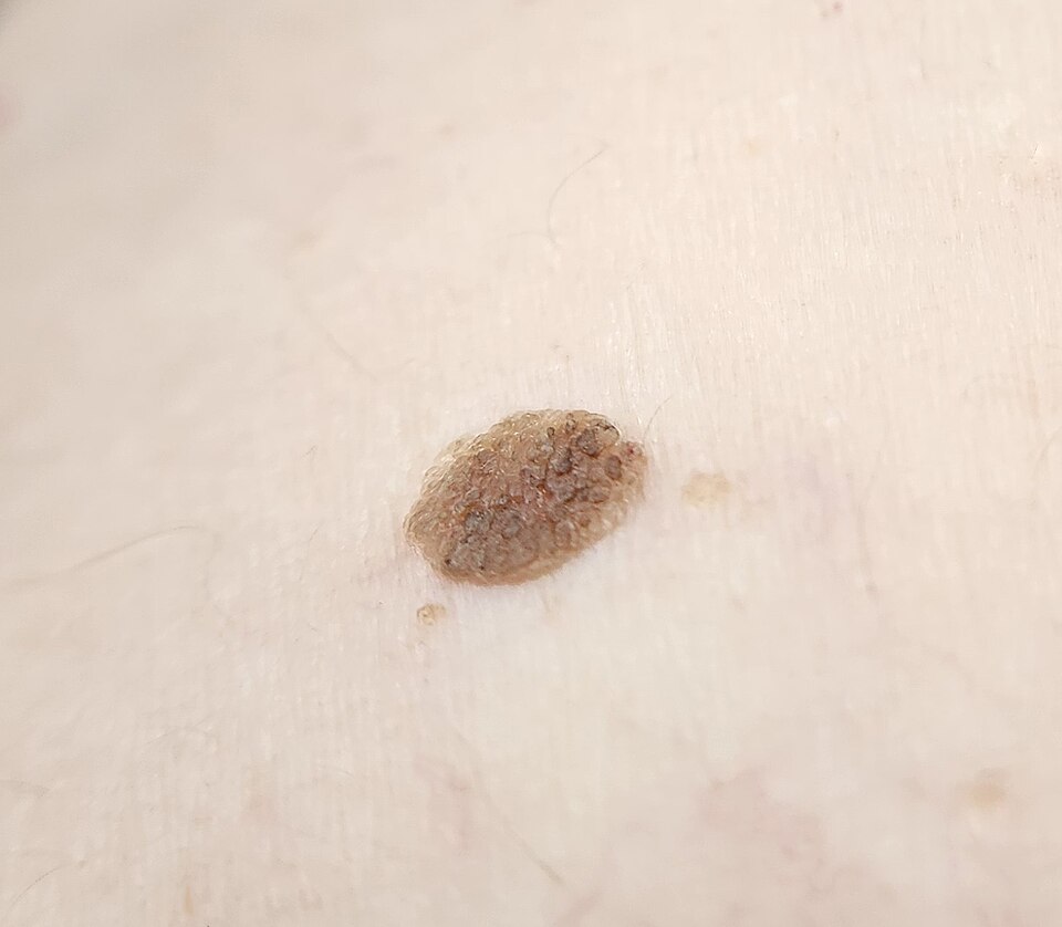
Seborrheic keratosis
Image Source: Wikimedia Commons (2020), ECG AVNRT, Licenced under a creative commons licence. Available for free at: https://commons.wikimedia.org/wiki/File:Seborrheic_keratosis_closup.jpg (Accessed: 20 May 2026).
Keratoacanthomas
- Presents as rapidly growing lesions over a few weeks to months, on sun-exposed areas, usually solitary and round papules that may progress to dome-shaped nodules with a shiny surface.
- 2-week wait referral as difficult to distinguish from SCC
- Excisional or deep biopsy required
A lipoma is a soft/firm smooth nodule, usually less than 5cm in diameter. They are typically situated in the deep fatty layer, are mobile and non-painful. Diagnosis is based on clinical appearance.
Pyogenic granulomas are solitary red or purple papules/nodules which vary in size. They have a polypoid appearance and typically develop a ‘stalk’ at their base. Typically seen to bleed, crust or ulcerate. They are fast-growing and most commonly occur on the head, neck or extremities.
Management
Cherry angiomas are benign lesions and typically require no treatment. If removal is required, usually for cosmetic reasons, treatment options include curettage, electrocautery, or excision.
Actinic keratosis:
- Prevention and education: limit sun exposure, vigilant for any suspicious features
- Topical:
- 5-fluorouracil: individual lesions
- Imiquimod 5%: multiple lesions
- Diclofenac gel: thin areas
- Tirbanibulin 1%: used focally or in broad areas
- Cryotherapy: single or multiple discrete lesions
- Photodynamic therapy: more effective than cryotherapy and associated with better cosmetic results
- Curettage or excisional surgery: discrete hyperkeratotic lesions
Sebaceous hyperplasia is benign and does not require treatment unless for cosmetic reasons. If so, curettage or cryotherapy may be used.
Seborrheic keratosis:
- Irritated/itchy: corticosteroids
- Raised: curettage or cautery
- Flat: cryotherapy
Keratoacanthomas
- Complete excision, if not amenable to surgery, topical treatments such as retinoids and intralesional methotrexate may be used
Lipomas
- Usually not removed
- Complete excision is sometimes performed in those with fast-growing lipomas or for cosmetic reasons.
Pyogenic granulomas are typically treated due to bleeding. Options include:
- Topical:
- Imiquimod 5%
- Timolol gel 0.5%
- Topical beta-blockers
- Intralesional steroid injection
- Curettage and cautery
- Excision with primary closure
References
- https://dermnetnz.org/cme/dermoscopy-course/dermoscopy-of-seborrhoeic-keratosis
- https://patient.info/doctor/seborrhoeic-wart
- https://patient.info/doctor/epidermoid-and-pilar-cysts-sebaceous-cysts-pro
- https://patient.info/doctor/keratoacanthoma-pro
- https://bestpractice.bmj.com/topics/en-gb/617/pdf/617/Seborrhoeic%20keratosis.pdf
Complete Guide to Clinical Genetics ST3 Application and Recruitment (2027)
Written by Medibuddy. Last updated: October 2026. Clinical Genetics is

Complete Guide to Clinical Radiology ST3 Application and Recruitment (2027)
Recruitment paused: NHS England is not running Clinical Radiology ST3+ recru

Complete Guide to Paediatric Surgery ST3 Application and Recruitment in United Kingdom (2026)
Written by Medibuddy. Last updated: October 2026. If you’re planning


Take your subscriptions with you
Our mobile app allows you to access your interview and exam question banks wherever you are.

