


 Close
Close
 14th July 2026
Diagnosis
Cherry angiomas are usually found on the trunk or on upper extremities and are typically asymptomatic.
Actinic keratosis
- Presents as a small, scaly plaque
- Risk of malignant change
- Clinical diagnosis and can be classified as follows:
- Grade 1: slightly palpable
- Grade 2: moderately thick
- Grade 3: very thick, hyperkeratotic
- 2-week wait referral warranted if any suspicious features of SCC:
- Recent growth, bleeding/ulceration, nodular lesion
Sebaceous hyperplasia is mainly found on the face and presents as white/yellow papules. On dermoscopy, sebaceous hyperplasia is typically associated with group white globules with crown vessels.
Seborrheic keratosis presents as flat lesions that appear ‘stuck’ on the skin. They are usually pigmented and have a well-defined border. Typically, lesions are asymptomatic; however, they may easily become inflamed or itchy. Dermoscopy reveals thickened epidermis with multiple milia-like cysts and comedo-like openings, fingerprint-like structures and gyri and sulci (Figure 6). On immunohistochemistry, seborrheic keratosis typically presents as uniform hairpin (looped) vessels.
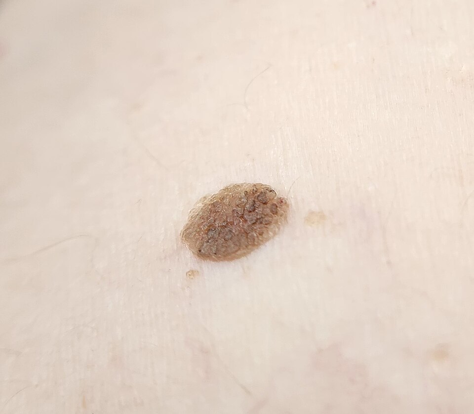
Seborrheic keratosis
Image Source: Wikimedia Commons (2020), ECG AVNRT, Licenced under a creative commons licence. Available for free at: https://commons.wikimedia.org/wiki/File:Seborrheic_keratosis_closup.jpg (Accessed: 20 May 2026).
Keratoacanthomas
- Presents as rapidly growing lesions over a few weeks to months, on sun-exposed areas, usually solitary and round papules that may progress to dome-shaped nodules with a shiny surface.
- 2-week wait referral as difficult to distinguish from SCC
- Excisional or deep biopsy required
A lipoma is a soft/firm smooth nodule, usually less than 5cm in diameter. They are typically situated in the deep fatty layer, are mobile and non-painful. Diagnosis is based on clinical appearance.
Pyogenic granulomas are solitary red or purple papules/nodules which vary in size. They have a polypoid appearance and typically develop a ‘stalk’ at their base. Typically seen to bleed, crust or ulcerate. They are fast-growing and most commonly occur on the head, neck or extremities.
Complete Guide to Clinical Genetics ST3 Application and Recruitment (2027)
Written by Medibuddy. Last updated: October 2026. Clinical Genetics is

Complete Guide to Clinical Radiology ST3 Application and Recruitment (2027)
Recruitment paused: NHS England is not running Clinical Radiology ST3+ recru

Complete Guide to Paediatric Surgery ST3 Application and Recruitment in United Kingdom (2026)
Written by Medibuddy. Last updated: October 2026. If you’re planning


Take your subscriptions with you
Our mobile app allows you to access your interview and exam question banks wherever you are.

