


 Close
Close
 14th July 2026
Malignant Melanoma
Background
Malignancy arises from melanocytes and is one of the most common forms of cancer in young adults. Typical presentation includes a pigmented skin lesion in sun-exposed regions. Most commonly seen in those with Fitzpatrick type I skin. Diagnosis is confirmed with biopsy. Management options include surgical excision and, in those cases where surgery is not indicated, topical chemotherapy or radiotherapy.
Epidemiology
Melanoma is amongst the most common forms of cancer and causes of cancer-related deaths. Men and those with Fitzpatrick type 1 skin are more likely to develop melanoma.
Aetiology
Exposure to UV radiation, history of sunburn during childhood, family history of melanoma, Fitzpatrick type I and II skin (Table 1), genetic syndromes (xeroderma pigmentosum), the proportion of congenital and acquired melanocytic nevi and genetic susceptibility. Melanoma arises from melanocytes. The CDKN2A gene is associated with familial melanoma. The MAPK pathway activates the BRADF and NRAS oncogenes and the CDKN2/RB1 pathway. BRAF mutations are present in around 40 to 50% of melanomas.
| Type | Characteristics |
|---|---|
| I | Always burns, pale white skin |
| II | Burns easily, tans minimally, white skin |
| III | Burns moderately, tans uniformly, light brown skin |
| IV | Burns minimally, always tans well, moderate brown skin |
| V | Rarely burns, tans profusely, dark brown skin |
| VI | Never burns, deeply pigmented dark brown to black skin |
Table 1. Fitzpatrick skin types
Risk factors for metastasis in those with stage I or II:
- > 55 years of age
- Head and neck tumour
- Breslow’s thickness > 4mm
- BRAF mutations
- Vascular invasion
Diagnosis
Subtypes of melanoma:
- Superficial spreading (Figure 5): most common, usually found on the torso, incidence increases with age
- Nodular: second most common, typically associated with a late diagnosis and subsequent poor prognosis
- Lentigo maligna melanoma: more common in older population and on sun-damaged skin, typically slow growing. Dermoscopy findings include annular granular pigmentation, atypical globules, atypical network, rhomboidal structures, and blue-white veil.
- Acral lentiginous: ~1-3% of melanomas, most commonly seen in those with Fitzpatrick type V or VI.
- Rate variants: amelanotic (non-pigmented), spitzoid, desmoplastic
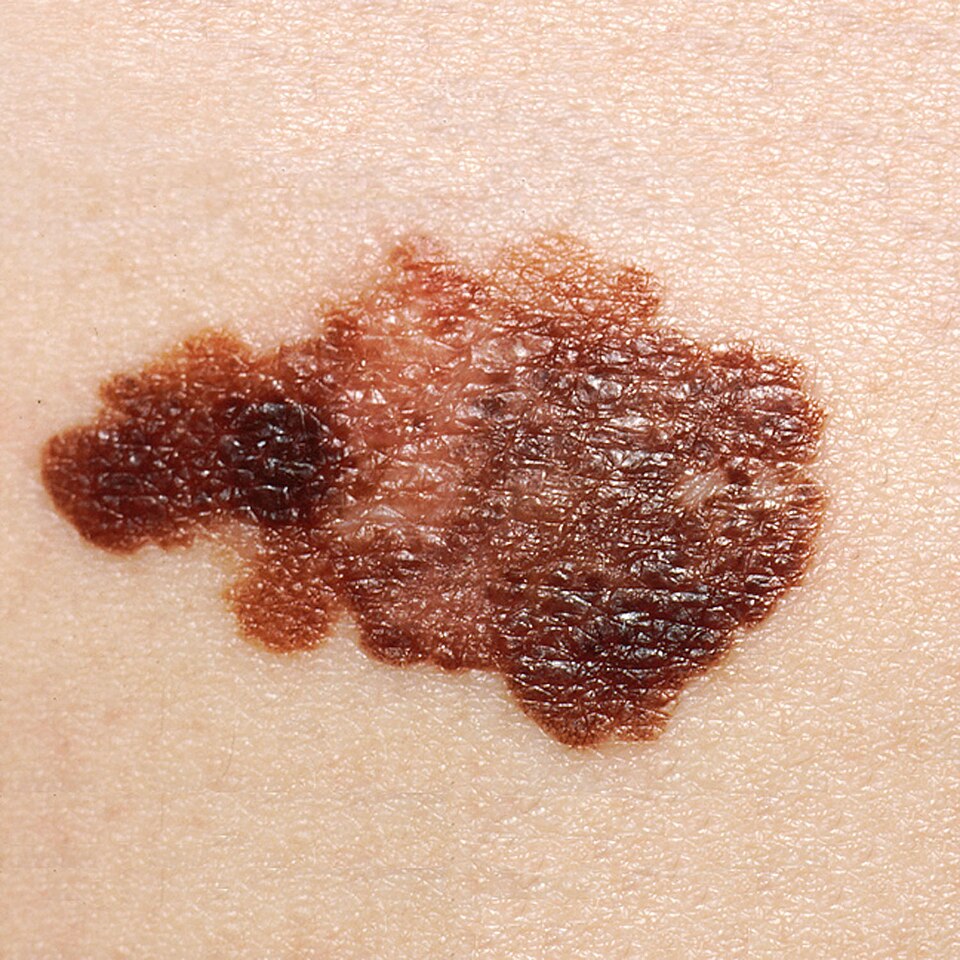
Superficial spreading melanoma
Image Source: Wikimedia Commons (2020), ECG AVNRT, Licenced under a creative commons licence. Available for free at: https://commons.wikimedia.org/wiki/File:Melanoma_(2).jpg (Accessed: 20 May 2026).
History and examination
History includes questions around size, shape, colouration, or ulceration of the lesion. Bleeding or pruritic lesions should have a high clinical suspicion for melanoma.
ABCDE is a useful way to assess lesions:
- A: asymmetry
- B: border irregularity
- C: colour
- D: diameter > 6mm
- E: evolution
Investigations
A vitamin D level should be checked at diagnosis in all patients with melanoma
Dermoscopy: atypical globules and dots, atypical network, blue-white veil
Biopsy and histopathology:
- Biopsy: full-thickness excision of lesion with 1-3mm margin
- Prognostic indicators: Breslow’s thickness (depth from granular cell layer to deepest tumour penetration in dermis), ulceration, mitotic count, lymphovascular invasion, microscopic satellites, serum LDH
- Abnormal and severe melanocytic proliferation in epidermis +/- dermis, asymmetrical growth pattern
Immunohistochemistry: markers used in analysis include Melan-A/MART-1, S-100, HMB-45, SOX10, MiTF and tyrosinase.
BRAF mutation testing should be performed in those with stage IIIB onwards.
Staging of cutaneous melanoma:
- Localised disease (stage I-III)
- Regional disease (stage III)
- Distant metastatic disease (stage IV)
Clark’s level
- Level 1: in-situ melanoma
- Level 2: invasion within papillary dermis
- Level 3: invasion filling papillary dermis
- Level 4: invasion within reticular dermis
Management
Dependent on subtype of melanoma and stage of cancer.
In patients where surgery is not indicated (i.e., multi-comorbid patients or difficult tumour location), topical chemotherapy (imiquimod), or radiotherapy may be used.
In those with melanoma in situ, topical imiquimod should be considered if removal of 5mm margins would cause disfigurement.
Unresectable advanced stage (stage III or IV):
- Systemic therapy (nivolumab and ipilimumab, nivolumab or pembrolizumab)
- Where surgery cannot be performed for metastasis, talimogene laherparepvec or electrochemotherapy may be indicated in some patients.
Surgical management
Early stage (melanoma in situ, stage I and stage II):
- Wide local excision with 5-10 mm margins (5mm for lentigo maligna).
- Mohs’ surgery can be considered in those with minimally invasive tumours on sites such as the ear or face.
- Sentinel lymph node biopsy (SNLB) may be used in those with stage I or stage II, and is an important prognostic factor in this patient group.
Resectable advanced stage (stage III or IV):
- Wide local excision PLUS adjuvant systemic therapy (nivolumab, pembrolizumab or dabrafenib and trametinib).
Monitoring
Early stage (I/IIA): follow-up recommended (up to four times in first year after treatment completion)
Those with stage IB-IIB should be followed up every 3 months for the first three years after treatment completion, followed by every six months for the next two years, with the aim of discharge after five years.
Advanced disease (stage III or IV) and receiving systemic therapy: regular imaging every 3 months to assess progression
Complications
High likelihood: adverse effects of treatment, SNLB complications (post-operative lymphodema), sequelae of metastatic disease
Low likelihood: local surgical complications (bleeding/infection), SNLB complications, local recurrence,
References
Complete Guide to Clinical Genetics ST3 Application and Recruitment (2027)
Written by Medibuddy. Last updated: October 2026. Clinical Genetics is

Complete Guide to Clinical Radiology ST3 Application and Recruitment (2027)
Recruitment paused: NHS England is not running Clinical Radiology ST3+ recru

Complete Guide to Paediatric Surgery ST3 Application and Recruitment in United Kingdom (2026)
Written by Medibuddy. Last updated: October 2026. If you’re planning


Take your subscriptions with you
Our mobile app allows you to access your interview and exam question banks wherever you are.

