


 Close
Close
 14th July 2026
Background
Squamous cell carcinoma (SCC) occurs in keratinocytes, located in the epidermis. SCC is the second most common skin cancer, secondary to basal cell carcinoma. Risk factors include UV exposure and immunosuppression. Treatment includes both non-surgical options (cryotherapy, topical and systemic chemotherapy) and surgical options (surgical excision, Mohs surgery).
Epidemiology
The second most common non-melanoma skin cancer, after basal cell carcinoma. Frequently seen in those >40 years of age, women, and in those with Fitzpatrick skin type I and II.
Aetiology
Solar UV exposure, ionising radiation, arsenic exposure, burns, previous psoralen, UV-A light therapy, HPV and immunocompromised states are known to increase risk of SCC. SCC occurs in keratinocytes, which have proliferated uncontrollably due to mutations and malignant transformation of the cells.
Risk factors can be separated into low risk, high risk and very-high risk as per the British Association of Dermatologists guidelines:
- Low risk:
- Patient factors: no lymphovascular or perineural invasion, Breslow thickness ≤ 4mm, tumour diameter ≤ 20mm
- Tumour factors: immune-competent
- High risk:
- Patient factors: immunosuppression or biological therapies, HIV therapy stabilised on HAART
- Tumour factors: lymphovascular invasion, poorly differentiated histology, tumour site, Breslow thickness of >4-6mm
- Very high risk:
- Patient factors: history of chronic lymphocytic leukaemia or myelofibrosis, solid organ transplant recipients
- Tumour factors: bone invasion, Breslow thickness >6mm, diameter >40mm
Diagnosis
SCC can be classified into four types, namely actinic keratosis (precursor lesion), SCC in situ (otherwise known as Bowen’s disease), invasive and metastatic.
SCC variants:
- Keratoacanthoma: rapidly growing dome-shaped nodule with keratin-filled central crater, grows over weeks and then shrinks after months.
- Verrucous carcinoma: fungating nodules or plaques, locally destructive, rarely metastasises
- Marjolin ulcer: aggressive, ulcerating, high rate of metastasis
History and examination
- Typically present with multiple actinic keratoses (erythematous, poorly defined, and irregularly shaped macules or plaques on sun-exposed areas of the skin).
- Most commonly seen on head and neck, and on upper extremities.
- Lesions are friable and can bleed easily.
- Lesions often have a scale or haemorrhagic crust.
SCC in situ (Bowen’s disease) is non-melanocytic and is characterised by a slow-growing and longstanding erythematous thin scaly patch/plaque with irregular and well-demarcated borders. (Figure 1)
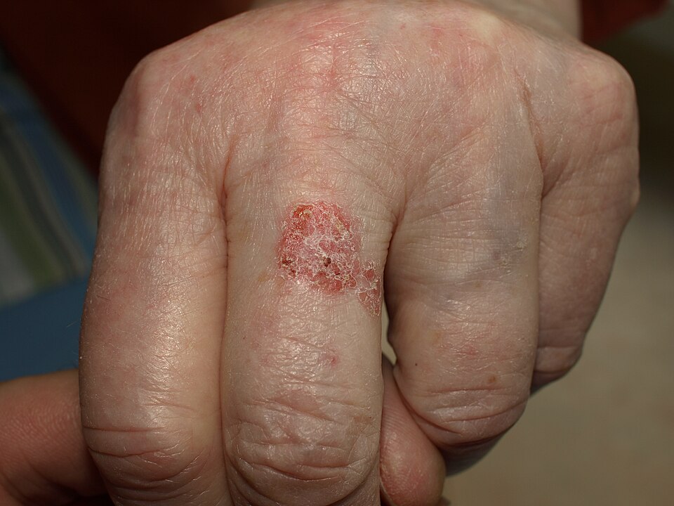
Bowen’s disease (cutaneous SCC in situ)
Image Source: Wikimedia Commons (2020), ECG AVNRT, Licenced under a creative commons licence. Available for free at: https://en.wikipedia.org/wiki/Cutaneous_squamous-cell_carcinoma#/media/File:Bowen11.jpg (Accessed: 25 May 2026).
SCCs typically are painful and fast-growing.
Invasive SCC presents as exophytic (outward-growing) and ulcerated tumours.
Regional lymph nodes are involved in 85% of metastatic SCCs.
Clinical history and examination are key to a diagnosis, although a skin biopsy is needed to confirm diagnosis.
Investigations
Dermoscopy:
- Actinic keratosis: erythematous pseudonetwork, surface scale, linear waxy vessels
- Bowen’s disease: erythema, keratosis, and glomerular vessels.
- Invasive: ulceration, keratinisation, irregular groups of white follicular cells
Histology typically shows nests of squamous cells with keratinisation, keratin pearls, large vesicular nuclei, and scant cytoplasm (Figure 2):
- Bowen’s disease: full-thickness keratinocyte atypia, confined to epidermis with an intact basement membrane
- Invasive SCC: extend beyond basement membrane
Imaging may be warranted for high-risk SCC: CT/PET/MRI
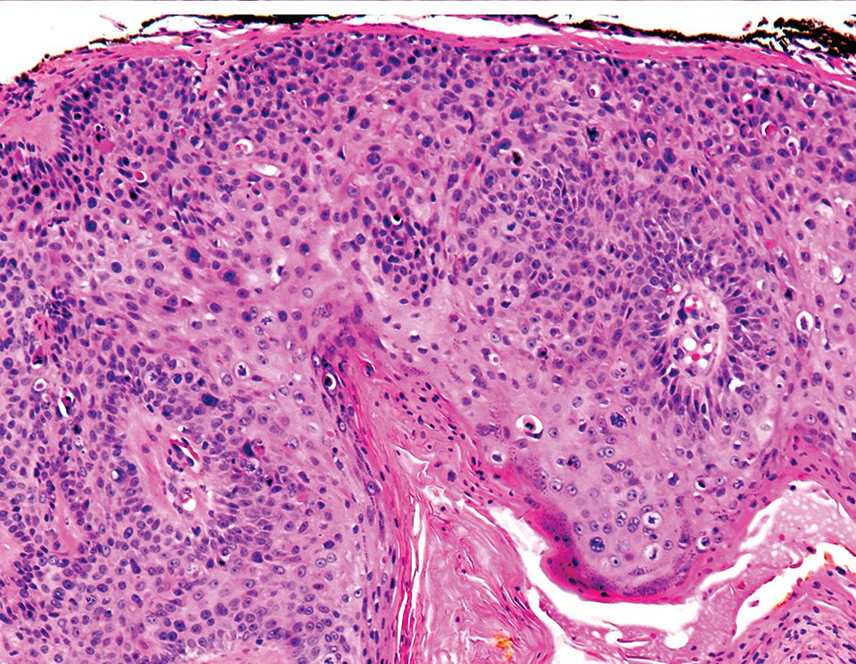
Figure 2: Histology of SCC
Image Source: Wikimedia Commons (2020), ECG AVNRT, Licenced under a creative commons licence. Available for free at: https://commons.wikimedia.org/wiki/File:Micrograph_of_squamous_cell_carcinoma_in_situ_-_100x.jpg (Accessed: 20 May 2026).
Management
Management differs depending on subtype of SCC:
- Bowen’s disease:
- Cryotherapy: liquid nitrogen leads to the delay in formation of vesicles or bullae.
- Topical chemotherapy: fluorouracil 5% (twice daily for 3-6 weeks) or imiquimod 5% (one daily, 2-3 times a week for 3-6 weeks).
- Second line:
- Surgical excision or Mohs: for tumours that do not respond
- Radiotherapy: for unresectable lesions or poor surgical candidates
- Invasive
- Conventional surgical excision (non-cosmetically sensitive), <2cm in diameter, and
- Excision with a ≥4mm if low risk, ≥6mm margin if high risk or ≥10mm margin if very high risk
- Mohs surgery (if cosmetically sensitive, i.e., face), >2cm in diameter
- Radiotherapy: may be indicated in aggressive subtypes
- Conventional surgical excision (non-cosmetically sensitive), <2cm in diameter, and
- Metastatic
- Surgical excision (non-cosmetically sensitive) PLUS radiotherapy
- Mohs surgery (cosmetically sensitive), >2cm PLUS radiotherapy
Complications
High likelihood: scarring
Medium likelihood: deformities due to local destruction,
References
Complete Guide to Clinical Genetics ST3 Application and Recruitment (2027)
Written by Medibuddy. Last updated: October 2026. Clinical Genetics is

Complete Guide to Clinical Radiology ST3 Application and Recruitment (2027)
Recruitment paused: NHS England is not running Clinical Radiology ST3+ recru

Complete Guide to Paediatric Surgery ST3 Application and Recruitment in United Kingdom (2026)
Written by Medibuddy. Last updated: October 2026. If you’re planning


Take your subscriptions with you
Our mobile app allows you to access your interview and exam question banks wherever you are.

