


 Close
Close
 4th June 2026
Investigations
Bloods
- FBC
- Us&Es
- LFTs
- Coagulation profile
- Troponin – If there has been a MI secondary to the dissection this may be raised
- Lactate – A raised lactate can indicate tissue ischaemia
- Group and save
- Crossmatch
- D-dimer – This will be elevated in most cases. Whilst a negative result significantly lowers the likelihood, it does not exclude a dissection.
- ABG
- Smooth muscle myosin heavy chain
Imaging
- ECG – Common to see ischaemic changes on an ECG and may mimic an MI in up to 40%. Roughly 30% of ECGs can be normal in an acute dissection.
- CXR – About 60% show a widened mediastinum (>8cm). It may also show an abnormal aortic contour, a globular heart (haemopericardium) or pleural effusion (L>R), which represents a haemothorax. A CXR may be normal in confirmed aortic dissection.
- CT angiography (whole aorta) – Gold standard and first-line in an acute setting. Sensitivity is 95-100%, and it is quicker than an MRI. However unable to assess for AR or LVF. Two lumens are identified with a visible intimal flap for diagnosis. An initial tear may be visualised and evidence of aortic dilatation or acute rupture. Any poor perfusion of end organs will also be seen.
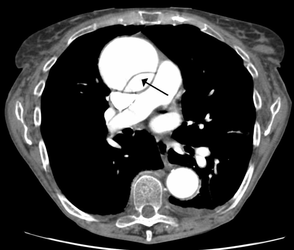
CT scan showing a dissection of the ascending aorta with an arrow pointing to the false lumen
Image Source: Wikimedia Commons (2020), ECG AVNRT, Licenced under a creative commons licence. Available for free at: https://commons.wikimedia.org/wiki/File:DissectionCT.png (Accessed: 27 April 2026)
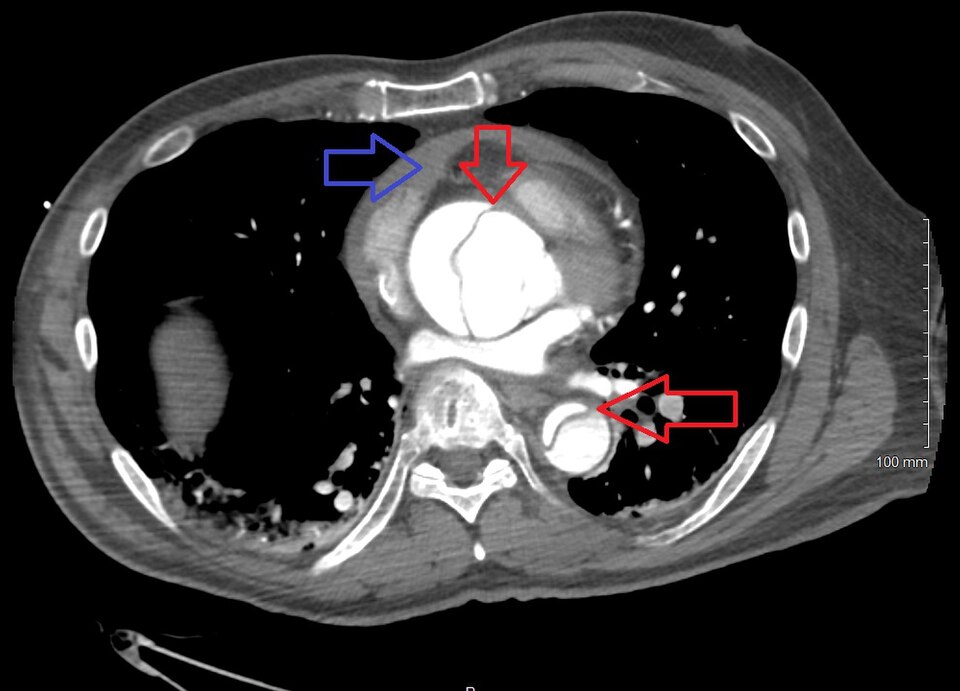
CT showing a Type A dissection with a pericardial effusion as a result. Red arrows show the ascending and descending thoracic aorta. The blue arrow highlights the pericardial effusion.
Image Source: Wikimedia Commons (2020), ECG AVNRT, Licenced under a creative commons licence. Available for free at: https://commons.wikimedia.org/wiki/File:Dissection2018WithPericardial.jpg (Accessed: 27 April 2026)
- Transthoracic echocardiogram (TTE) – Useful for identifying complications such as cardiac tamponade or aortic regurgitation. Poor sensitivity for type B dissections. Useful as an adjunct to CT.
- Transoesophageal echocardiogram (TOE) – Can be undertaken at the bedside. TOE has a sensitivity of roughly 98% and can easily view the extent of dissection.
- Magnetic resonance angiography (MRA) – High sensitivity, however, limited by its availability and difficulties in continuous monitoring whilst undergoing the MRA.
The Aortic Dissection Detection Risk Score (ADD-RS)
The ADD-RS is a risk stratification tool to assist with the low-to-moderate risk patient where a diagnosis of acute aortic dissection is suspected.
Low risk – score of 0, intermediate risk if the score is 1, and high risk if the score is >1.
A higher risk score indicates a higher likelihood of aortic dissection
Table 2: ADD-RS Scoring Criteria
| Clinical history | Points | |
|---|---|---|
| Anyone with these high-risk conditions | Family history of aortic disease, Marfan syndrome, known thoracic aortic aneurysm, Recent aortic manipulation, known aortic valve disease | +1 |
| Anyone with these high-risk pain features | Chest, abdominal or back pain that is described as: 1. Abrupt onset or 2. Severe intensity or 3. Tearing/ripping in nature | +1 |
| Anyone with these high-risk examination features | New murmur of aortic insufficiency (with any pain associated), hypotension or shock, Evidence of perfusion deficit | +1 |
Complete Guide to Clinical Genetics ST3 Application and Recruitment (2027)
Written by Medibuddy. Last updated: October 2026. Clinical Genetics is

Complete Guide to Clinical Radiology ST3 Application and Recruitment (2027)
Recruitment paused: NHS England is not running Clinical Radiology ST3+ recru

Complete Guide to Paediatric Surgery ST3 Application and Recruitment in United Kingdom (2026)
Written by Medibuddy. Last updated: October 2026. If you’re planning


Take your subscriptions with you
Our mobile app allows you to access your interview and exam question banks wherever you are.

