


 Close
Close
 5th June 2025
Pityriasis Versicolor
Pityriasis versicolour (PV) also known as tinea versicolor, is a common superficial fungal infection of the skin. Patients with this condition present with scaly macules that are hypo- or hyperpigmented.
Cause
It is caused by the lipid dependent Malassezia yeasts and therefore found in areas abundant in sebaceous glands- such as scalp, flexures and upper trunk. This fungus is part of the normal skin flora in over 90% of adults. However, in this condition the Malassezia transforms from yeast cells to a pathogenic mycelial form.
Factors leading to this pathological form include high temperature, hyperhidrosis, malnutrition, immunodeficiency, immunosuppression and application of occlusive creams or clothing to skin.
Prevalence
PV occurs worldwide but is most common in tropical climates, where prevalence can be as high as 50%. It usually affects adolescents and young adults as sebaceous glands are more active from hormonal changes.
Diagnosis
The diagnosis is usually made clinically through its characteristic features. There is normally several oval to round macules and confluent patches seen. In lighter skin tone, lesions fail to tan in the summer making it more apparent and in darker skin tone the affected skin is paler. The evoked scale sign is pathognomonic for PV and describes the fine scale on the affected skin becoming more obvious when the skin Is stretched or scraped.
From the history the skin lesions are asymptomatic, but mild pruritus occurs in some cases. The presenting complaint is usually the cosmetic disfigurement from pigmentary changes associated with the condition. Also, patients will describe a relapsing/recurring history with active flares during increase in weather temperatures.
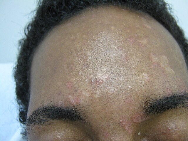
Image Source: Wikimedia Commons (2011). Pityriasis Versicolor Frontal Originale, Licensed under a creative commons licence. Available for free at: https://commons.wikimedia.org/w/index.php?search=Pityriasis_versicolor_frontal_originale.jpg&title=Special:MediaSearch&type=image (Accessed 20 May 2025).
Investigations
Microscopic confirmation using potassium hydroxide on skin scrapings can be used to diagnose PV in uncertain instances. The slides should resemble ‘spaghetti and meatballs’ as many short hyphae and spores will be revealed.
The skin can be examined under a Wood light in a dark room as lesions of PV may fluoresce a characteristic bright yellow or gold colour. The advantage of the test is limited as it is only positive in a third of cases. Only M furfur is fluorescent under Wood light and most infections are from non-fluorescent species of Malassezia.
A skin biopsy may be done to exclude conditions with similar presentation such as pityriasis alba, seborrheic dermatitis, confluent and reticulated papillomatosis, and vitiligo.
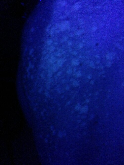
Image Source: Wikimedia Commons (2013). Tinea Versicolor (Wood’s Lamp), Licensed under a creative commons licence. Available for free at: https://commons.wikimedia.org/wiki/File:Tinea_Versicolor_(Wood%27s_Lamp).JPG (Accessed 20 May 2025).
Medical Management
The preferred first line treatment for PV is topical therapy. The first line treatments for extensive areas include ketoconazole 2% shampoo for 5 days or selenium sulphide for 7 days. These are applied to the affected area for around 10 minutes before being washed off in the shower. The first line treatment for a small area is imidazole cream applied for 3 weeks. These agents are normally well tolerated with the most common adverse effects being skin irritation and contact allergy. Systemic treatment can be used if extensive skin disease or topical treatment has not been effective. This includes oral itraconazole for 7 days or oral fluconazole for 2-4 weeks.
Spontaneous remission of PV is uncommon, and the infection will persist for many years if not treated. With treatment the prognosis is very good however recurrence is common especially in hot climates. Also, it usually takes 2-3 months for skin pigmentation to fully clear after beginning antifungal treatment, but can persist longer in some cases.
Prevention
Episodes of PV can recur in humid and warm environments so prophylactic treatment is recommended before exposure such as travel abroad. For this ketoconazole shampoo can be used 3 days before exposure. Oral agents are reserved in patients with extensive disease recurrence that is refractory to topical treatment. For this oral itraconazole can be used.
Complete Guide to Clinical Genetics ST3 Application and Recruitment (2027)
Written by Medibuddy. Last updated: October 2026. Clinical Genetics is

Complete Guide to Clinical Radiology ST3 Application and Recruitment (2027)
Recruitment paused: NHS England is not running Clinical Radiology ST3+ recru

Complete Guide to Paediatric Surgery ST3 Application and Recruitment in United Kingdom (2026)
Written by Medibuddy. Last updated: October 2026. If you’re planning


Take your subscriptions with you
Our mobile app allows you to access your interview and exam question banks wherever you are.

