


 Close
Close
 5th June 2025
Metabolic causes of hyperpigmentation
Cirrhosis
Cirrhosis is the diffuse pathological process characterised by fibrosis and change of normal liver architecture into structurally abnormal nodules.
Hyperpigmentation is a common clinical manifestation of cirrhosis. Skin changes that may be seen in some patients include a grey complexion and black discolouration in distal extremities, the pathogenesis of this is not fully understood and may be multifactorial due to bilirubinaemia, hormone changes associated with cirrhosis, or association with haemochromotosis. Also, due to extravasation of red blood cells and deposition of hemosiderin from lower extremity oedema and obstruction of venous reflux, pigmentation may be seen on the tibia.
In patients presenting with hyperpigmentation that may be suggestive of cirrhosis, investigations including blood tests, imaging and referral to gastroenterology for ongoing management should be considered. Management of hyperpigmentation related to cirrhosis requires the underlying causes of cirrhosis to be treated and managed. For patients with pruritus, non-sedating antihistamines, topical ammonium lactate and cholestyramine may be helpful.
Haemochromatosis
Haemochromatosis is a multisystem autosomal recessive disorder caused by a defect in the ability to downregulate iron absorption leading to iron overload and deposition in multiple body tissues.
It is most commonly due to a genetic condition from gene mutations but other causes include iron loading anaemias, parenteral iron overload, chronic liver disease and dysmetabolic iron overload syndrome.
It can lead to hyperpigmentation of the skin, but this is a rare and late finding. The pigmentation that occurs is distinctive and looks like skin bronzing. The finding can be confused with a tan.
Other clinical features include:
- Fatigue, depression, poor memory
- Arthralgia
- Loss of libido, impotence, early menopause, changes to menstrual cycle
- Loss of body hair
- Cardiomyopathy
- Diabetes
- Liver dysfunction
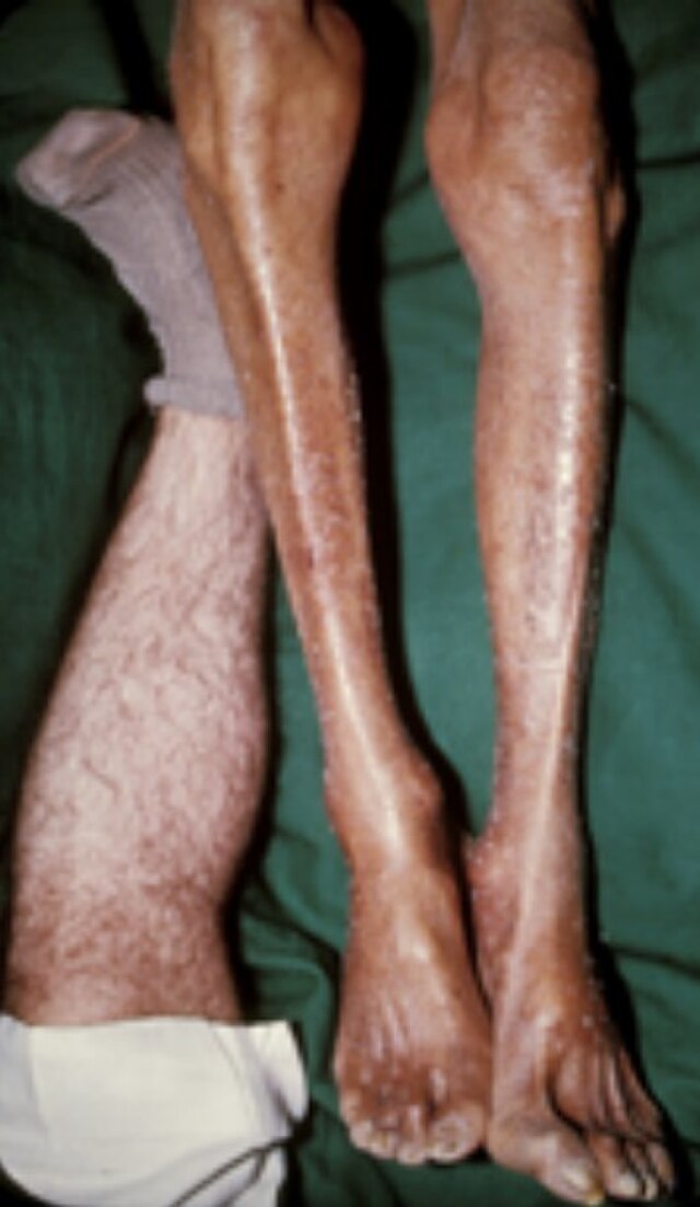
Image Source: Wikimedia Commons (2008). Hemochromatosis Skin, Licensed under a creative commons licence. Available for free at: https://commons.wikimedia.org/w/index.php?search=HemochromatosisSkin.jpg&title=Special:MediaSearch&type=image (Accessed 20 May 2025).
Haemochromatosis is suggested by raised transferrin saturation and ferritin on blood tests. Other tests include liver MRI, liver biopsy, LFT, fasting blood glucose, ECG, echocardiogram, bone densitometry and measuring sex hormones.
Management involves preventing iron overload in early- stage disease and removing excess iron from body stores in late-stage disease. Treatment includes:
- Phlebotomy regimen- removal of blood
- Avoid iron/ iron containing supplements
- Avoid vitamin C/ vitamin C containing supplements
- Prevention of worsening liver disease: Avoid excess alcohol, hepatitis vaccination/treatment, liver transplant
Complications include cirrhosis, hepatocellular cancer, arthropathy, diabetes and heart disease.
Porphyria Cutanea Tarda
Porphyria cutanea tarda is an acquired metabolic disorder of haem biosynthesis. It can be inherited but this is rarer. It occurs when there is significant deficiency of the enzyme uroporphyrinogen decarboxylase in the liver. The substrates of the deficient enzyme, which are porphyrinogens, accumulate in the liver and are oxidised to porphyrins and transferred to the skin causing cutaneous symptoms and signs. It is usually seen in middle-aged men and women.
Factors that contribute to susceptibility of porphyria cutanea tarda include hepatitis C, alcohol use, haemochromatosis, oestrogen treatment, HIV, smoking and uroporphyrinogen decarboxylase (UROD) mutations.
Signs and symptoms:
- Skin hyperpigmentation
- Photosensitivity
- Blistering and crusted skin lesion on sun exposed sites
- Hypertrichosis
- Sclerodermatous thickening
- Dark/ reddish urine
- Skin fragility
- Blister formation from minor trauma
- Liver damage
Investigations to confirm porphyria cutanea tarda involves finding significant increase in porphyrins and other related haem precursors in the urine, blood, and faeces. Genetic testing can be considered for UROD and haemochromatosis mutations for genetic counselling and treatment options too.
Treatment depends on the cause. In iron overload, regular phlebotomy is advised. Management options may also include reducing sun exposure, alcohol, smoking, oestrogen treatment and iron supplements. Specifically in iron overload or haemochromatosis regular venesection is advised. If there is no haemochromatosis or in those who cannot tolerate phlebotomy, low dose hydroxychloroquine or chloroquine is an option.
Renal Failure
Renal failure is defined as abnormalities in kidney structure or function. The most common causes for renal failure include diabetes mellitus and hypertension. Other causes include cystic kidney disorders, obstructive uropathy, glomerular nephrotic and nephritic syndrome.
Hyperpigmentation on the skin and nails is a feature in renal failure and is due to increase in skin melanin from excessive beta-melanocyte stimulating hormone. The hyperpigmentation may also be persistent and progressive despite dialysis as beta-melanocyte-stimulating hormones are poorly dialysed. Hyperpigmentation may also be from causes of renal failure such as diabetes mellitus complications. In diabetes mellitus, acanthosis nigricans may be present which is thickened, velvety, hyperpigmented plaques found on neck folds, axillae, and groin. Another cause of renal failure is neurofibromatosis which leads to renal artery stenosis, infarction, and aneurysm. The hyperpigmentation of this disease include café au lait macules which are flat hyperpigmented macules.
Other signs and symptoms:
- Haematuria
- Proteinuria
- Reduced GFR with elevated serum creatinine
- Pulmonary oedema- dyspnoea and orthopnoea
- Uraemia- fatigue, anorexia, nausea, vomiting, restless leg, pruritus, cognition abnormalities, seizures, coma
- Hypertension
- Peripheral oedema (from sodium retention and hypoalbuminaemia)
- Pallor due to anaemia
- Diabetic or hypertensive retinopathy
The following investigation are used in renal failure:
- Urea and electrolytes
- Urinalysis and protein/creatinine ration or albumin/creatinine ratio
- Full blood count
- Renal ultrasound with duplex, MRI
- Renal biopsy
The mainstay management of the dermatological manifestation of renal failure treatment of the underlying renal failure, with adjuncts such as antihistamines and topical creams being useful for some patients.
Complete Guide to Clinical Genetics ST3 Application and Recruitment (2027)
Written by Medibuddy. Last updated: October 2026. Clinical Genetics is

Complete Guide to Clinical Radiology ST3 Application and Recruitment (2027)
Recruitment paused: NHS England is not running Clinical Radiology ST3+ recru

Complete Guide to Paediatric Surgery ST3 Application and Recruitment in United Kingdom (2026)
Written by Medibuddy. Last updated: October 2026. If you’re planning


Take your subscriptions with you
Our mobile app allows you to access your interview and exam question banks wherever you are.

