


 Close
Close
 5th August 2024
Secondary headache syndromes
Overview
Secondary headaches occur as a symptom of underlying disease. It is a common symptom therefore the list of causes is extensive. This section will focus on: medication overuse headache, idiopathic intracranial hypertension, subarachnoid haemorrhage, cerebral venous sinus thrombosis, giant cell arteritis, intracranial neoplasia and head injury. A full list can be found in the International Classification of Headache Disorders-3.
Medication overuse headache
Medication overuse headache occurs in patients with a pre-existing headache disorder who overuse analgesia on a regular basis. It presents as a change in their usual headache. Patients will report having a headache most days (at least 15 days/month). It should be considered in those who regularly take paracetamol, aspirin, NSAIDs, triptans, opioids or ergots. The treatment is to stop the overused medications.
Idiopathic intracranial hypertension (IIH)
IIH is a disorder of raised intracranial pressure in patients with a normal ventricular system. The cause remains unknown, however we do know that it predominantly occurs in overweight females of childbearing age.
- Symptoms include a throbbing headache, transient loss of vision, blurring of vision, pulse -synchronous tinnitus, photophobia and nausea.
- Cranial nerve exam may show signs of raised intracranial pressure including papilloedema, visual field deficits and cranial nerve VI paresis.
- The Modified Frisen Scale is used to grade papilledema. Those with grade 3 to 5 papilloedema are at high risk of visual loss. The major criteria for each grade are shown below:
| Grade | Key feature |
|---|---|
| Grade 0 | Normal optic disc |
| Grade 1 | C-shaped halo with a temporal gap obscuring the retinal details |
| Grade 2 | A circumferential halo |
| Grade 3 | Obscuration of 1 or more segments of major blood vessels leaving the disc |
| Grade 4 | Total obscuration of a segment of a major blood vessel on the disc |
| Grade 5 | Obscuration of all vessels both on the disc and leaving the disc |
- The modified Dandy criteria states that CSF pressure must be >25 cmH2O on lumbar puncture for a diagnosis of IIH to be made.
- MRI brain may show an empty sella or flattening of the posterior globe.
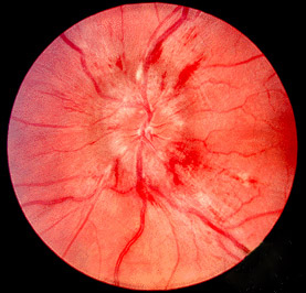
Image Source: Wikimedia Commons (2011). Papilledema, Licensed under a creative commons licence. Available for free at: https://commons.wikimedia.org/wiki/File:Papilledema.jpg (Accessed: 16 January 2024)
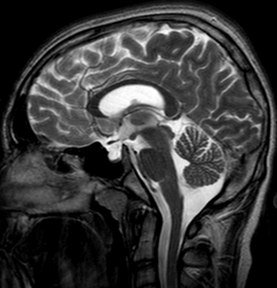
Image Source: Wikimedia Commons (2011). Empty SellaMRT T2 sag 002, Licensed under a creative commons licence. Available for free at: https://commons.wikimedia.org/wiki/File:Empty_Sella_MRT_T2_sag_002.jpg (Accessed: 16 January 2024)
- Initial treatment is with weight reduction, a low salt diet, fluid restriction, analgesia and stopping any medications which are known to increase intracranial pressure (for example, tetracyclines, retinoids and vitamin A). In patients with moderate papilloedema, acetazolamide or furosemide may also be started.
- If symptoms do not respond to medical treatment then optic nerve sheath fenestration or CSF shunting may be performed. The prognosis varies and symptoms can be transient or chronic. Evidence shows that with treatment about half of patients have significant improvement in their vision.
Subarachnoid haemorrhage
SAH refers to bleeding into the subarachnoid space, usually as the result of a ruptured intracranial aneurysm. Those with hypertension, autosomal dominant polycystic kidney disease, a smoking history or a family history of SAH are at increased risk of developing SAH.
- Patients report a severe, sudden ‘thunderclap’ headache associated with vomiting and meningism. In severe cases, it can present with coma or sudden death.
- A diagnosis can be confirmed by CT brain or lumbar puncture. Studies have shown that a CT performed within 6 hours of symptom onset can detect almost all cases, showing hyperdensity in the subarachnoid space. This sensitivity decreases over time, therefore in the majority of cases a lumbar puncture should also be performed if the scan is negative. This should be done at least 12 hours after the onset of a headache so that the xanthochromia can be accurately detected.
- Patients should be warned about the risk of developing a headache post lumbar puncture. The risk of this can be reduced by replacing the stylet before removing the LP needle.
- Note that hyponatraemia and hyperglycaemia can be features of SAH.
- All cases of confirmed SAH should immediately be discussed with the neurosurgery team. The use of nimodipine has been shown to improve outcomes.
- Around 30% of patients die within 30 days of a SAH and many survivors experience cognitive impairment.
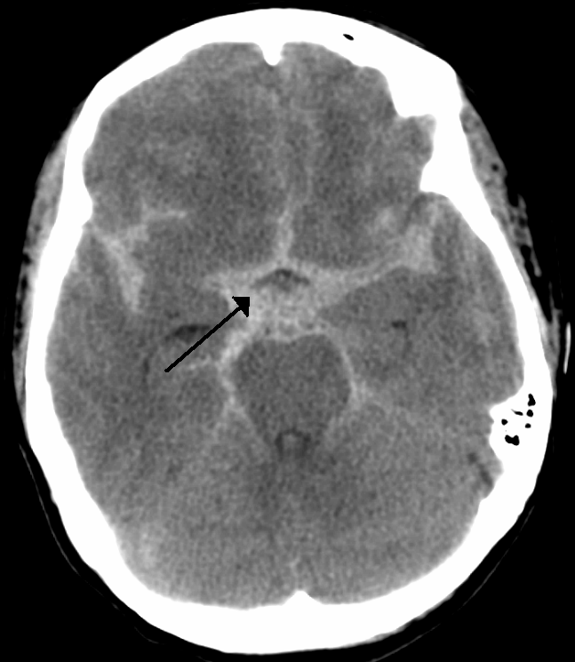
Image Source: Wikimedia Commons (2010). SubarachnoidP, Licensed under a creative commons licence. Available for free at: https://commons.wikimedia.org/wiki/File:SubarachnoidP.png (Accessed: 16 January 2024)
A condition that presents similarly to SAH is reversible cerebral vasoconstriction syndrome. This condition is caused by reversible constriction of the cerebral arteries and presents with recurrent thunderclap headaches. Some people also experience focal neurological deficits. Triggers such as stress, valsalva manoeuvres or exertion are often reported. The characteristic neuroimaging finding is multifocal segmental cerebral artery vasoconstriction on cerebral angiography. There may also be evidence of subarachnoid haemorrhage or ischaemia on imaging.
Cerebral venous sinus thrombosis
The pathophysiology of CVST (cerebral venous sinus thrombosis) is not fully understood, however it is thought to be caused by the thrombosis of cerebral veins or dural sinuses leading to blockages in the drainage of blood in the brain. Reduced CSF absorption in the brain is also thought to play a role. These mechanisms ultimately lead to an increase in intracranial pressure.
Risk factors are female sex, prothrombotic conditions, pregnancy and the puerperium, oral contraceptives, cancer, ENT infections and head injury.
It can be difficult to diagnose as it can present in a variety of ways. The headache can be acute, subacute or chronic and does not follow a specific pattern. Diagnosis relies on an MRI brain and MR venogram or CT brain and venogram.
The three key clinical presentations to look for are:
- Intracranial hypertensive syndrome: headache, visual changes and papilloedema.
- Focal neurological deficits: seizures, hemiparesis or dysphasia.
- Encephalopathy: delirium, multiple neurological deficits or coma.
It should also be noted that isolated thrombosis of the different sinuses or veins will present differently:
- Cavernous sinus thrombosis presents with ocular signs and is generally preceded by an ear, sinus or tooth infection.
- Lateral sinus thrombosis, a rare complication of untreated otitis media, usually presents with a headache or isolated intracranial hypertension but focal neurological deficits can be present (usually CN [cranial nerve] VI palsy).
- Sagittal sinus thrombosis presents with motor deficits or seizures. It is associated with the use of oral contraceptives and is also a rare complication of meningitis.
After diagnosis, antithrombotic treatment with low molecular weight heparin is started. Most patients are then given dabigatran or warfarin for a minimum of 3 months.
With treatment, the prognosis is usually quite good, with studies reporting a complete recovery in around 80% of patients. However about 5% unfortunately die during the acute phase of the disease.
Giant cell arteritis (GCA)
GCA is a vasculitis which affects medium and large vessels, especially branches of the external carotid artery. It is more common in those aged over 50 years.
Symptoms include headache, scalp tenderness, jaw claudication, fever and loss of vision. It is also commonly associated with polymyalgia rheumatica. In severe cases, patients can develop permanent loss of vision.
Blood tests will show raised CRP and ESR. Diagnosis is confirmed with vascular ultrasonography and/or temporal artery biopsy, however temporal biopsy has been shown to give a high number of false negative results. The ultrasound finding which is most suggestive of GCA is a circumferential hypoechoic vessel wall thickening which does not disappear on compression. This is known as the halo sign.
Treatment is with high-dose steroids, which should be started on suspicion of the diagnosis, rather than awaiting a temporal artery biopsy.
Intracranial neoplasia
Brain tumours can occur as primary intracranial neoplasms or as a result of metastatic malignancies.
Around 20% of those with a brain tumour experience headache. This headache is generally progressive, worse in the mornings, exacerbated by valsalva manoeuvres and associated with nausea and vomiting.
On examination, neurological deficits may be present, which are usually progressive.
CT brain with contrast will identify the site and size of the tumour. A biopsy is usually required to confirm the diagnosis.
Management will depend on the patient and the nature of the tumour. Dexamethasone is used to manage symptomatic oedema.
The most common primary brain tumours are gliomas, meningiomas and pituitary adenomas and the most common cause of brain metastases are lung and breast cancer. The typical neuroimaging findings of these tumours are described below:
| Tumour | Imaging findings |
|---|---|
| High-grade glioma | Hypointense masses on T1-weighted MRI, which enhance heterogeneously with contrast |
| Low-grade glioma | Hyperintense lesions on T2-weighted MRI. Most are non-enhancing |
| Meningioma | Extra-axial lesions which are hypointense (on T1 MRI) and enhance with contrast |
| Pituitary adenoma | Pituitary lesion which is isointense to grey matter. May extend into the suprasellar cistern and compress the optic chiasm |
| Brain metastases | Rounded, well circumscribed masses which enhance with contrast |
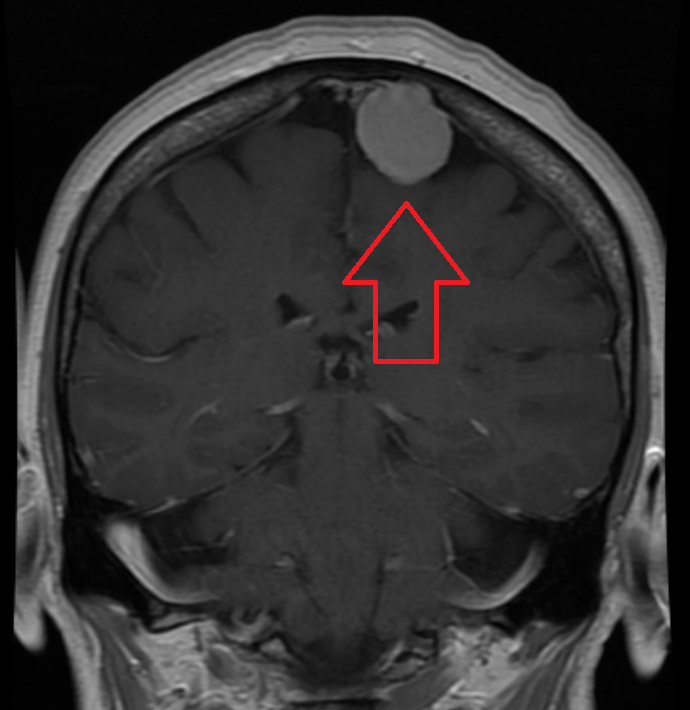
Image Source: Wikimedia Commons (2016). MRIMeningioma, Licensed under a creative commons licence. Available for free at: https://en.wikipedia.org/wiki/Meningioma#/media/File:MRIMeningioma.png (Accessed: 16 January 2024)

Image Source: Wikimedia Commons (2012). Brain MRI 131716 T2, Licensed under a creative commons licence. Available for free at: https://commons.wikimedia.org/wiki/File:Brain_MRI_131716_T2.png (Accessed: 16 January 2024)
It should be noted that pituitary adenomas can also present with a thunderclap headache in cases of pituitary apoplexy. This is when a bleed or infarction occurs in the pituitary gland, usually in the context of a pituitary adenoma. The possible complications include adrenal insufficiency, which should be treated with urgent corticosteroid replacement. The case should be urgently discussed with neurosurgery as transsphenoidal decompression should be considered.
Head injury
Head injuries are an extremely common A&E presentation, especially in teenagers, young adults and the elderly. Possible consequences of head injury include concussion, intracerebral haemorrhage, contusion, axonal injuries and skull fracture. Patients presenting with head injuries should be assessed using the ATLS (advanced trauma life support) guidelines. CT brain will identify most injuries. The guidelines as to when a CT head is indicated is shown in the table below.
| CT head to be performed within 1 hour if any of the following risk factors are present: | CT head to be performed within 8 hours if the patient has experienced some loss of consciousness or amnesia since the head injury and any of the: |
|---|---|
| GCS <13 on initial assessment or <15 2h post injury | Over 65 years old |
| Suspected opened or depressed skull fracture | History of bleeding or clotting disorders |
| Any sign of basal skull fracture | Those on anticoagulation therapy |
| Post-traumatic seizure | More than 30 minutes of retrograde amnesia |
| Focal neurological deficit | Dangerous mechanism of injury |
| More than 1 episode of vomiting |
Concussion is the most common injury seen in head trauma, however these microscopic changes cannot be seen on CT. The diagnosis is therefore clinical and symptoms include headache, dizziness and confusion.
Subdural haematomas usually occur in motor-vehicle accidents or falls. They are more common in those with cerebral atrophy or coagulopathies. Patients may develop confusion or focal neurological deficits (Figure 8).
Extradural haematomas usually occur after direct force to the skull and can be associated with fractures of the temporal bone. Patients may experience a lucid interval before a subsequent deterioration in GCS (Figure 9).

Image Source: Wikimedia Commons (2012). Subduralandherniation, Licensed under a creative commons licence. Available for free at: https://en.wikipedia.org/wiki/Subdural_hematoma#/media/File:Subduralandherniation.PNG (Accessed: 16 January 2024)
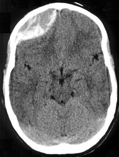
Image Source: Wikimedia Commons (2012). Traumatic acute epidual hematoma, Licensed under a creative commons licence. Available for free at: https://en.wikipedia.org/wiki/Epidural_hematoma#/media/File:Traumatic_acute_epidual_hematoma.jpg (Accessed: 16 January 2024)
Notable differentials
There are many conditions that can present with headache. Below are a few differentials to consider.
| Condition | Presentation and key information |
|---|---|
| Sinusitis | Nasal congestion and sinus tenderness. |
| Temporomandibular joint (TMJ) dysfunction | TMJ pain, TMJ click and limited mandibular movement. |
| Dental caries | Pain on consumption of sweet, hot or cold food or drink. Caries may be visible. |
| Otitis media | Ear pain. A bulging tympanic membrane may be present on otoscopy. |
| Acute angle-closure glaucoma | Eye pain, blurred vision, redness of the sclera and vomiting. |
| Hypertensive encephalopathy | Headache, nausea, change in mental status or visual changes in the context of a raised blood pressure. There may be evidence of end-organ damage on examination. |
| Pre-eclampsia | Usually presents after 20 weeks gestation. Patients will have a raised BP and proteinuria. The presence of a headache is suggestive of severe disease. If not treated the patient can develop eclampsia. |
| Carbon monoxide poisoning | Morning headaches, dizziness, ataxia and nausea. Oxygen saturations are usually normal but ABG will show raised carboxyhaemoglobin levels. |
| Malaria | Malaria and other vector-borne infections such as Dengue fever, Chikungunya and Zika virus can present with headache. |
| Stroke | Very rarely strokes can present with headache. |
References
- Classification Committee of the International Headache Society. The International Classification of Headache Disorders [Online]. 3rd edition. London, UK: Sage Publications Ltd, 2018. [Cited 11 Dec. 2021]. Available from: https://ichd-3.org/classification-outline/
- National Institute for Health and Care Excellence. Headaches. In: NICE Pathways. [Online]. National Institute for Health and Care Excellence, 2021. [Cited 11 Dec. 2021]. Available from: https://pathways.nice.org.uk/pathways/headaches#content=view-quality-statement%3Aquality-statements-classification-of-headache-type
- Neblett, M. Assessment of acute headache in adults. In: BMJ Best Practice. [Online]. London, UK: BMJ Publishing Group, 2021. [Cited 11 Dec. 2021]. Available from: https://bestpractice.bmj.com/topics/en-gb/9
- Manji, H., Connolly, S., Kitchen, N., Lambery, C., Mehta, A. Oxford Handbook of Neurology [Online]. 2nd edition. Oxford, UK: Oxford University Press, 2014. [Cited 4 Dec. 2021] Available from: https://oxfordmedicine.com/view/10.1093/med/9780199601172.001.0001/med-9780199601172
- Wall, M. and Mughal, M. Idiopathic intracranial hypertension. In BMJ Best Practice. [Online]. London, UK: BMJ Publishing Group, 2021. [Cited 11 Dec. 2021]. Available from: https://bestpractice.bmj.com/topics/en-gb/1070
- Ferro, J., Canhão, P. Cerebral venous thrombosis: Etiology, clinical features and diagnosis. In: Kasner, S., Goddeau, R., editors. UpToDate [Online]. Waltham (MA): UpToDate Inc: 2021. [Cited 12 Dec 2021]. Available from: https://www.uptodate.com/contents/cerebral-venous-thrombosis-etiology-clinical-features-and-diagnosis
- Ferro, J., Canhão, P. Cerebral venous thrombosis: Treatment and prognosis. In: Kasner, S., Goddeau, R., Nordli, D., editors. UpToDate [Online]. Waltham (MA): UpToDate Inc; 2021. [Cited 12 Dec. 2021]. Available from: https://www.uptodate.com/contents/cerebral-venous-thrombosis-treatment-and-prognosis
- Jones, M., Power, R. Subarachnoid haemorrhage. In: BMJ Best Practice. [Online]. London, UK: BMJ Publishing Group, 2021. [Cited 12 Dec. 2021]. Available from: https://bestpractice.bmj.com/topics/en-gb/3000106
- Singer, R., Ogilvy, C., Rordorf, G. Aneurysmal subarachnoid haemorrhage: Clinical manifestations and diagnosis. In: Biller, J., Rabinstein, A., Edlow, J., Goddeau, R., editors. UpToDate [Online]. Waltham (MA): UpToDate Inc; 2021. [Cited 12 Dec. 2021]. Available from: https://www.uptodate.com/contents/aneurysmal-subarachnoid-hemorrhage-clinical-manifestations-and-diagnosis
- Singer, R., Ogilvy, C., Rordorf, G. Aneurysmal subarachnoid haemorrhage: Treatment and prognosis. In: Biller, J., Rabinstein, A., Edlow, J., Goddeau, R., editors. UpToDate [Online]. Waltham (MA): UpToDate Inc; 2021. [Cited 12 Dec. 2021]. Available from: https://www.uptodate.com/contents/aneurysmal-subarachnoid-hemorrhage-treatment-and-prognosis
- Lay, C., Sun-Edelstein, C. Brain tumour headache. In: Swanson, J., Eichler, A., editors. UpToDate [Online]. Waltham (MA): UpToDate Inc; 2021. [Cited 12 Dec. 2021]. Available from: https://www.uptodate.com/contents/brain-tumor-headache
- Haydel, M. Assessment of traumatic brain injury, acute. In: BMJ Best Practice. [Online]. London, UK: BMJ Publishing Group, 2021. [Cited 12 Dec. 2021]. Available from: https://bestpractice.bmj.com/topics/en-gb/515
- National Institute for Health and Care Excellence. Head injury. In: NICE Pathways. [Online]. National Institute for Health and Care Excellence, 2021. [Cited 12 Dec. 2021]. Available from: https://pathways.nice.org.uk/pathways/head-injury/investigation-for-clinically-important-brain-injuries-in-patients-with-head-injury
- Williamson, C., Rajajee, V., Traumatic brain injury: Epidemiology, classification, and pathophysiology. In: Aminoff, M., Wilterdink, J., editors. UpToDate [Online]. Waltham (MA): UpToDate Inc; 2021. [Cited 12 Dec. 2021]. Available from: https://www.uptodate.com/contents/traumatic-brain-injury-epidemiology-classification-and-pathophysiology
- Mukhyar, C. Giant cell arteritis. In: BMJ Best Practice. [Online]. London, UK: BMJ Publishing Group, 2021. [Cited 20 Jan. 2022]. Available from: https://bestpractice.bmj.com/topics/en-gb/3000249
- Brook, I., Townsend, G. Brain abscesses and other focal pyogenic infections of the central nervous system. In: Cohen, J., Powderly, W., Opal, S., Infectious Diseases [Online]. 3rd edition. Elsevier Inc., 2010. [Cited 6 March 2022]. Available from: https://www.sciencedirect.com/topics/medicine-and-dentistry/lateral-sinus-thrombosis
- Absher, J. Cerebrovascular disease. In: Ramachandran, V., Encyclopaedia of the Human Brain [Online]. Elsevier Ltd, 2002. [Cited 6 March 2022]. Available from: https://www.sciencedirect.com/topics/medicine-and-dentistry/sagittal-sinus-thrombosis
- Chehade, J. Assessment of pituitary mass. In: BMJ Best Practice. [Online]. London, UK: BMJ Publishing Group, 2020. [Cited 6 March 2022]. Available from: https://bestpractice.bmj.com/topics/en-gb/1124/urgent-considerations
- Singhal, A. Reversible cerebral vasoconstriction syndrome. In: Kasner, S., Rabinstein, A., Tung, G. editors. UpToDate [Online]. Waltham (MA): UpToDate Inc; 2022. [Cited 6 March 2022]. Available from: https://www.uptodate.com/contents/reversible-cerebral-vasoconstriction-syndrome
- Wong, E., Wu, J. Overview of the clinical features and diagnosis of brain tumours in adults. In: Loeffler, J., Wen, P. editors. UpToDate [Online]. Waltham (MA): UpToDate Inc; 2021. [Cited 7 March 2022]. Available from: https://www.uptodate.com/contents/overview-of-the-clinical-features-and-diagnosis-of-brain-tumors-in-adults
- Snyder, P. Causes, presentation and evaluation of sellar masses. In: Cooper, D. editors. UpToDate [Online]. Waltham (MA): UpToDate Inc; 2021. [Cited 7 March 2022]. Available from: https://www.uptodate.com/contents/causes-presentation-and-evaluation-of-sellar-masses
Complete Guide to Clinical Genetics ST3 Application and Recruitment (2027)
Written by Medibuddy. Last updated: October 2026. Clinical Genetics is

Complete Guide to Clinical Radiology ST3 Application and Recruitment (2027)
Recruitment paused: NHS England is not running Clinical Radiology ST3+ recru

Complete Guide to Paediatric Surgery ST3 Application and Recruitment in United Kingdom (2026)
Written by Medibuddy. Last updated: October 2026. If you’re planning


Take your subscriptions with you
Our mobile app allows you to access your interview and exam question banks wherever you are.

