


 Close
Close
 5th August 2024
Head injury
Head injuries are an extremely common A&E presentation, especially in teenagers, young adults and the elderly. Possible consequences of head injury include concussion, intracerebral haemorrhage, contusion, axonal injuries and skull fracture. Patients presenting with head injuries should be assessed using the ATLS (advanced trauma life support) guidelines. CT brain will identify most injuries. The guidelines as to when a CT head is indicated is shown in the table below.
| CT head to be performed within 1 hour if any of the following risk factors are present: | CT head to be performed within 8 hours if the patient has experienced some loss of consciousness or amnesia since the head injury and any of the: |
|---|---|
| GCS <13 on initial assessment or <15 2h post injury | Over 65 years old |
| Suspected opened or depressed skull fracture | History of bleeding or clotting disorders |
| Any sign of basal skull fracture | Those on anticoagulation therapy |
| Post-traumatic seizure | More than 30 minutes of retrograde amnesia |
| Focal neurological deficit | Dangerous mechanism of injury |
| More than 1 episode of vomiting |
Concussion is the most common injury seen in head trauma, however these microscopic changes cannot be seen on CT. The diagnosis is therefore clinical and symptoms include headache, dizziness and confusion.
Subdural haematomas usually occur in motor-vehicle accidents or falls. They are more common in those with cerebral atrophy or coagulopathies. Patients may develop confusion or focal neurological deficits (Figure 8).
Extradural haematomas usually occur after direct force to the skull and can be associated with fractures of the temporal bone. Patients may experience a lucid interval before a subsequent deterioration in GCS (Figure 9).

Image Source: Wikimedia Commons (2012). Subduralandherniation, Licensed under a creative commons licence. Available for free at: https://en.wikipedia.org/wiki/Subdural_hematoma#/media/File:Subduralandherniation.PNG (Accessed: 16 January 2024)
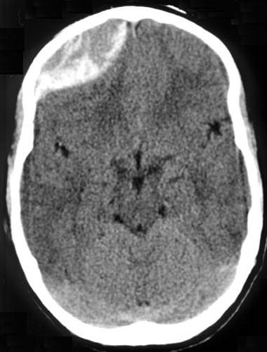
Image Source: Wikimedia Commons (2012). Traumatic acute epidual hematoma, Licensed under a creative commons licence. Available for free at: https://en.wikipedia.org/wiki/Epidural_hematoma#/media/File:Traumatic_acute_epidual_hematoma.jpg (Accessed: 16 January 2024)
Complete Guide to Clinical Genetics ST3 Application and Recruitment (2027)
Written by Medibuddy. Last updated: October 2026. Clinical Genetics is

Complete Guide to Clinical Radiology ST3 Application and Recruitment (2027)
Recruitment paused: NHS England is not running Clinical Radiology ST3+ recru

Complete Guide to Paediatric Surgery ST3 Application and Recruitment in United Kingdom (2026)
Written by Medibuddy. Last updated: October 2026. If you’re planning


Take your subscriptions with you
Our mobile app allows you to access your interview and exam question banks wherever you are.

