


 Close
Close
 5th August 2024
Intracranial neoplasia
Brain tumours can occur as primary intracranial neoplasms or as a result of metastatic malignancies.
Around 20% of those with a brain tumour experience headache. This headache is generally progressive, worse in the mornings, exacerbated by valsalva manoeuvres and associated with nausea and vomiting.
On examination, neurological deficits may be present, which are usually progressive.
CT brain with contrast will identify the site and size of the tumour. A biopsy is usually required to confirm the diagnosis.
Management will depend on the patient and the nature of the tumour. Dexamethasone is used to manage symptomatic oedema.
The most common primary brain tumours are gliomas, meningiomas and pituitary adenomas and the most common cause of brain metastases are lung and breast cancer. The typical neuroimaging findings of these tumours are described below:
| Tumour | Imaging findings |
|---|---|
| High-grade glioma | Hypointense masses on T1-weighted MRI, which enhance heterogeneously with contrast |
| Low-grade glioma | Hyperintense lesions on T2-weighted MRI. Most are non-enhancing |
| Meningioma | Extra-axial lesions which are hypointense (on T1 MRI) and enhance with contrast |
| Pituitary adenoma | Pituitary lesion which is isointense to grey matter. May extend into the suprasellar cistern and compress the optic chiasm |
| Brain metastases | Rounded, well circumscribed masses which enhance with contrast |
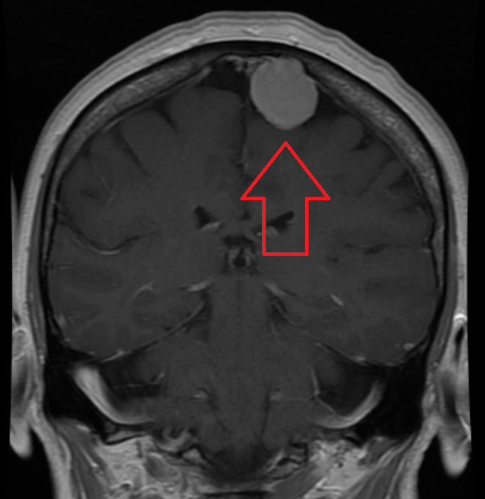
Image Source: Wikimedia Commons (2016). MRIMeningioma, Licensed under a creative commons licence. Available for free at: https://en.wikipedia.org/wiki/Meningioma#/media/File:MRIMeningioma.png (Accessed: 16 January 2024)

Image Source: Wikimedia Commons (2012). Brain MRI 131716 T2, Licensed under a creative commons licence. Available for free at: https://commons.wikimedia.org/wiki/File:Brain_MRI_131716_T2.png (Accessed: 16 January 2024)
It should be noted that pituitary adenomas can also present with a thunderclap headache in cases of pituitary apoplexy. This is when a bleed or infarction occurs in the pituitary gland, usually in the context of a pituitary adenoma. The possible complications include adrenal insufficiency, which should be treated with urgent corticosteroid replacement. The case should be urgently discussed with neurosurgery as transsphenoidal decompression should be considered.
Complete Guide to Clinical Genetics ST3 Application and Recruitment (2027)
Written by Medibuddy. Last updated: October 2026. Clinical Genetics is

Complete Guide to Clinical Radiology ST3 Application and Recruitment (2027)
Recruitment paused: NHS England is not running Clinical Radiology ST3+ recru

Complete Guide to Paediatric Surgery ST3 Application and Recruitment in United Kingdom (2026)
Written by Medibuddy. Last updated: October 2026. If you’re planning


Take your subscriptions with you
Our mobile app allows you to access your interview and exam question banks wherever you are.

