


 Close
Close
 19th June 2026
Hypertension
Hypertension
Background
Hypertension is a persistent elevation of blood pressure, defined as systolic blood pressure (SBP) ≥140/90 mmHg and/or diastolic blood pressure >90 mmHg. It is associated with an increased risk of cardiovascular disease (CVD; encompassing coronary heart disease, heart failure, stroke, myocardial infarction, atrial fibrillation and peripheral artery disease), stroke and renal failure.
Epidemiology
Hypertension is a significant contributor to the global burden of disease; it is the leading single contributor to all-cause mortality and disability worldwide. It affects more than 1 billion people and contributes to cardiovascular death more than any other risk factor globally. Certain ethnic groups are disproportionately affected, such as Black individuals, who are in turn more likely to experience complications. The World Health Organisation estimates that hypertension is responsible for nearly 7.5 million deaths worldwide. In the UK, the National Health Survey 2019 reported that hypertension affects approximately 1 in 4 adults and is a leading cause of morbidity and mortality.
The following are recognised risk factors for hypertension:
- Age
- Family history
- Obesity
- Physical inactivity
- High salt intake
- Alcohol consumption
- Smoking
- Stress
- Diabetes
- High cholesterol
- Sleep apnoea
Aetiology and pathophysiology
The mechanism underlying hypertension is multifactorial, displaying a complex interplay between genetic, environmental and lifestyle factors. Hypertension can be categorised into primary and secondary causes.
Primary hypertension (or essential hypertension) accounts for 90-95% of cases of hypertension and has no identifiable cause.
Secondary hypertension refers to hypertension with a known cause. These are summarised in Table 1.
Table 1. Secondary causes of hypertension
| Condition | |
|---|---|
| Cardiac | Aortic coarctation |
| Atherosclerotic renovascular disease | |
| Congenital heart defects | |
| Heart failure | |
| Endocrine causes | Cushing’s syndrome |
| Hyperthyroidism | |
| Hypothyroidism | |
| Primary aldosteronism | |
| Pheochromocytoma | |
| Renal | Chronic kidney disease |
| Glomerulonephritis | |
| Polycystic kidney disease | |
| Renal artery stenosis | |
| Other | Medications, such as NSAIDs, oral contraceptives, corticosteroids, ciclosporin and atypical antipsychotics |
| Obstructive sleep apnoea | |
| Pregnancy |
Clinical manifestations
The large majority of patients with hypertension are asymptomatic. Headaches are a common feature, classically occipital. Other associated features give useful clues to the underlying secondary cause of hypertension, for example, weight gain, muscle weakness and thinning skin in Cushing’s syndrome.
Malignant hypertension may be associated with:
- Hypertensive retinopathy
- Visual disturbance
- Cardiac symptoms, e.g. chest pain
- Confusion (cerebral oedema)
- Oliguria or polyuria
Diagnosis and staging
The evaluation of patients with hypertension involves a combination of clinical examination, laboratory tests, and imaging to assess for evidence of target-organ damage (for example, left ventricular hypertrophy and hypertensive retinopathy) and presence of comorbidities.
NICE guidelines recommend that initial investigations should include
- Measurement of blood pressure in both arms
- Physical examination to identify secondary causes of hypertension
- Assessment of cardiovascular risk. Formal assessment may involve using risk calculators such as the Framingham Risk Calculator 1991 or the Q-RISK2
Table 2. Directions by NICE for diagnosing hypertension
| Blood pressure measurement | Direction |
|---|---|
| Clinic blood pressure |
|
| Ambulatory blood pressure monitoring (ABPM) – used to confirm the diagnosis of hypertension |
|
| Home blood pressure monitoring (HBPM): offer if ABPM is unsuitable or the patient is unable to tolerate it |
|
In those diagnosed or whilst awaiting confirmation of diagnosis:
- Albumin-creatinine ratio (ACR) and urinalysis: to assess for renal impairment, as indicated by elevated ACR and proteinuria/haematuria
- Fundoscopy: to assess for hypertensive retinopathy
- 12-lead ECG: to assess for abnormalities in heart rhythm, structure or function
- Bloods: HbA1c, U&Es, total cholesterol and HDL cholesterol
Hypertension is confirmed if:
- Clinic blood pressure is ≥ 140/90 mmHg.
- ABPM or HBPM provide average values of ≥ 135/85 mmHg.
Table 3. Diagnostic criteria of hypertension by NICE
| Hypertension category | Clinic reading (mmHg) | ABPM/HBPM (mmHg) |
|---|---|---|
| Stage 1 | ≥140/90 | ≥135/85 |
| Stage 2 | ≥160/100 | ≥150/95 |
| Stage 3 (severe hypertension) | SBP ≥ 180 or DBP ≥ 120 Carry out same-day referral |
|
| Accelerated (malignant) hypertension | ≥180/120 with signs of retinal damage, haemorrhage or papilloedema. Usually associated with new or progressive target-organ damage. | |
| Isolated systolic hypertension | SBP ≥ 160 |
NICE, National Institute for Health and Care Excellence; ABPM, Ambulatory Blood Pressure Monitoring; HBPM, Home Blood Pressure Monitoring; SBP, systolic blood pressure; DBP, diastolic blood pressure
Table 4. Investigations for hypertension
| Investigation | Notes |
|---|---|
| Blood pressure measurement | All patients 24-hour ABPM or HBPM in suspected white coat hypertension or masked hypertension |
| Fasting blood glucose and lipid profile | Assessment of cardiovascular risk profile |
| Serum U&Es | Assessment of renal function |
| Endocrine investigations (e.g. renin/aldosterone ratio, TFTs) | Suspected secondary hypertension |
| Urinalysis | Assessment of proteinuria and haematuria |
| ECG | Assessment of abnormalities in heart rhythm, structure or function |
| Echocardiogram | |
| Renal artery imaging | Suspected renal artery stenosis |
ABPM, Ambulatory Blood Pressure Monitoring; HBPM, Home Blood Pressure Monitoring; U&Es, urea and electrolytes; TFTs, thyroid function tests
Cardiovascular disease risk assessment
Assessment of cardiovascular disease (CVD) risk is an important component in the management of hypertension. The NICE guidelines recommend using the QRISK3 risk assessment tool to calculate the 10-year risk of developing CVD in people with hypertension who are not already known to be at high risk.
The QRISK3 tool takes into account various factors, including age, sex, ethnicity, smoking status, systolic blood pressure, total cholesterol to HDL ratio, body mass index, family history of coronary heart disease, and other medical conditions such as diabetes, atrial fibrillation, and chronic kidney disease.
For individuals with a 10-year CVD risk of 10% or greater, NICE recommends offering statin therapy for primary prevention, in addition to lifestyle modifications and appropriate antihypertensive treatment. This emphasises the importance of considering overall cardiovascular risk, rather than treating hypertension in isolation.
Management
Management of hypertension involves a combination of lifestyle interventions and pharmacological therapy, which aim to reduce blood pressure and prevent complications.
Lifestyle interventions are the first-line treatment for suspected or diagnosed hypertension. This includes:
- Dietary changes: reducing sodium intake, increasing potassium intake, and following a Mediterranean diet
- Regular physical activity
- Weight loss for overweight/obese individuals
- Reducing caffeine
- Limiting alcohol consumption
- Smoking cessation
The choice of pharmacological therapy depends on factors such as age, ethnicity, comorbidities and contraindications. Figure 1 below outlines the patient groups who should be offered antihypertensive treatment. Figure 2 shows the stepped-care treatment protocol recommended by NICE, with medications added or changed based on a patient’s response to treatment.
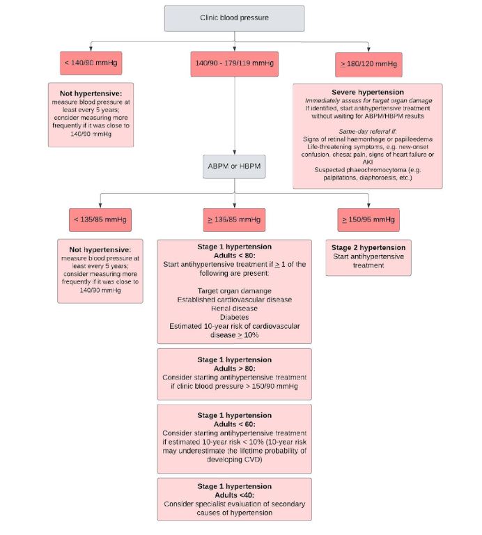
Summary of NICE guidelines on the initiation of antihypertensive drug treatment. Adapted from NICE guidelines (2019)
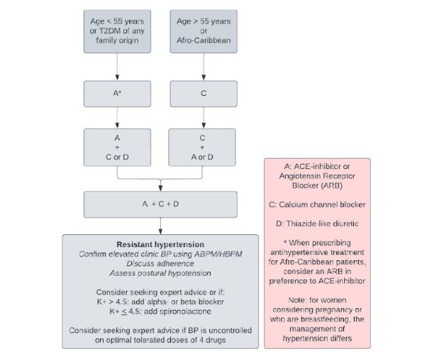
Stepped-care treatment protocol for the initiation of antihypertensives by NICE and the BIHS. Adapted from NICE guidelines (2019)
Table 5. NICE recommendations for blood pressure targets in patients with hypertension
| Patient group | Target blood pressure using ABPM/HBPM (mmHg) | Target blood pressure in clinic (mmHg) |
|---|---|---|
| Adults < 80 | < 135/85 | < 140/90 |
| Adults ≥ 80 | < 145/85 | < 150/90 |
Gestational hypertension
Background
Gestational hypertension (GH) is hypertension (blood pressure ≥ 140/90 mmHg on two occasions measured at least 4 hours apart) that develops after 20 weeks’ gestation in previously normotensive women in the absence of proteinuria. GH is a common complication in pregnancy and affects 6-10% of pregnancies globally.
Risk factors
- Age > 40 years
- Pre-existing hypertension, including essential hypertension, preeclampsia or gestational hypertension
- Pre-existing renal or vascular disease
- Obesity
- Diabetes
- Nulliparous
- Multiple pregnancy
- Pregnancy interval > 10 years
- Family history of preeclampsia
- Black or Hispanic ethnicity
Clinical features
- Most women are asymptomatic
- Headache that is persistent and severe
- Visual disturbances
- Women should be asked about symptoms that suggest pre-eclampsia, e.g. weight gain, peripheral oedema, headaches, and upper abdominal pain
Diagnosis and investigations
Table 6. Investigations for gestational hypertension
| Investigation | Notes |
|---|---|
| Blood pressure measurement |
|
| Urine protein |
|
| FBC |
|
| LFTs |
|
| Renal function tests |
|
| Coagulation studies |
|
| Foetal surveillance, e.g. assessing foetal movement or biophysical profile testing |
|
| Other |
|
Severity classification of gestational hypertension:
- Hypertension: systolic BP ≥ 140/90 mmHg or diastolic BP ≥ 90 mmHg, or both
- Severe hypertension: BP persistently ≥ 160/100 mmHg for a minimum of 4 hours
Complications
- Pre-eclampsia: a severe form of GH, characterised by hypertension, proteinuria and end-organ damage
- Placental abruption: the placenta separates from the uterine wall, causing heavy bleeding and foetal distress
- Intrauterine growth restriction: leads to low birth weight and other complications
- Pre-term delivery: may be necessary if GH is severe or if there are other complications
Management
The management of GH depends on the severity of the condition, and ranges from close monitoring of BP and urine protein levels in mild GH to admission to hospital and antihypertensive treatment in the context of high or persistently elevated BP (summarised in Table 2).
Important considerations
- Lifestyle modifications: the same advice on rest, exercise and work to women with chronic hypertension
- Antihypertensive treatment: Oral labetalol is 1st line (avoid in asthma). Alternatives include:
- Nifedipine if worsening hypertension previously controlled with oral labetalol
- Methyldopa, if labetalol or nifedipine are not suitable; avoid postpartum due to risk of depression, and change to an alternative within 2 days of delivery if started.
- Planned early delivery. Indications include the following:
- Preterm labour or premature rupture of membranes
- Vaginal bleeding
- Abnormal foetal testing
- Intrauterine growth restriction
- Development of pre-eclampsia or eclampsia
- Evidence of end-organ damage (e.g. neurological, hepatic or renal dysfunction)
- Corticosteroids (e.g. betamethasone), if planned early delivery < 37 weeks’ gestation is necessary to promote foetal lung maturation, as indicated
- Magnesium sulphate if planned early delivery < 34 weeks’ gestation, for neuroprotection, as indicated
- Low-dose aspirin (PO 75mg) in subsequent pregnancies to reduce the risk of pregnancy-induced hypertension and pre-eclampsia
Table 7. Management of gestational hypertension
| Management | Mild GH | Severe GH |
|---|---|---|
| Admit to hospital | Not routinely recommended | Admit to hospital |
| BP monitoring | Once or twice per week until BP ≤ 135/85 mmHg | Every 15-30 mins until BP < 160/100 mmHg |
| Urine protein levels | Once or twice per week | Daily |
| Blood tests | FBC, LFTs and renal function at presentation, then weekly PIGF-based testing on 1 occasion if there is suspicion of pre-eclampsia |
|
| Foetal assessment | Foetal heart auscultation at every antenatal appointment | |
| Foetal US at diagnosis – if normal, repeat every 2-4 weeks if indicated | Foetal US at diagnosis – if normal, repeat every 2 weeks if severe HTN persists | |
| CTG only if indicated | CTG at diagnosis; repeat only if indicated | |
| Antihypertensive medication | Not routinely recommended, but may be considered by BP remains elevated (> 140/90 mmHg) despite lifestyle modifications, or if there are concerns regarding maternal/foetal well-being Target BP: ≤ 135/85 mmHg |
1st line options: Labetalol PO/IV, Nifedipine PO, Hydralazine IV Target BP: ≤ 135 /85 mmHg |
| Delivery | Do not offer planned early delivery < 37 weeks’ gestation unless indications for early delivery are met. | Indicated if severe hypertension, end-organ damage or signs of foetal compromise. Timing should balance the risks of prematurity against the risks of continuing the pregnancy |
Postnatal investigation, monitoring and treatment
- Continue monitoring the mother’s BP. Measure BP daily for the first 2 days after birth, at least once between day 3 and day 5, and as clinically indicated afterwards.
- Continue antihypertensive treatment if required. Start treatment if BP ≥ 150/100 mmHg and they were not treated during pregnancy. Reduce treatment dosages if BP falls < 130/80 mmHg
- Follow-up with GP or specialist 6-8 weeks after birth, or 2 weeks after transfer to community care for women who remain on antihypertensive treatment
Pre-eclampsia and eclampsia
Background
Pre-eclampsia is a systemic syndrome in pregnancy that usually occurs after 20 weeks’ gestation, characterised by hypertension and proteinuria. The mechanism underlying pre-eclampsia has not been fully elucidated; however, it is thought to be related to abnormal placentation and inadequate trophoblastic invasion of the uterine spiral arteries, leading to vasoconstriction, increased systemic vascular resistance, and decreased organ perfusion. Eclampsia is a complication of pre-eclampsia, characterised by seizures.
Epidemiology
Nearly 2-8% pregnancies worldwide are implicated by pre-eclampsia. Eclampsia is a rare complication, with an incidence of 1 in 2,000 pregnancies.
Risk factors
- Nulliparity
- Previous maternal history or family history
- BMI > 30
- Maternal age > 40 years
- Multiple pregnancy
- > 10-year interval since previous pregnancy
- Sub-fertility
- Gestational hypertension
- Pre-existing diabetes
- Pre-existing renal or vascular disease
- PCOS
- Autoimmune disease
Clinical features
- The key clinical features of pre-eclampsia are hypertension and proteinuria after 20 weeks’ gestation
- Women may be asymptomatic and diagnosed at a routine clinic visit
- Headaches (usually frontal): indicates severe pre-eclampsia
- Visual disturbances
- Peripheral oedema
- Upper abdominal pain (usually right upper quadrant)
- Nausea and vomiting
- Breathlessness (due to pulmonary oedema): one of the main causes of maternal mortality
- Seizures: requires prompt admission to ICU, stabilisation and delivery
- Oliguria
HELLP syndrome is a variant of severe pre-eclampsia characterised by haemolysis, elevated liver enzymes, and low platelets.
Diagnosis and investigations
Diagnosis is based on the presence of the following after 20 weeks’ gestation:
- Hypertension: SBP ≥ 140 mmHg and/or a DBP ≥ 90 mmHg on 2 occasions at least 4 hours apart
- Proteinuria: ≥ 30 mg/mmol on PCR or ≥ 0.3g/24 hours or ≥ 1+ on a urine dipstick
After diagnosing pre-eclampsia, further testing should be performed, including foetal assessment for complications and maternal testing for systemic involvement. A summary of the investigations can be found in Table 1.
Table 8. Investigations for pre-eclampsia
| Investigation | Notes |
|---|---|
| Blood pressure | At each antenatal visit |
| Urine dipstick for proteinuria | At each antenatal visit NICE recommends not routinely testing for 24-hour urine collection to quantify proteinuria in pregnancy. Alternative spot tests should be used, e.g. PCR |
| FBC | Anaemia can exacerbate hypertension Thrombocytopaenia is a potential complication of pre-eclampsia |
| LFTs | Hepatic dysfunction is a potential complication of pre-eclampsia Elevated liver enzymes may prompt further investigations |
| Renal function tests | Renal impairment is a potential complication of pre-eclampsia |
| Coagulation studies | Coagulopathy is a potential complication of pre-eclampsia |
| Serum uric acid | May be elevated in pre-eclampsia |
| Serum calcium | May be decreased in pre-eclampsia |
| Assessment of fetal well-being |
|
| Other |
|
PCR, protein: creatinine ratio; PIGF, placental growth factor
Severity classification of pre-eclampsia:
- Hypertension: BP of 140/90 – 159/109 mmHg
- Severe hypertension: BP of ≥ 160/110 mmHg
Complications
Maternal complications
- Seizures (eclampsia)
- Placental abruption
- Pulmonary oedema
- Pregnancy-associated stroke
- Cardiovascular disease
- Renal failure
- Death
Foetal complications
- IUGR
- Pre-term delivery
- Intrauterine death
Management
Managing pre-eclampsia requires attention to the following critical considerations:
- Monitoring
- Delivery: date and method
- Controlling blood pressure
- Controlling seizures
- Postnatal fluid management
Important considerations
- Antihypertensive treatment: choice should be based on pre-existing treatment, possible side effects, maternal vs foetal risk, and the woman’s preference. Combination therapy may be required.
- Corticosteroids (e.g. betamethasone) to promote foetal lung maturation, as indicated
- Magnesium sulphate if planned early delivery. It is the treatment of choice for eclampsia. Monitor for signs of magnesium toxicity (e.g. respiratory depression and loss of reflexes); calcium gluconate may be used as a reversal agent.
- Delivery: the definitive management of pre-eclampsia, however it may take a few days to resolve completely. Indications for planned early delivery < 37 weeks may include the following:
- Inability to control maternal BP despite ≥ 3 classes of antihypertensives
- Maternal pulse oximetry < 90%
- Progressive deterioration in blood results
- Ongoing neurological features, e.g. severe intractable headache, repeated visual scotomata
- Placental abruption
- Reversed end-diastolic flow in umbilical artery doppler velocimetry, a non-reassuring CTG, or stillbirth
Table 9. Management of pre-eclampsia
| Management | Hypertension | Severe hypertension |
|---|---|---|
| Admit to hospital | Admit to hospital if clinical concerns for maternal or foetal wellbeing, e.g. impending eclampsia, pulmonary oedema or suspected foetal compromise Admit to hospital. If BP falls < 160/110 mmHg, manage as for hypertension |
|
| Risk prediction models like fullPIERS or PREP-S have been validated and can be used alongside a comprehensive clinical assessment to help guide decisions about admission and thresholds for intervention | ||
| BP monitoring | At least every 48 hours (more frequently if an inpatient) Every 15-30 mins until BP < 160/100 mmHg, then ≥ 4 times daily while an inpatient |
|
| Urine protein levels | Only repeat if clinically indicated, e.g. if new symptoms or signs develop, or if there is uncertainty over diagnosis | |
| Blood tests | FBC, LFTs and renal function 2x / week | FBC, LFTs and renal function 3x / week |
| Foetal assessment | Foetal heart auscultation at every antenatal appointment Foetal US at diagnosis – if normal, repeat every 2 weeks CTG at diagnosis, then only if clinically indicated |
|
| Antihypertensive medication | 1st line: Labetalol 2nd line: Nifedipine 3rd line: Methyldopa |
|
| Offer if BP > 140/90 mmHg Target BP: ≤ 135/85 mmHg |
Offer to all women Target BP: ≤ 135/85 mmHg |
|
| Timing of delivery | < 34 weeks: continue surveillance unless there are indications for planned early birth. Offer IV magnesium sulphate and corticosteroids 34-36+⁶ weeks: continue surveillance unless there are indications for planned early birth. Consider corticosteroids ≥ 37 weeks: initiate birth within 24-48 hours |
|
fullPIERS, Pre-eclampsia Integrated Estimate of RiSk; PREP-S, Prediction of complications in early-onset pre-eclampsia (using a survival model).
Postnatal investigation, monitoring and treatment
- Measure BP at least once between day 3 and 5 after birth, then on alternate days until normal, if BP was abnormal
- Continue antihypertensive treatment if required. Start treatment if BP ≥ 150/100 mmHg and they were not treated during pregnancy. Reduce treatment dosages if BP falls < 130/80 mmHg
- Follow-up with GP or specialist 6-8 weeks after birth, or 2 weeks after transfer to community care for women who remain on antihypertensive treatment
- The main risk to the mother in this period is fluid overload; therefore, fluid management is crucial. NICE guidelines fluid restriction to 80 ml/hour
Hypertensive emergency
Background
Hypertensive emergency is defined as a significantly elevated blood pressure which usually results in end-organ damage. Generally, this is a systolic blood pressure greater than 180mmHg and a diastolic blood pressure of 120mmHg. End-organ damage may include acute kidney injury, ischaemic or haemorrhagic stroke, myocardial ischaemia or heart failure.
Epidemiology
1-2% of patients with hypertension will suffer from a hypertensive emergency in their lives.
Risk factors
Hypertensive emergency occurs largely due to undiagnosed as well as poorly controlled essential hypertension. It may also be due to secondary hypertension caused by a systemic condition such as phaeochromocytoma or hyperaldosteronism.
Poor lifestyle choices such as increased salt intake and obesity, as well as certain medications such as steroids, sympathomimetics and NSAIDs, may also predispose a hypertensive emergency.
Clinical features
A patient with hypertensive emergency may present with no symptoms and may incidentally find a very high blood pressure. Patients, however, may present with a variety of symptoms seen in the table below.
| Symptoms | Signs | |
|---|---|---|
| Neurological | Headache, dizziness, blurry vision, weakness | Abnormal cognition, motor function, sensation, gait, cranial nerve function |
| Cardiac | Chest pain, shortness of breath, palpitations, dizziness, leg swelling | Murmur, peripheral oedema, S3, raised JVP |
| Renal | Change in urine output | |
| Fundoscopy | Retinal haemorrhages, exudates, papilledema |
Diagnosis and investigations
Diagnosis is generally based on a systolic blood pressure >180mmHg and a diastolic pressure >120mmHg.
Investigations may include:
- CT head: This may reveal a hemorrhagic stroke, but will not rule out an ischaemic stroke
- Fundoscopy: to look for signs of papilloedema
- Urine dipstick: to look for blood and protein in the urine
- Renal profile: deranged renal function may explain
- ECG: to look for left ventricular hypertrophy and rule out ischaemia
- Cardiac enzymes: to rule out myocardial infarction
- Urine drug screen: illicit drugs
- Thyroid function tests
- Plasma renin and aldosterone levels may point towards hyperaldosteronism as a cause
- Urine metanephrines for phaeochromocytoma
- 24-hour urinary cortisol for Cushing’s syndrome
- CXR
- CT aortogram if aortic dissection is suspected.
- Renal ultrasound / MRA
Complications
- Heart failure
- Cerebral ischaemia, haemorrhage, encephalopathy
- Acute kidney injury
Management
Management of hypertensive emergencies should not be delayed whilst investigating end-organ damage and underlying cause. Patients are usually kept on a cardiac monitor and commenced on parenteral anti-hypertensive medication with the aim of gradual reduction in blood pressure to avoid renal, cerebral or cardiac ischaemia. This is with a few exceptions, as detailed below.
Generally, it is recommended to reduce blood pressure by no more than 25% in the first hour. A further reduction is done in the next 4-6 hours with an aim of reaching a normal blood pressure within 24-48 hours, and weaning parenteral infusion.
There are a few exceptions:
- Patients suffering from an ischaemic stroke should not have an aggressive reduction in blood pressure and may benefit from maintaining a very high blood pressure to maintain cerebral perfusion.
- On the other hand, patients with ischaemic stroke who are candidates for thrombolysis should have a gentle reduction in their blood pressure <185/110 before thrombolysis.
- Patients suffering from an aortic dissection should have aggressive reduction in blood pressure within the first hour to a systolic less than 120mmHg.
- Patients with pre-eclampsia, eclampsia or phaeochromocytoma crisis should have a reduction in their blood pressure within the first hour to a systolic <140mmHg.
Specific emergencies
Malignant hypertension
Malignant hypertension is a subcategory of hypertensive emergency, which includes either grade III or IV retinopathy. Hypertensive encephalopathy refers to neurological symptoms caused by severely elevated blood pressure and typically improves with blood pressure correction. Intracranial haemorrhage may occur with malignant hypertension.
Management:
- 1st line – Labetalol infusion
- 2nd line – Nicardipine or nitroprusside if no evidence of raised intracranial pressure
- 3rd line – Fenoldopam
Aim to reduce mean arterial pressure by 20-25% within the first 2-4 hours, then to 160/100-110 mmHg within the next 2-6 hours [2].
Myocardial Ischaemia or Infarction
Management:
- 1st line – Esmolol + GTN infusion
- 2nd line – Labetalol + GTN infusion
Heart failure / pulmonary oedema
IV nitroglycerin or nitroprusside may be considered as an adjuvant to diuretic therapy. Due to the potential of significant hypotension, nitroprusside should be used with invasive haemodynamic monitoring, typically in the intensive care setting.
Aortic Dissection
Aortic dissection requires particularly aggressive blood pressure management to reduce the risk of propagation of the dissection.
Aortic dissection is classified based on its location and duration:
- Stanford Classification:
- Type A: Involves the ascending aorta, with or without the involvement of the aortic arch and descending aorta
- Type B: Involves only the descending aorta
- DeBakey Classification:
- Type I: Originates in ascending aorta, propagates at least to the aortic arch
- Type II: Originates in and is confined to the ascending aorta
- Type III: Originates in descending aorta, extends distally or proximally
Management:
- 1st line – Combination of IV beta-blocker (e.g., labetalol or esmolol) and vasodilator (e.g., nicardipine or nitroprusside)
- If using labetalol alone, higher doses may be required
Targets:
- Reduce systolic BP to <120 mmHg within the first hour of presentation
- Maintain systolic BP < 120 mmHg or to the lowest BP that maintains adequate end-organ perfusion
- Target heart rate of 60 to 80 bpm
Definitive treatment depends on the type of dissection:
- Type A typically requires urgent surgical intervention
- Uncomplicated Type B can often be managed medically, with endovascular repair considered in certain cases
Remember, in all cases, the key principle is to lower blood pressure gradually to avoid organ hypoperfusion, while being mindful of the specific risks associated with each condition.
References
- Forouzanfar, M. H. et al. Global, regional, and national comparative risk assessment of 79 behavioural, environmental and occupational, and metabolic risks or clusters of risks, 1990–2015: a systematic analysis for the Global Burden of Disease Study 2015. Lancet 388, 1659–1724 (2016).
- World Health Organization. Hypertension. https://www.who.int/news-room/fact-sheets/detail/hypertension. Accessed on 24 March 2023.
- NHS Digital. Health Survey for England 2019: Hypertension. https://digital.nhs.uk/data-and-information/publications/statistical/health-survey-for-england/2019/part-6-cardiovascular-disease-and-risk-factors/hse19-cardiovascular-disease-and-risk-factors-hypertension. Accessed on 24 March 2023.
- Public Health England. Health profile for England: 2021. https://www.gov.uk/government/publications/health-profile-for-england. Accessed on 24 March 2023.
- National Institute for Health and Care Excellence (NICE). Hypertension in adults: diagnosis and management. Updated 2019. Available at: https://www.nice.org.uk/guidance/ng136. Accessed March 24, 2023.
- Abalos, E., Cuesta, C., Grosso, A. L., Chou, D., Say, L., & Global Maternal Mortality Working Group. (2013). Global and regional estimates of preeclampsia and eclampsia: a systematic review. European journal of obstetrics, gynecology, and reproductive biology, 170(1), 1-7.
- American College of Obstetricians and Gynecologists. Gestational hypertension and preeclampsia: ACOG practice bulletin summary, number 222. Obstet Gynecol. 2020 Jun;135(6):1492-5. Abstract (http://www.ncbi.nlm.nih.gov/pubmed/32443077?tool=bestpractice.bmj.com)
- National Institute for Health and Care Excellence. (2019). Hypertension in pregnancy: diagnosis and management. Retrieved from https://www.nice.org.uk/guidance/ng133.
- BMJ Best Practice. (2021). Hypertensive disorders of pregnancy. Retrieved from https://bestpractice.bmj.com/topics/en-gb/528.
- Jeyabalan A. Epidemiology of preeclampsia: impact of obesity. Nutr Rev. 2013 Oct;71(suppl 1):S18-25. Full text (https://academic.oup.com/nutritionreviews/article/71/suppl_1/S18/1834571) Abstract (http://www.ncbi.nlm.nih.gov/pubmed/24147919?tool=bestpractice.bmj.com)
- National Institute for Health and Care Excellence. (2019). Hypertension in pregnancy: diagnosis and management. Retrieved from https://www.nice.org.uk/guidance/ng133.
- BMJ Best Practice. (2021). Hypertensive disorders of pregnancy. Retrieved from https://bestpractice.bmj.com/topics/en-gb/528.
- https://bestpractice.bmj.com/topics/en-gb/27
- Whelton PK, Carey RM, Aronow WS, Casey DE Jr, Collins KJ, Dennison Himmelfarb C, et al. 2017 ACC/AHA/AAPA/ABC/ACPM/AGS/APhA/ASH/ASPC/NMA/PCNA Guideline for the Prevention, Detection, Evaluation, and Management of High Blood Pressure in Adults: A Report of the American College of Cardiology/American Heart Association Task Force on Clinical Practice Guidelines. J Am Coll Cardiol. 2018 May 15;71(19):e127-e248
- Isselbacher EM, Preventza O, Hamilton Black J 3rd, Augoustides JGT, Beck AW, Bolen MA, et al. 2022 ACC/AHA Guideline for the Diagnosis and Management of Aortic Disease: A Report of the American Heart Association/American College of Cardiology Joint Committee on Clinical Practice Guidelines. J Am Coll Cardiol. 2022;80(24)
- Heidenreich PA, Bozkurt B, Aguilar D, Allen LA, Byun JJ, Colvin MM, et al. 2022 AHA/ACC/HFSA Guideline for the Management of Heart Failure: A Report of the American College of Cardiology/American Heart Association Joint Committee on Clinical Practice Guidelines. J Am Coll Cardiol. 2022 May 3;79(17):e263-e421.
- Keith NM, Wagener HP, Barker NW. Some different types of essential hypertension: their course and prognosis. Am. J. Med. Sci. 1974 Dec;268(6):336-45
Complete Guide to Clinical Genetics ST3 Application and Recruitment (2027)
Written by Medibuddy. Last updated: October 2026. Clinical Genetics is

Complete Guide to Clinical Radiology ST3 Application and Recruitment (2027)
Recruitment paused: NHS England is not running Clinical Radiology ST3+ recru

Complete Guide to Paediatric Surgery ST3 Application and Recruitment in United Kingdom (2026)
Written by Medibuddy. Last updated: October 2026. If you’re planning


Take your subscriptions with you
Our mobile app allows you to access your interview and exam question banks wherever you are.

