


 Close
Close
 20th February 2025
Interstitial Lung Disease
Idiopathic interstitial pneumonias
Overview of idiopathic interstitial pneumonias
Idiopathic interstitial pneumonias (IIP) are a group of lung diseases affecting the interstitium of the lung. This is comprised of support tissues, including the alveolar epithelium, pulmonary capillary endothelium, basement membrane, perivascular and perilymphatic tissues.
The most common IIP is idiopathic pulmonary fibrosis (IPF). Others are rarer and this chapter will review them briefly.
Idiopathic pulmonary fibrosis (IPF)
Overview of IPF
Idiopathic pulmonary fibrosis is the most prevalent IIP. The pathogenesis is poorly understood, but the characteristic histological pattern is usual interstitial pneumonia (UIP). It is more common in older men.
Signs and symptoms include:
- Progressive exertional dyspnoea
- Cough (typically dry)
- Bi-basal, fine, end-inspiratory crackles which do not clear on coughing
- Clubbing
Diagnosis of IPF
Key investigations include:
- Blood tests (mostly to exclude alternative diagnoses): ANA, RF (rheumatoid factor), anti-centromere, anti-Ro, anti-La, anti-RNP, anti-dsDNA, anti-smith, anti tRNA synthetase, anti-PM-Scl, anti-Th/To, anti-U3 RNP, ANCA panel, anti-topoisomerase (Scl-70)
- Lung function tests: classically show a restrictive pattern and reduced transfer factor (TLCO). Should be performed at the time of diagnosis, 6 and 12 months
- Chest X-ray: bilateral, peripheral and basal interstitial reticulation. Sometimes, honeycombing is visible
- High-resolution CT (HRCT) thorax: the investigation of choice. Features include bilateral, subpleural, predominantly basal reticulation and honeycombing (must be present for a definite radiological diagnosis of UIP)
- Histology: UIP – alternating areas of normal lung, interstitial inflammation, fibroblast foci and honeycombing
NICE recommends that a bronchoalveolar lavage, transbronchial biopsy or surgical lung biopsy should be considered if the multidisciplinary team cannot make a confident diagnosis from clinical features, lung function tests and radiological findings. The risks and benefits should be discussed with the patient.
Management of IPF
General management includes:
- Pulmonary rehabilitation: assessment includes a 6-minute walk test and a quality-of-life assessment
- Anti-fibrotic agents (pirfenidone and nintedanib): there is no conclusive evidence to support the use of any drugs to increase survival of people with IPF. However, anti-fibrotic agents have been shown to slow the decline in FVC. NICE restricts their use to patients who fulfil certain criteria: for pirfenidone, FVC must be 50-80% of predicted, whereas for nintedanib, FVC should be ≥50% of predicted. Both drugs can cause LFT derangement, and therefore, LFTs and FBC should be monitored monthly for the first 6 months, then 3 monthly. Effectiveness is monitored through FVC and TLCO
- Ambulatory or long-term oxygen therapy may be needed
Lung transplantation may be considered.
However, ultimately, management often must trend towards best supportive care, including giving information and support, symptom relief, management of comorbidities, withdrawal of ineffective therapies, and holistic end of life care.
Acute exacerbations
These are defined as worsening or acute-onset dyspnoea (<30 days) in a patient with known IPF.
As well as the chronic UIP appearances of IPF, HRCT in this context may show more acute areas of ground glass and/or extensive consolidation.
Treatment usually involves high-dose steroids with broad-spectrum antibiotic cover, as it is difficult to confidently exclude infection. Invasive ventilation of patients with known IPF is often not appropriate as outcomes are very poor.
Non-specific interstitial pneumonia (NSIP)
Non-specific interstitial pneumonia refers to a histological pattern which is poorly understood and can be observed in a wide variety of clinical settings. It typically affects younger patients and has a better prognosis than IPF. It can be idiopathic or associated with connective tissue disease, infection, drugs or immunodeficiency.
HRCT shows basal ground-glass opacification, often without honeycombing. Relative subpleural sparing may be a helpful sign to differentiate NSIP from other IIPs. Histology is often necessary for diagnosis. Findings are variable and range from an inflammatory, cellular pattern with no fibrosis to a more fibrotic pattern.
NSIP responds better to steroids than IPF and these are the mainstay of treatment, followed by other immunosuppressants if steroids fail.
Acute interstitial pneumonia (AIP)
This form of IIP has an acute onset and is characterised by idiopathic, rapidly developing diffuse alveolar damage, possibly leading to fulminant respiratory failure.
It is often preceded by a viral-like illness. Examination reveals widespread crepitations. Profound hypoxia and respiratory failure are often present.
Investigations include:
- CXR: shows a non-specific pattern, often similar to ARDS with bilateral air-space diffuse opacities. This can progress to reticular shadowing and ground-glass appearance, often sparing the hila, heart borders and costophrenic angles
- HRCT: again, features are similar to ARDS in the initial stages with bilateral, diffuse ground glass and airspace consolidation. This can develop into traction bronchiectasis and cystic changes
- Lung biopsy is essential for diagnosis and histology typically shows diffuse alveolar damage
Management usually consists of high-dose steroids and antibiotics, as well as supportive measures such as high-flow O2 and often mechanical ventilation.
Cryptogenic organising pneumonia (COP)
This is a type of ILD where alveoli become congested with granulation tissue. Organising pneumonias can be cryptogenic (of uncertain origin) or secondary to other insults (infection, drugs, malignancy, etc).
Symptoms of breathlessness and cough typically develop in less than three months and patients may complain of an unresolved chest infection. Clubbing is not present on examination.
Diagnosis is usually made based on clinical features, HRCT and biopsy results.
- HRCT usually shows patchy consolidation with or without ground glass, in a predominantly subpleural and/or peribronchial distribution. Nodules or masses ranging in size might be seen, which can occasionally lead to an erroneous suspicious of malignancy
- Histology reveals patchy ‘plugging’ of alveolar spaces, and sometimes bronchioles, with granulation tissue
Prognosis is generally good and most patients respond well to steroids, which are the mainstay of treatment. Steroid courses of 6-12 months are usually indicated. If steroid therapy fails, other immunosuppressants such as azathioprine or cyclophosphamide might be used.
Lymphoid interstitial pneumonia (LIP)
This is a rare IIP characterised by lymphocytic infiltration of the interstitium and alveolar spaces. There is often an underlying cause (such as CTD [connective tissue disease], immunodeficiency, infection or autoimmune disease), but it can also be idiopathic.
Dyspnoea and cough develop over years.
Investigations include:
- Blood tests: a polyclonal (or occasionally monoclonal) IgM gammopathy in serum immunoglobulins
- HRCT: predominantly ground-glass change, with mid-to-lower-lobe predominance
- BAL: often reveals lymphocytosis
- Histology: lymphocytic infiltration of alveolar septa
Steroids are the mainstay of management.
Respiratory bronchiolitis-associated interstitial lung disease (RB-ILD)
This is a smoking-related form of ILD associated with the accumulation of pigment-laden macrophages in the bronchioles.
Symptoms include dyspnoea and cough. Examination reveals inspiratory crackles and clubbing is rare.
HRCT shows poorly defined centrilobular nodules, ground-glass opacities and other changes related to smoking, such as airway wall thickening and centrilobular emphysema. Diagnosis is made on clinical and HRCT findings, however biopsy or BAL may be needed to exclude differentials.
Management primarily involves smoking cessation, but steroids are sometimes used.
Desquamative interstitial pneumonia (DIP)
Often considered the end stage of RB-ILD, in DIP pigmented alveolar macrophages accumulate in alveolar spaces.
Dyspnoea and cough develop over weeks to months and clubbing may be seen on examination.
Investigations include:
- HRCT: ground-glass opacities, predominantly in the bases or peripheries
- Biopsy: usually required for diagnosis; diffuse accumulation of pigmented macrophages in alveolar spaces
As with RB-ILD, the mainstay of treatment is smoking cessation, but steroids may also be used.
Note on nintedanib
In November 2021, NICE recommended nintedanib as a treatment option for chronic progressive fibrosing interstitial lung diseases. This group of diseases includes but is not limited to IIP, autoimmune ILD and hypersensitivity pneumonitis.
References
- Natalini MEKJG. Idiopathic Pulmonary Fibrosis: BMJ Best Practice; 2021 [accessed 26/11/2021]. Available from: https://bestpractice.bmj.com/topics/en-gb/446.
- Raghu G, Weycker D, Edelsberg J, Bradford WZ, Oster G. Incidence and prevalence of idiopathic pulmonary fibrosis. Am J Respir Crit Care Med. 2006;174(7):810-6
- Mikolasch TA, Garthwaite HS, Porter JC. Update in diagnosis and management of interstitial lung disease. Clin Med (Lond). 2017;17(2):146-53
- Raghu G, Remy-Jardin M, Myers JL, Richeldi L, Ryerson CJ, Lederer DJ, et al. Diagnosis of Idiopathic Pulmonary Fibrosis. An Official ATS/ERS/JRS/ALAT Clinical Practice Guideline. Am J Respir Crit Care Med. 2018;198(5):e44-e68
- Rabeyrin M, Thivolet F, Ferretti GR, Chalabreysse L, Jankowski A, Cottin V, et al. Usual interstitial pneumonia end-stage features from explants with radiologic and pathological correlations. Ann Diagn Pathol. 2015;19(4):269-76
- NICE. Idiopathic pulmonary fibrosis in adults: diagnosis and management. National Institute for Health and Care Excellence; 2013 [accessed 27/11/2021]
- NICE. Nintedanib for treating progressive fibrosing interstitial lung diseases. National Institute for Health and Care Excellence, 2021 [accessed 18/03/2024]
- NICE. Nintedanib for treating idiopathic pulmonary fibrosis when forced vital capacity is above 80% predicted. National Institute for Health and Care Excellence; 2023 [accessed 18/03/2023]
- Chapman SJR, G. V.; Shrimanker, R. S.; Turnbull, C. D.; Wrightson, J. M. Oxford Handbook of Respiratory Medicine. Fourth Edition ed. Oxford, United Kingdom: Oxford University Press; 2021
Hypersensitivity pneumonitis
Overview of HP
Hypersensitivity pneumonitis (HP) is characterised by granulomatous lung inflammation due to various inhaled organic antigens. It involves immune complex and cell-mediated (types III and IV hypersensitivity reactions) tissue damage.
Exposure to organic antigens may result from various hobbies or occupations, which often provide the name for different subtypes of HP:
- Farmer’s lung – due to exposure to Saccharopolyspora rectivirgula in mouldy hay
- Bird-fancier’s lung – due to exposure to avian proteins on bird feathers and droppings
- Cheese-worker’s lung – due to exposure to Penicillium casei in cheese mould
- Malt-worker’s lung – due to exposure to Aspergillus clavatus in mouldy malt
- Mushroom-worker’s lung – due to exposure to thermophilic actinomycetes in mushroom compost
- Chemical-worker’s lung – due to exposure to trimellitic anhydride, diisocyanate and methylene diisocyanate in the manufacture of plastics, polyurethane foam and rubber
- Hot-tub lung – due to exposure to Mycobacterium avium in hot tubs
Traditionally, HP is divided into acute and chronic forms. The acute form, which develops after a short period of exposure to the antigen, causes pneumonitis. The chronic form, which requires longer exposure to the causative organism, tends to cause fibrosis and emphysema.
Signs and symptoms of HP
Acute (4-8 h after exposure): fever, dry cough, dyspnoea. Examination reveals crackles and squeaks on the chest and sometimes wheeze. If exposure ceases, symptoms often settle within a few days.
Chronic (weeks-months after exposure): cough, dyspnoea, weight loss, anorexia and reduced exercise tolerance. Cyanosis and clubbing may develop, as well as inspiratory crackles in the lower lung fields on auscultation. Chronic hypoxaemia may lead to pulmonary hypertension with right heart failure. Patients can experience acute-on-chronic exacerbations.
Diagnosis of HP
History is essential. Investigations include:
- Bloods: inflammatory markers may be raised (lymphocytosis; non specific). Certain IgG antibodies may support the diagnosis but are not specific. Eosinophilia is not usually present as HP is not a type I hypersensitivity reaction
- CXR: Acute – can be normal or may show diffuse micronodular interstitial shadowing, sometimes with ground-glass change. Chronic – reticular pattern of fibrosis with honeycombing, which can be more severe in the upper lobes. Volume loss and peribronchial thickening may also be visible.
- High-resolution CT: Acute – bilateral, patchy ground-glass opacity and numerous round centrilobular opacities may be seen. Areas of radiolucency may occur due to air trapping. Chronic – diffuse centrilobular nodules, ground-glass change and air trapping. Honeycombing and traction bronchiectasis (predominantly upper lobes)
- Lung function tests: typically restrictive with reduced gas transfer and lung volumes, but obstruction may also be noted, particularly in chronic forms
- Bronchoalveolar lavage: lymphocytosis and a CD4:CD8 ratio <1
Management of HP
Antigen exposure avoidance is key. Supportive therapies include oxygen and bronchodilators.
Corticosteroids may be useful to alleviate symptoms but do not have an effect on the long-term outcome of the condition.
Lung transplantation may be considered, although if re-exposure occurs, the patient may develop severe damage to the transplanted lung.
See note on nintedanib – this has now been approved as a treatment option for chronic progressive fibrosing interstitial lung diseases.
References
- Chandra D, Cherian SV. Hypersensitivity Pneumonitis. StatPearls. Treasure Island (FL)2021
- Spagnolo P, Rossi G, Cavazza A, Bonifazi M, Paladini I, Bonella F, et al. Hypersensitivity Pneumonitis: A Comprehensive Review. J Investig Allergol Clin Immunol. 2015;25(4):237-50; quiz follow 50
- Knott L. Hypersensitivity Pneumonitis 2020. Available from: https://patient.info/doctor/extrinsic-allergic-alveolitis-pro [accessed 04/01/2022]
- Ismail T, McSharry C, Boyd G. Extrinsic allergic alveolitis. Respirology. 2006;11(3):262-8
- Hirschmann JV, Pipavath SN, Godwin JD. Hypersensitivity pneumonitis: a historical, clinical, and radiologic review. Radiographics. 2009;29(7):1921-38
- Chapman SJR, G. V.; Shrimanker, R. S.; Turnbull, C. D.; Wrightson, J. M. Oxford Handbook of Respiratory Medicine. Fourth Edition ed. Oxford, United Kingdom: Oxford University Press; 2021
- Magee AL, Montner SM, Husain A, Adegunsoye A, Vij R, Chung JH. Imaging of Hypersensitivity Pneumonitis. Radiol Clin North Am. 2016;54(6):1033-46
- NICE. Nintedanib for treating progressive fibrosing interstitial lung diseases. National Institute for Health and Care Excellence, 2021 [accessed 18/03/2024]
Pneumoconiosis
Overview of pneumoconiosis
Pneumoconiosis, or occupational interstitial lung disease, refers to lung inflammation and fibrosis which occurs due to inhalation of mineral and metal dusts, often in the work environment. Due to the occupational nature of many of these diseases, financial compensation may be available for those affected.
Amongst the major types of pneumoconiosis are asbestosis, coal worker’s pneumoconiosis and silicosis.
Asbestosis
Overview and diagnosis of asbestosis
Asbestosis is interstitial fibrosis secondary to asbestos inhalation. Disease severity is correlated with length of exposure, type and quantity of asbestos.
Symptoms include gradual-onset shortness of breath and dry cough. Examination may reveal bi-basal inspiratory crackles and clubbing.
Diagnosis involves:
- Imaging: asbestosis can resemble IPF on CXR and HRCT, with reticulonodular shadowing (CXR) and subpleural fibrosis (CT). The presence of pleural thickening and plaques help to make the diagnosis of asbestosis
- Lung function tests: typically a restrictive pattern
- Biopsy: asbestos bodies may be seen
Management of asbestosis
There is no effective drug treatment for asbestosis. Supportive treatment including home oxygen may be required in advanced stages. Lung transplant is rare but can be considered.
As a high proportion of those with asbestosis die from malignancy, and cigarette smoking increases the severity and rate of progression of asbestosis, it may be appropriate to advise patients to stop smoking.
See note on nintedanib – this has now been approved as a treatment option for chronic progressive fibrosing interstitial lung diseases.
Coal worker’s pneumoconiosis
Overview and diagnosis of coal worker’s pneumoconiosis
This is an ILD caused by the inhalation and deposition of coal dust in the lung. Macrophages engulf coal dust particles and form coal macules. These accumulate throughout the lung and ultimately lead to focal emphysema.
This condition may be asymptomatic and respiratory examination is often normal.
Symptoms, if present, include gradual-onset shortness of breath (particularly on exertion), cough and wheeze. Patients may produce black-pigmented sputum.
History is an essential part of diagnosis to determine exposure.
Investigations include:
- CXR and HRCT: nodules, predominantly in the upper and middle zones, and there may be distortion of lung architecture and emphysema
- Lung function tests: a mixed obstructive and restrictive picture in severe disease
Management of coal worker’s pneumoconiosis
Management is mostly supportive. Avoidance of exposure or using personal protective equipment is of benefit. If there is evidence of COPD as a complication, bronchodilator therapy might be tried.
See note on nintedanib – this has now been approved as a treatment option for chronic progressive fibrosing interstitial lung diseases.
Caplan syndrome
Caplan syndrome describes lung fibrosis in the context of a history of exposure to coal dust on a background of rheumatoid arthritis. Symptoms include cough and shortness of breath as well as painful joints and morning stiffness. Signs include features of RA.
Treatment involves cessation of dust exposure and smoking. Steroids can be used after TB is excluded and DMARDs should be considered for the treatment of RA.
Silicosis
Overview and diagnosis of silicosis
Resulting from inhalation of silica particles, often used in industries such as stone masonry and ceramics, silicosis is a heavily fibrosing pneumoconiosis which often develops many years after exposure. There are various subtypes with different chronicities.
Symptoms include dry cough and shortness of breath.
There is an increased incidence of active TB in people with silicosis; this is described as silicotuberculosis. Symptoms such as haemoptysis or fever should prompt further investigation for TB.
Management of silicosis
There is no effective pharmacological treatment for silicosis. Smoking cessation should be advised due to increased risk of lung cancer, as silica itself is carcinogenic. Bronchodilators can help if there is an element of obstruction.
In severe cases, lung transplant can be considered.
See note on nintedanib – this has now been approved as a treatment option for chronic progressive fibrosing interstitial lung diseases.
References
- Cullinan P, Reid P. Pneumoconiosis. Prim Care Respir J. 2013;22(2):249-52
- DeLight N, Sachs H. Pneumoconiosis. StatPearls. Treasure Island (FL)2021
- Chapman SJR, G. V.; Shrimanker, R. S.; Turnbull, C. D.; Wrightson, J. M. Oxford Handbook of Respiratory Medicine. Fourth Edition ed. Oxford, United Kingdom: Oxford University Press; 2021
- Rosenman K. Pneumoconioses: BMJ best practice; 2020. Available from: https://bestpractice.bmj.com/topics/en-gb/1112 [accessed 05/01/2022]
- Caplan A. Rheumatoid disease and pneumoconiosis (Caplan’s syndrome). Proc R Soc Med. 1959;52:1111-3
- Draper R. Caplan’s Syndrome: Patient; 2011. Available from: https://patient.info/doctor/Caplan’s-Syndrome.htm [accessed 05/01/2022]
- NICE. Nintedanib for treating progressive fibrosing interstitial lung diseases. National Institute for Health and Care Excellence, 2021 [accessed 18/03/2024]
Sarcoidosis
Overview of sarcoidosis
Sarcoidosis is a multisystem inflammatory disease characterised by the formation of non-caseating granulomas in various organs. This chapter will focus on its respiratory manifestations while briefly mentioning other organ involvement.
The aetiology of sarcoidosis is unknown. It is most common in northern European/Scandinavian and African-American adults under the age of 50.
Signs and symptoms of sarcoidosis
Clinical features can be system specific or constitutional:
- Constitutional: fatigue, fevers, weight loss
- Respiratory: dyspnoea, non-productive cough
- Skin: erythema nodosum (Figure 1), lupus pernio (Figure 2)
- Eyes: granulomatous uveitis, dry eyes and glaucoma (can appear after other symptoms have resolved)
- Neurological: any part of the central or peripheral nervous systems can be affected. The most common manifestations are Bell’s palsy and lymphocytic meningitis. Other symptoms include paraesthesia, visual defects, facial weakness or numbness, polydipsia and hearing impairment
- Cardiac: symptoms of heart failure due to cardiomyopathy
- Joints: inflammatory arthritis
- Hypercalcaemia: granulomas cause increased conversion of vitamin D to its active form. This can result in symptomatic hypercalcaemia
- Lymphadenopathy
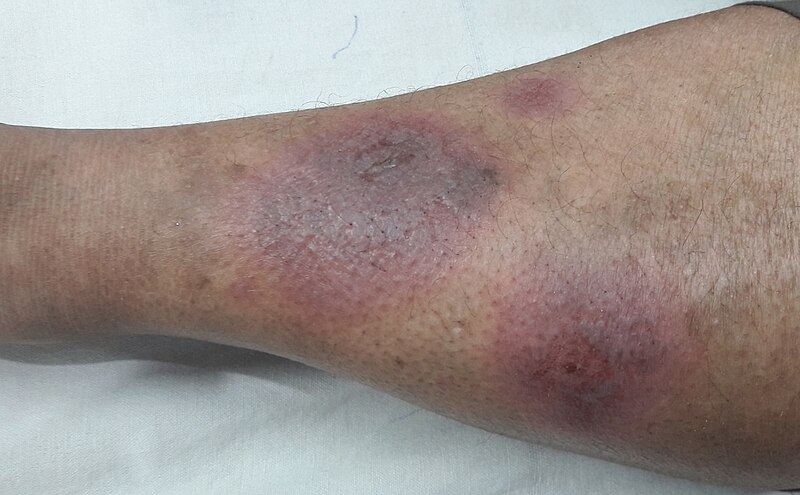
Image Source: Wikimedia Commons (2018). Erythema Nodosum, Licensed under a creative commons licence. Available for free at: https://commons.wikimedia.org/wiki/File:Erythema_Nodosum.jpg#/media/File:Erythema_Nodosum.jpg (Accessed: 17 July 2024).
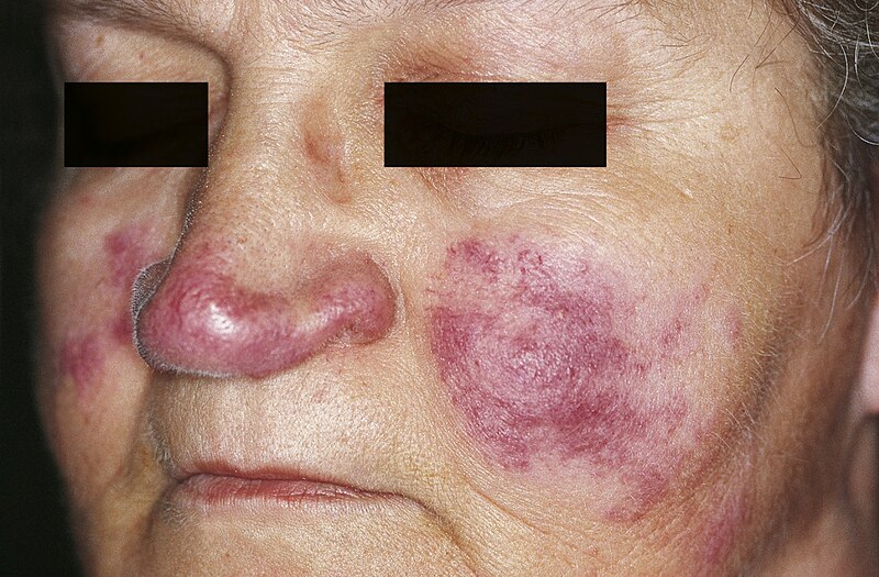
Image Source: Wikimedia Commons (2010). Lupus pernio, Licensed under a creative commons licence. Available for free at: https://commons.wikimedia.org/wiki/File:Lupus_pernio_01.jpg#/media/File:Lupus_pernio_01.jpg (Accessed: 17 July 2024).
Syndromes associated with sarcoidosis
Sarcoidosis can present as certain syndromes:
- Löfgren syndrome: bilateral hilar lymphadenopathy, erythema nodosum, fever and polyarthralgia. This is an acute form of sarcoidosis and carries a very good prognosis
- Heerfordt syndrome: parotid enlargement, fever and uveitis secondary to sarcoidosis
Diagnosis of sarcoidosis
Investigation findings include:
- Urine: hypercalciuria
- Bloods: possible abnormalities include abnormal FBC (may show anaemia, lymphopenia, eosinophilia or raised white cell count), raised ESR, raised calcium, raised phosphate, raised ALP, deranged U&E (although renal impairment is rare), deranged LFTs, elevated serum ACE (in around 60% of patients with acute disease)
- CXR aids with staging:
- Stage 0 – Normal CXR
- Stage 1 – Bilateral hilar lymphadenopathy
- Stage 2 – Bilateral hilar lymphadenopathy and interstitial infiltrates
- Stage 3 – Diffuse interstitial infiltrates
- Stage 4 – Diffuse fibrosis
- HRCT: may show lymph node enlargement or calcification, fibrosis or nodules. A minority of patients have a UIP pattern on CT and this carries a worse prognosis
- Lung function tests: typically a restrictive defect with decreased TLCO and VC
- FDG-PET: used to assess inflammatory activity and aids prognostication
- Bronchoscopy: bronchoalveolar lavage may show increased lymphocytes (raised CD4:CD8 ratio)
- Biopsy: endobronchial ultrasound-guided or transbronchial biopsy of suspicious lesions shows non-caseating granulomas. Other lesions (such as skin or lymph nodes) may be biopsied to aid diagnosis
Management of sarcoidosis
Oral glucocorticoids are indicated in certain contexts:
- With significant reduction in pulmonary function tests
- With eye, heart or neurological involvement
Second-line immunosuppression may be considered, with agents such as methotrexate, mycophenolate, leflunomide and azathioprine. This is indicated when steroid therapy is not appropriate or ineffective, e.g. if there is progression of pulmonary disease or an unacceptable symptom burden despite steroids, inability to taper steroids below 10-15 mg per day, inability to tolerate steroids, in the presence of comorbidities that would be adversely affected by steroids therapy, or if a patient refuses to take steroids.
Biological therapy is a third-line option only available through tertiary centres. This includes TNF inhibitors.
Lung transplantation may be considered.
See note on nintedanib – this has now been approved as a treatment option for chronic progressive fibrosing interstitial lung diseases.
References
- Dempsey OJ, Paterson, E.W., Kerr, K.M., et al. Sarcoidosis. BMJ. 2009;28339:b3206
- Tidy C. Sarcoidosis: Patient; 2016. Available from: https://patient.info/doctor/sarcoidosis-pro# [accessed 04/01/2022]
- Thillai M, Atkins, C. A. et al. BTS Clinical Statement on pulmonary sarcoidosis. Thorax. 2020;0:1-17
- Chapman SJR, G. V.; Shrimanker, R. S.; Turnbull, C. D.; Wrightson, J. M. Oxford Handbook of Respiratory Medicine. Fourth Edition ed. Oxford, United Kingdom: Oxford University Press; 2021
- NICE. Nintedanib for treating progressive fibrosing interstitial lung diseases. National Institute for Health and Care Excellence, 2021 [accessed 18/03/2024]
Connective tissue disease-associated interstitial lung disease
Overview of connective tissue disease-associated ILD
Several connective tissue diseases (CTD) have respiratory complications, including systemic sclerosis (SSc), rheumatoid arthritis (RA), polymyositis/dermatomyositis (PM/DM), Sjögren’s syndrome, systemic lupus erythematosus (SLE), ankylosing spondylitis and mixed connective tissue disease. This chapter will briefly describe pulmonary involvement in some of the major rheumatological conditions, particularly focusing on interstitial lung disease.
The majority of these diseases cause lower zone fibrosis with the notable exception being ankylosing spondylitis, which causes upper zone fibrosis.
Symptoms and signs are often those associated with the underlying CTD such as rashes, joint pain, constitutional symptoms, ulcers, synovitis, joint deformity, dysphagia, sclerodactyly (particularly in SSc), dry mucosa (present in Sjögren’s), Gottron’s papules (seen in dermatomyositis), and proximal muscle weakness (for example in inflammatory myopathies).
In addition, respiratory features such as dyspnoea, cough and crackles may be noted.
Bloods should be sent to screen for CTD, including ANA, anti-dsDNA, anti-ribonucleoprotein antibody, anti-smith antibody, anti-Scl-70, anti-Ro, anti-La, rheumatoid factor, anticyclic citrullinated peptide, anti-Jo-1 antibody, creatine kinase, aldolase, ESR, CRP.
Lung function tests in most of these conditions show a restrictive pattern with a low KCO.
HRCT usually shows a non-specific interstitial pneumonia pattern although variation between conditions will apply.
Management is driven by a multidisciplinary team approach. There is a low threshold for starting treatment in the hope of preventing pulmonary fibrosis. In most CTD-associated ILD, the mainstay of treatment is steroids and adjuvant immunosuppressive agents.
See note on nintedanib – this has now been approved as a treatment option for chronic progressive fibrosing interstitial lung diseases.
Rheumatoid arthritis
RA can affect the lung in a multitude of ways, including pleuritis, pleural effusion, pulmonary fibrosis, Caplan syndrome and vasculitis. We will focus on pulmonary fibrosis in this section.
Pulmonary fibrosis in RA is often asymptomatic. It is most common in seropositive patients, those with high ANA titres, nodules and multisystem disease. If present, symptoms include dyspnoea and cough. Crackles are mostly bibasal.
Management options are steroids and immunosuppressive therapy, however prognosis is poor and these therapies are often not effective.
Systemic lupus erythematosus
As in RA, SLE may affect the lung in multiple ways. We will focus on interstitial involvement: diffuse ILD and acute lupus pneumonitis.
Diffuse ILD occurs in the majority of patients but is often asymptomatic.
Acute lupus pneumonitis can result in very severe disease with features such as cough, dyspnoea, fever, pain and hypoxia. Examination reveals widespread crepitations. Widespread infiltrates may be seen on CXR.
Management is with steroids and cytotoxic drugs. It can later develop into chronic pneumonitis.
Idiopathic inflammatory myopathies
This group of diseases includes polymyositis (PM) and dermatomyositis (DM).
ILD features often in patients with these conditions, more commonly in those with anti-synthetase antibodies (including anti-Jo) or anti-MDA5 antibodies.
Clinical features include dyspnoea, cough, joint pain and fevers, with fine bibasal crackles on examination.
Early treatment is recommended in ILD associated with PM/DM to prevent disease progression. Steroids are often used. Other options include rituximab, however this should be discussed with a rheumatology specialist.
Systemic sclerosis
Most patients with SSc will have some evidence of ILD and the presence of anti-topoisomerase antibodies (Scl-70) appears to be strongly associated with development of significant lung disease.
Features of systemic sclerosis associated with dyspnoea should prompt suspicion of associated ILD. On examination, bibasal crackles might be noted.
Low-dose steroids should be used if required as high doses carry the risk of renal crisis. Other options for management include cyclophosphamide or mycophenolate mofetil.
Ankylosing spondylitis
Notably, ankylosing spondylitis causes bilateral apical fibrosis, as opposed to the bibasal pattern seen in most other CTD-associated ILD. Cysts or cavities may also develop.
Lung function tests typically show a restrictive pattern, however, it should be noted that this may be partly related to spinal deformity.
References
- Vij R, Strek ME. Diagnosis and treatment of connective tissue disease-associated interstitial lung disease. Chest. 2013;143(3):814-24
- Oliveira RP, Ribeiro R, Melo L, Grima B, Oliveira S, Alves JD. Connective tissue disease-associated interstitial lung disease. Pulmonology. 2020
- Bradley B, Branley HM, Egan JJ, Greaves MS, Hansell DM, Harrison NK, et al. Interstitial lung disease guideline: the British Thoracic Society in collaboration with the Thoracic Society of Australia and New Zealand and the Irish Thoracic Society. Thorax. 2008;63 Suppl 5:v1-58
- Chapman SJR, G. V.; Shrimanker, R. S.; Turnbull, C. D.; Wrightson, J. M. Oxford Handbook of Respiratory Medicine. Fourth Edition ed. Oxford, United Kingdom: Oxford University Press; 2021
- NICE. Nintedanib for treating progressive fibrosing interstitial lung diseases. National Institute for Health and Care Excellence, 2021 [accessed 18/03/2024]
Complete Guide to Clinical Genetics ST3 Application and Recruitment (2027)
Written by Medibuddy. Last updated: October 2026. Clinical Genetics is

Complete Guide to Clinical Radiology ST3 Application and Recruitment (2027)
Recruitment paused: NHS England is not running Clinical Radiology ST3+ recru

Complete Guide to Paediatric Surgery ST3 Application and Recruitment in United Kingdom (2026)
Written by Medibuddy. Last updated: October 2026. If you’re planning


Take your subscriptions with you
Our mobile app allows you to access your interview and exam question banks wherever you are.

