


 Close
Close
 20th February 2025
Sarcoidosis
Overview of sarcoidosis
Sarcoidosis is a multisystem inflammatory disease characterised by the formation of non-caseating granulomas in various organs. This chapter will focus on its respiratory manifestations while briefly mentioning other organ involvement.
The aetiology of sarcoidosis is unknown. It is most common in northern European/Scandinavian and African-American adults under the age of 50.
Signs and symptoms of sarcoidosis
Clinical features can be system specific or constitutional:
- Constitutional: fatigue, fevers, weight loss
- Respiratory: dyspnoea, non-productive cough
- Skin: erythema nodosum (Figure 1), lupus pernio (Figure 2)
- Eyes: granulomatous uveitis, dry eyes and glaucoma (can appear after other symptoms have resolved)
- Neurological: any part of the central or peripheral nervous systems can be affected. The most common manifestations are Bell’s palsy and lymphocytic meningitis. Other symptoms include paraesthesia, visual defects, facial weakness or numbness, polydipsia and hearing impairment
- Cardiac: symptoms of heart failure due to cardiomyopathy
- Joints: inflammatory arthritis
- Hypercalcaemia: granulomas cause increased conversion of vitamin D to its active form. This can result in symptomatic hypercalcaemia
- Lymphadenopathy
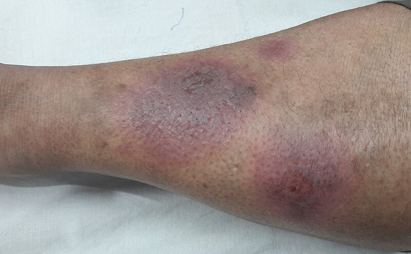
Image Source: Wikimedia Commons (2018). Erythema Nodosum, Licensed under a creative commons licence. Available for free at: https://commons.wikimedia.org/wiki/File:Erythema_Nodosum.jpg#/media/File:Erythema_Nodosum.jpg (Accessed: 17 July 2024).
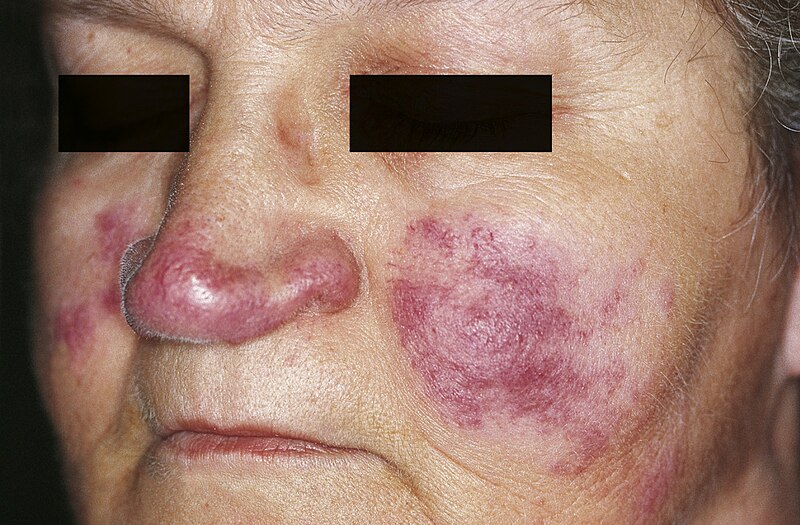
Image Source: Wikimedia Commons (2010). Lupus pernio, Licensed under a creative commons licence. Available for free at: https://commons.wikimedia.org/wiki/File:Lupus_pernio_01.jpg#/media/File:Lupus_pernio_01.jpg (Accessed: 17 July 2024).
Syndromes associated with sarcoidosis
Sarcoidosis can present as certain syndromes:
- Löfgren syndrome: bilateral hilar lymphadenopathy, erythema nodosum, fever and polyarthralgia. This is an acute form of sarcoidosis and carries a very good prognosis
- Heerfordt syndrome: parotid enlargement, fever and uveitis secondary to sarcoidosis
Diagnosis of sarcoidosis
Investigation findings include:
- Urine: hypercalciuria
- Bloods: possible abnormalities include abnormal FBC (may show anaemia, lymphopenia, eosinophilia or raised white cell count), raised ESR, raised calcium, raised phosphate, raised ALP, deranged U&E (although renal impairment is rare), deranged LFTs, elevated serum ACE (in around 60% of patients with acute disease)
- CXR aids with staging:
- Stage 0 – Normal CXR
- Stage 1 – Bilateral hilar lymphadenopathy
- Stage 2 – Bilateral hilar lymphadenopathy and interstitial infiltrates
- Stage 3 – Diffuse interstitial infiltrates
- Stage 4 – Diffuse fibrosis
- HRCT: may show lymph node enlargement or calcification, fibrosis or nodules. A minority of patients have a UIP pattern on CT and this carries a worse prognosis
- Lung function tests: typically a restrictive defect with decreased TLCO and VC
- FDG-PET: used to assess inflammatory activity and aids prognostication
- Bronchoscopy: bronchoalveolar lavage may show increased lymphocytes (raised CD4:CD8 ratio)
- Biopsy: endobronchial ultrasound-guided or transbronchial biopsy of suspicious lesions shows non-caseating granulomas. Other lesions (such as skin or lymph nodes) may be biopsied to aid diagnosis
Management of sarcoidosis
Oral glucocorticoids are indicated in certain contexts:
- With significant reduction in pulmonary function tests
- With eye, heart or neurological involvement
Second-line immunosuppression may be considered, with agents such as methotrexate, mycophenolate, leflunomide and azathioprine. This is indicated when steroid therapy is not appropriate or ineffective, e.g. if there is progression of pulmonary disease or an unacceptable symptom burden despite steroids, inability to taper steroids below 10-15 mg per day, inability to tolerate steroids, in the presence of comorbidities that would be adversely affected by steroids therapy, or if a patient refuses to take steroids.
Biological therapy is a third-line option only available through tertiary centres. This includes TNF inhibitors.
Lung transplantation may be considered.
See note on nintedanib – this has now been approved as a treatment option for chronic progressive fibrosing interstitial lung diseases.
References
- Dempsey OJ, Paterson, E.W., Kerr, K.M., et al. Sarcoidosis. BMJ. 2009;28339:b3206
- Tidy C. Sarcoidosis: Patient; 2016. Available from: https://patient.info/doctor/sarcoidosis-pro# [accessed 04/01/2022]
- Thillai M, Atkins, C. A. et al. BTS Clinical Statement on pulmonary sarcoidosis. Thorax. 2020;0:1-17
- Chapman SJR, G. V.; Shrimanker, R. S.; Turnbull, C. D.; Wrightson, J. M. Oxford Handbook of Respiratory Medicine. Fourth Edition ed. Oxford, United Kingdom: Oxford University Press; 2021
- NICE. Nintedanib for treating progressive fibrosing interstitial lung diseases. National Institute for Health and Care Excellence, 2021 [accessed 18/03/2024]
Complete Guide to Clinical Genetics ST3 Application and Recruitment (2027)
Written by Medibuddy. Last updated: October 2026. Clinical Genetics is

Complete Guide to Clinical Radiology ST3 Application and Recruitment (2027)
Recruitment paused: NHS England is not running Clinical Radiology ST3+ recru

Complete Guide to Paediatric Surgery ST3 Application and Recruitment in United Kingdom (2026)
Written by Medibuddy. Last updated: October 2026. If you’re planning


Take your subscriptions with you
Our mobile app allows you to access your interview and exam question banks wherever you are.

