


 Close
Close
 24th February 2025
Gout
Overview of gout
Gout is a common, chronic condition characterised by acute, severe flares of joint pain, classically affecting the first metatarsophalangeal (MTP) joints (known as podagra). If risk factors are not managed over time, it may cause a destructive and deforming arthropathy. It manifests due to deposition of urate crystals in the joint space, typically due to raised plasma urate levels.
Risk factors for gout include male gender, advancing age, obesity, hypertension (partly due to the medications used to treat), diabetes mellitus, CKD (due to impaired urate excretion), trauma and any condition causing rapid cell turnover (such as tumour lysis syndrome classically, or indeed any condition causing haemolysis). Historically, it is associated with a diet rich in meat, fish and alcohol. There are a host of drugs associated with gout; these include: salicylates, diuretics (particularly furosemide but also bendroflumethiazide), ciclosporin, levodopa and the tuberculosis drugs pyrazinamide and ethambutol.
The acute management typically involves anti-inflammatory drugs such as NSAIDs, colchicine or corticosteroids, while the chronic management focuses on urate-lowering therapies such as allopurinol, a xanthine oxidase inhibitor (as well as managing any underlying cause).
Signs and symptoms of gout
The typical presentation for an attack of gout is a sudden-onset (over hours), severely painful, erythematous, swollen joint (typically a single joint) and, in cases of podagra (see Figure 6), exquisite difficulty in weight bearing. It typically affects joints in the lower limbs rather than the upper limbs (and almost never the spinal column). Over time, it becomes more likely that attacks are polyarticular rather than the usual monoarticular presentation. Urate crystals eventually become deposited in the soft tissues (such as the ear pinna and Achilles tendon), forming subcutaneous tophi (see Figure 7), which are a marker of chronically raised urate levels and can diagnostically be helpful.
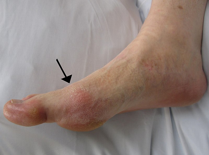
Image source: Wikimedia Commons (2019). Gout2010, Licensed under a Creative Commons licence. Available for free at: https://commons.wikimedia.org/wiki/File:Gout2010.JPG (Accessed: 19 January 2024).
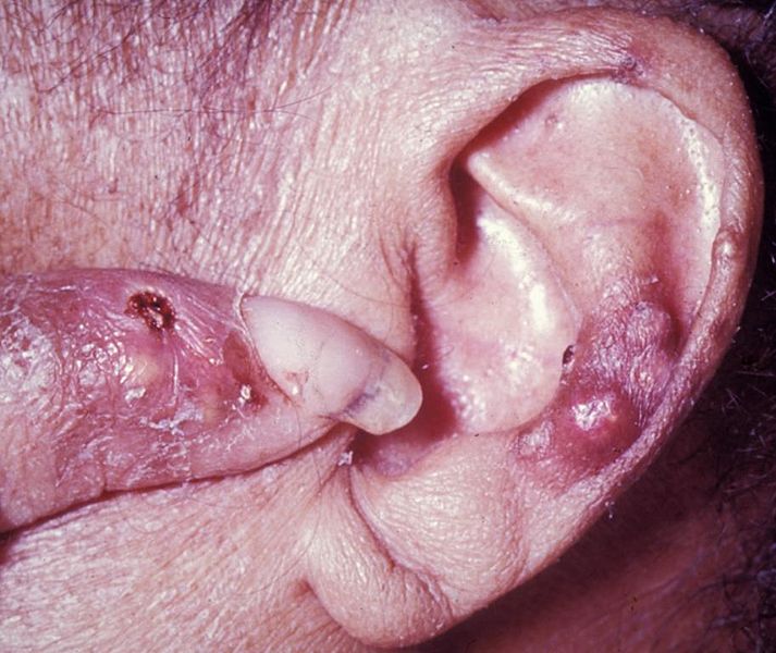
Image source: Wikimedia Commons (2010). Case 30-top, Licensed under a Creative Commons licence. Available for free at: https://en.m.wikipedia.org/wiki/File:Case_30-top.jpg (Accessed: 19 January 2024).
Investigation of gout
The diagnosis is typically suspected based on a previous history, having identifiable risk factors (especially if there is a recent temporal relationship) and clinical examination.
Gout can be confirmed by needle aspiration of synovial fluid during a flare, showing negatively birefringent needle-shaped crystals on microscopy (though their absence does not exclude the diagnosis). X-rays may highlight punched-out erosions and sclerotic margins, as well as soft tissue swelling and joint effusion (see Figure 8), and ultrasound may show tophaceous deposits.
Other differential diagnoses to consider include pseudogout, OA, RA, psoriatic arthritis, as well as both reactive arthritis and, crucially, septic arthritis in the immunosuppressed. There is no role for measuring urate levels in the acute phase to confirm or refute the diagnosis; it should only be used to confirm chronically elevated levels and the response to therapy. Note, however, that a raised serum urate may be detected in an asymptomatic patient without any previous episodes of gout and does not necessarily predict future episodes of gout; treatment in this circumstance remains controversial.
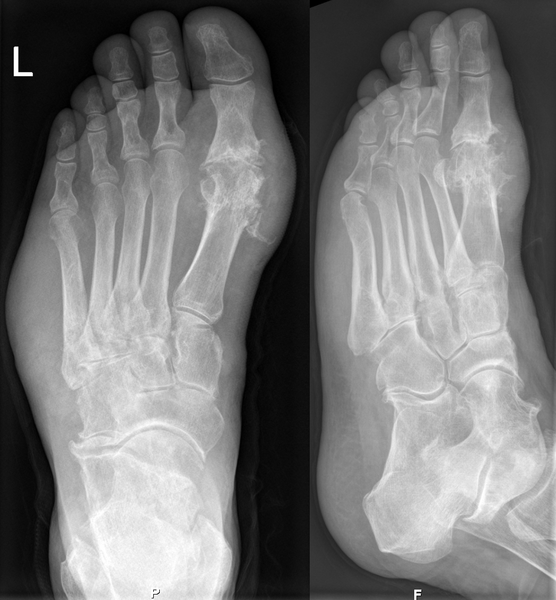
Image source: Wikimedia Commons (2010). Gichtfuss im Roentgenbild 002, Licensed under a Creative Commons licence. Available for free at: https://commons.wikimedia.org/wiki/File:Gichtfuss_im_Roentgenbild_002.png (Accessed: 19 January 2024).
Management of gout
The management can be divided into acute and chronic.
Acute treatment relies on supportive measures such as rest, ice and elevation combined with a short course of NSAIDs, colchicine (0.5 mg, 2-4 times a day) or corticosteroids, either oral (if multiple joints affected) or intra-articular (if a single joint is affected). In this circumstance, urate-lowering drugs such as allopurinol should be continued, although if this is the first presentation, their initiation should be delayed until the flare resolves. The anti-IL-1𝛃 drugs, such as anakinra, can be initiated in repeated and/or refractory gout or where other medications are contraindicated.
The chronic management involves urate-lowering drugs, such as allopurinol, aiming for a reduction in serum urate levels <360 µmol/L. Febuxostat is an alternative from the same family of drugs, used if allopurinol does not achieve the recommended urate levels, and Rasburicase can be used in frequent, severe gout not controlled by the previous medications mentioned. Care should be used in the initiation of uricosuric drugs such as probenecid in patients who suffer with nephrolithiasis. The modification of lifestyle factors is of vital importance; weight loss and dietary modification should be encouraged. The management of other conditions triggering attacks should also be optimised, and medications such as diuretics should be continued at the lowest effective dose.
Complete Guide to Clinical Genetics ST3 Application and Recruitment (2027)
Written by Medibuddy. Last updated: October 2026. Clinical Genetics is

Complete Guide to Clinical Radiology ST3 Application and Recruitment (2027)
Recruitment paused: NHS England is not running Clinical Radiology ST3+ recru

Complete Guide to Paediatric Surgery ST3 Application and Recruitment in United Kingdom (2026)
Written by Medibuddy. Last updated: October 2026. If you’re planning


Take your subscriptions with you
Our mobile app allows you to access your interview and exam question banks wherever you are.

