


 Close
Close
 24th February 2025
Rheumatoid arthritis (RA)
Overview of RA
RA is an autoimmune disease that typically affects patients from their fifth decade and, like most autoimmune diseases, has a 3:1 female-to-male predominance.
The pathophysiology is due to an immune response triggered between both B and T lymphocytes and fibroblasts of the synovial membranes, generating an inflammatory cascade mediated by TNF-ɑ and IL-6. The deposition of immune complexes causes cartilage degradation and, thus, joint pain. In certain cases, the inflammatory cascade is so pronounced that multi-system extra-articular manifestations can occur and so close monitoring for complications is required.
The treatment consists of combinations of immunosuppressive therapies, known as disease-modifying antirheumatic drugs (DMARDs, such as methotrexate) and biologics (first line are anti-TNF-ɑ) to dampen down the immune response and thus the symptomology, though they all require close monitoring and do not come without their own side effects.
Signs and symptoms of RA
RA is typically polyarticular, symmetrical and may be fleeting and migratory. The intra-articular features are pain, erythema, swelling and early morning stiffness of the small joints, typically of the hands, causing significant loss of function and deformity over time. The classic skeletal deformities include ulnar deviation of the wrist, swan-neck and Boutonnière deformities of the fingers (see Figures 3-4) and atlanto-axial subluxation of the cervical spine.
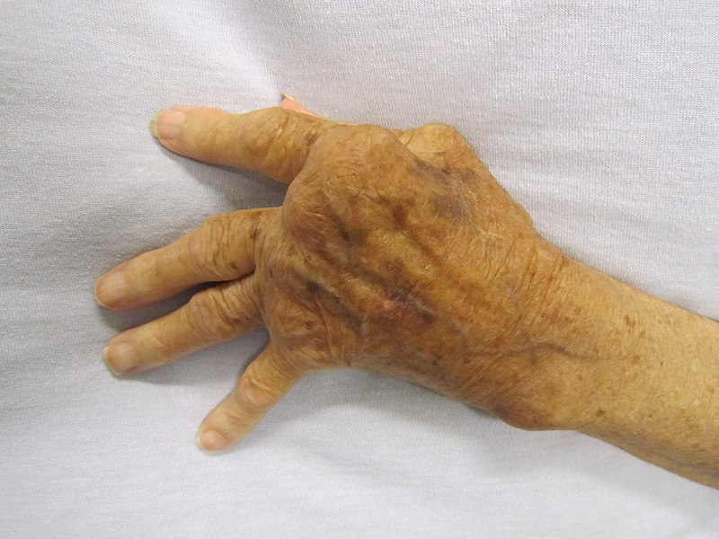
Image source: Wikimedia Commons (2010). Rheumatoid Arthritis, Licensed under a Creative Commons licence. Available for free at: https://commons.wikimedia.org/wiki/File:Rheumatoid_Arthritis.JPG (Accessed: 17 January 2024).
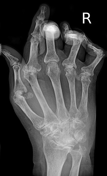
Image source: Wikimedia Commons (2015). RheumatoideArthritisAP, Licensed under a Creative Commons licence. Available for free at: https://commons.wikimedia.org/wiki/File:RheumatoideArthritisAP.jpg (Accessed: 17 January 2024).
Constitutional symptoms may include fatigue, malaise, weight loss and rashes. Specific extra-articular manifestations can be classified by system:
| System | Manifestation |
|---|---|
| Cardiac/cardiovascular | Amyloid, pericarditis, vasculitis |
| Respiratory | Pulmonary fibrosis, pulmonary nodules, exudative pleural effusions, pleurisy, bronchiolitis obliterans |
| Ophthalmic | Sicca syndrome, episcleritis, scleritis, corneal ulcers |
| Neurological | Radiculopathy due to joint laxity causing nerve impingement, mononeuritis multiplex |
| Miscellaneous | Felty’s syndrome (RA, splenomegaly and neutropenia), lymphadenopathy |
Table 1 Extra-articular manifestations of RA (not an exhaustive list)
Complications of established RA include anaemia of chronic disease, osteoporosis, septic arthritis (due to immunosuppressive therapies), cardiovascular sequelae due to chronic inflammation (think of Virchow’s triad) and consequential mental health issues of living with this chronic, often relapsing and remitting condition. Interestingly, malignancy rates (particularly Hodgkin’s lymphoma) are elevated in those with RA.
Given the varied and widespread possible signs and symptoms with RA, a thorough clinical history and examination is crucial in making the diagnosis and a high index of suspicion is required, especially in the context of pre-existing autoimmune disease.
Investigation of RA
RA is essentially a clinical diagnosis; however, specific auto-antibodies are useful investigations which can confirm or refute the provisional diagnosis: rheumatoid factor (RF; 69% sensitivity, 85% specificity) and anti-cyclic citrullinated peptide (anti-CCP; 67% sensitivity, 95% specificity). If both RF and anti-CCP are positive, there is a 100% positive predictive value for a diagnosis of RA. Interestingly, a diagnosis of seronegative RA can be made without positive auto-antibodies if the clinical features and other investigations are suggestive of RA. This can be challenging as other diagnoses, such as spondyloarthritis (including psoriatic arthritis), need to be considered and are often later thought to be the actual diagnosis.
Inflammatory markers such as CRP and ESR are often useful adjuncts and can be repeated to assess the response to therapy. An FBC can be useful to look at the haemoglobin (anaemia of chronic disease) and white cell count (inflammation/infection), and LFTs and U&Es to get a baseline (as these are required prior to and for ongoing monitoring of most DMARDs). X-rays (joint erosion and fusion; see Figure 5) and ultrasound (synovitis and effusions) are useful in assessing painful joints, and bone density scanning (DEXA, dual-energy X-ray absorptiometry) to assess bone mineral health (particularly in the context of repeated corticosteroid courses).
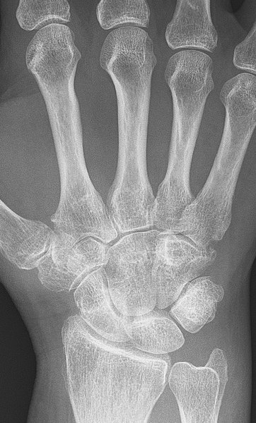
Left: Image source: Wikimedia Commons (2017). Rheumatoid arthritis with unaffected carpal bones 2009, Licensed under a Creative Commons licence. Available for free at: https://commons.wikimedia.org/wiki/File:Rheumatoid_arthritis_with_unaffected_carpal_bones_2009.jpg (Accessed: 17 January 2024).
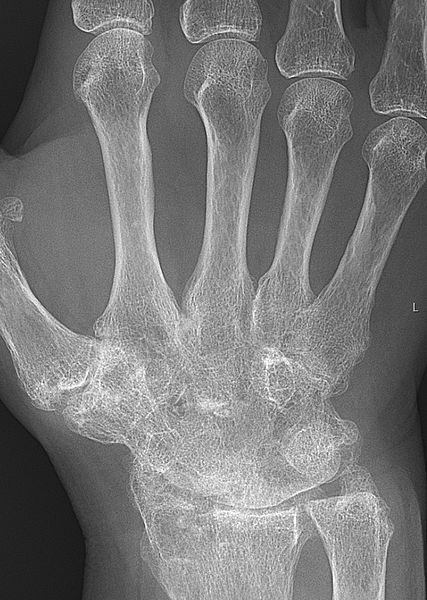
Right: Image source: Wikimedia Commons (2017). Rheumatoid arthritis with carpal ankylosis 2017, Licensed under a Creative Commons licence. Available for free at: https://commons.wikimedia.org/wiki/File:Rheumatoid_arthritis_with_carpal_ankylosis_2017.jpg (Accessed: 17 January 2024).
Alongside clinical judgement, the ACR/EULAR (American College of Rheumatology/European League Against Rheumatism) Classification Criteria (2010) can be used to identify those with early RA who are likely to develop chronic RA. It cannot be used to exclude RA, and indeed, over time, the score may change as the condition evolves. In those who have clinical synovitis in one joint or more, which is not better explained by another pathology, a score of six or more is suggestive of definite RA (points scored for joint involvement, serology, acute phase reactants and duration of symptoms).
Management of RA
The key treatment aim in RA is to intensively pursue remission to prevent deformity and disability. Tools such as the Disease Activity Score 28 Joints (DAS-28) are used to assess the treatment response over time in a patient-centred way.
The initial step of the treatment ladder usually consists of a DMARD and a corticosteroid (whilst the DMARD takes effect). Typically, methotrexate is used first-line. If a clinical response is not seen within 3-6 months, or if it is not tolerated, then it will be substituted for one of either hydroxychloroquine, sulfasalazine or leflunomide. The following table details the monitoring required and the potential side effects of each of these agents:
| DMARD | Monitoring | Side effects | Contraindications |
|---|---|---|---|
| Corticosteroids | BP, blood sugar, lipids | GI ulceration, osteoporosis, mood disturbance, delayed wound healing, weight gain, diabetes, hypertension | |
| Methotrexate (co-prescribed with folate) | FBC, LFTs, U&Es | GI disturbance, hepatitis, myelosuppression, pneumonitis | Pregnancy, breastfeeding |
| Hydroxychloroquine | Eye screening | Retinal toxicity, maculopathy | |
| Sulfasalazine | FBC, LFTs, U&Es | GI disturbance, pancytopenia, aplastic anaemia | |
| Leflunomide | FBC, LFTs, U&Es, BP | GI disturbance, hypertension | Pregnancy, breastfeeding |
Table 2 DMARDs used in RA with their monitoring requirements and side effects (not an exhaustive list)
The next step, if remission is not induced at the maximum tolerated doses of two different DMARDs combined (one of which is usually methotrexate), is the addition of a biologic agent to the DMARD (ideally methotrexate). The first-line anti-TNF-ɑ drugs are adalimumab, etanercept and infliximab. If remission is still not achieved, then an alternative biologic is tried, such as anti-CD-20 drug (rituximab) or anti-IL-6 drug (tocilizumab).
Again, these drugs require careful monitoring and, because they are powerful immunosuppressants, require certain investigations prior to their initiation. This normally includes excluding tuberculosis (risk stratifying, chest X-ray and IgRA, interferon-gamma release assay), hepatitis, HIV and ANA positivity (can cause an SLE-like syndrome when started on anti-TNF-ɑ drugs). Additionally, demyelinating disorders, congestive cardiac failure, interstitial lung disease and uveitis require caution when considering initiation, as all of these conditions can be exacerbated by anti-TNF-ɑ drugs. Interestingly, anti-TNF-ɑ therapies have also been associated with the recurrence of previous malignancies and de novo skin cancers (but not solid tumours). The decision to initiate should be made on an individual risk/benefit basis.
Once remission has been induced, the corticosteroid is gradually weaned. Patients on these medications should be carefully counselled about seeking medical help when feeling unwell as they are prone to infections with opportunistic or rare organisms such as herpes zoster (for which immunoglobulin should be given) or fungi. In the context of suspected sepsis, they require aggressive broad-spectrum antimicrobial therapy and require their DMARDs and biologics to be held and their corticosteroids (if they are on them) to be up-titrated during their illness, as they are at risk of adrenal insufficiency in times of physiological stress.
Effective analgesia usually includes paracetamol and ibuprofen (often with gastroprotection).
References
- Clunie, G, Wilkinson N, Nikiphorou E, Jadon D. Oxford Handbook of Rheumatology. Fourth Edition. Oxford: Oxford University Press; 2018
- Yazici Y. Rheumatoid Arthritis [Internet]. BMJ Best Practice. BMJ Publishing Group; 2023 [Cited 2023 August 17]. Available from: https://bestpractice.bmj.com/topics/en-gb/105
Complete Guide to Clinical Genetics ST3 Application and Recruitment (2027)
Written by Medibuddy. Last updated: October 2026. Clinical Genetics is

Complete Guide to Clinical Radiology ST3 Application and Recruitment (2027)
Recruitment paused: NHS England is not running Clinical Radiology ST3+ recru

Complete Guide to Paediatric Surgery ST3 Application and Recruitment in United Kingdom (2026)
Written by Medibuddy. Last updated: October 2026. If you’re planning


Take your subscriptions with you
Our mobile app allows you to access your interview and exam question banks wherever you are.

