


 Close
Close
 13th February 2025
Loss of Vision and Diplopia
Acute vision loss
Overview of acute vision loss
The cause of acute vision loss can be categorised into ophthalmological, inflammatory (most notably uveitis), vascular, neurovascular and oncological. Migraine should be considered as a differential diagnosis for acute, transient vision loss, but with caution in those with no prior history of aura.
Ophthalmological
Cornea
Corneal disease is the most common ophthalmological cause for acute vision loss. It often presents as a flare of an underlying corneal disease. These include:
- Keratoconus
- Dry eye
- Corneal ulcer
- Corneal hydrops (a complication of corneal disorders causing corneal swelling)
Glaucoma
Acute angle closure glaucoma can present with acute vision loss alongside other symptoms and signs.
Optic nerve
Disorders affecting the optic nerve include:
Optic neuritis
This inflammation of the optic nerve. It results in unilateral, painful vision loss over hours or days. Examination will likely reveal reduced visual acuity, visual field loss, red desaturation (dyschromatopsia), painful eye movements and a relative afferent pupillary defect.
Investigations should include MRI as it has a high sensitivity and specificity for detecting inflammatory changes in the optic nerve. Management is with high-dose corticosteroids to accelerate the speed of visual recovery.
Optic neuritis is the initial presentation of multiple sclerosis in 25% of cases, and of those with multiple sclerosis, 70% will be affected by optic neuritis.
Papilloedema
This is optic nerve swelling due to raised intracranial pressure (ICP). Therefore, papilloedema is always bilateral but not necessarily symmetrical. Symptoms include nausea or vomiting and headaches which are worse when leaning forward. Possible underlying conditions include an intracerebral mass or haemorrhage, hydrocephalus, meningitis or idiopathic intracranial hypertension (IIH). Management relies on correcting the underlying cause to prevent optic nerve damage and irreversible vision loss.
Leber’s hereditary optic neuropathy
An inherited mitochondrial disease which predominantly affects young males. Symptoms typically begin at age 10-30 years with unilateral progressive optic neuropathy. The contralateral eye also becomes affected months to years later. Diagnosis is confirmed with genetic testing.
Infectious optic neuropathy
This is uncommon but can be caused by tuberculosis, hepatitis B, syphilis and cytomegalovirus.
Drug-induced optic neuropathy
- Alcohol – chronic use can lead to vitamin B12 and folate deficiency.
- Ethambutol – effects are irreversible and dependent on dose and duration. Patients taking ethambutol who are asymptomatic should have regular eye screening to monitor for optic nerve compromise.
- Amiodarone – causes optic neuropathy through nerve demyelination. Visual field defects are variable and nerve damage is not dose related.
- Vigabatrin – visual field defects can be bilateral and concentric and are predominantly nasal.
Retina
Retinal detachment presents with the 4 ‘Fs’: floaters, flashes, visual field loss and fall in acuity. Risk factors for detachment include high myopia (short sightedness) and recent trauma or surgery.
Patients require urgent referral for surgery, which can include vitrectomy and gas or oil tamponade.
Vascular
Retinal vein occlusion and retinal artery occlusion
Retinal vein occlusion is more common than retinal artery occlusion and less sudden. Both need to be managed as an emergency. Note that branch retinal vein or artery occlusions can occur and typically present with vision loss to a lesser extent, and the patient may not even be aware of the defect.
| Central retinal vein occlusion (CRVO) | Central retinal artery occlusion (CRAO) | |
|---|---|---|
| Cause | Atherosclerosis | Emboli (most common), thrombus or vasculitis |
| Risk factors | Hypertension
Diabetes mellitus Smoking Hyperlipidaemia Polycythaemia Renal failure Glaucoma |
Older age
Male Hypertension Diabetes mellitus Smoking Hyperlipidaemia Cardiovascular disease Periocular aesthetic injections (rare) |
| Symptoms | Painless monocular loss of vision | Painless monocular loss of vision over seconds |
| Fundoscopy appearance | Multiple retinal haemorrhages – the ‘stormy sunset’ appearance (Figure 1)
Cotton wool spots Optic disc swelling |
Pale retina with cherry red spot (Figure 2) |
| Management | Monitor for secondary complications including retinal neovascularisation and macular oedema | Patients should be reviewed by a Stroke team urgently. There is no evidence to demonstrate any treatment improves vision |
Table 1 Clinical features and management of CRVO and CRAO
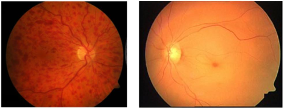
Image Source: Wikimedia Commons (2019). Fondo de ojo Ostrucción Vena central, Licensed under a creative commons licence. Available for free at: https://commons.wikimedia.org/wiki/File:Fondo_de_ojo_Ostrucción_Vena_central.png (Accessed: 18 January 2024).
Image Source: Wikimedia Commons (2014). Cherry Red Spot Fiess, Licensed under a creative commons licence. Available for free at: https://commons.wikimedia.org/wiki/File:Cherry_Red_Spot_Fiess.jpg, via Wikimedia Commons svg (Accessed: 18 January 2024).
Retinal vein occlusions can be clinically characterised into two types: ischaemic (25%) and non-ischaemic (75%) according to the area of non-perfusion in the posterior pole. Ischaemic retinal vein occlusions present with more severe vision loss, relative afferent pupillary defect and more marked fundoscopy findings. Prognosis is poorer and secondary complications are more common in ischaemic presentations.
Amaurosis fugax
Amaurosis fugax describes transient, painless monocular loss of vision caused by a carotid artery atheroma or embolus into the central retinal artery. It is considered as a retinal transient ischaemic attack (TIA) and hence the precursor to a stroke. Management should address any underlying cardiac risk factors and consider a carotid endarterectomy.
Vitreous haemorrhage
Vitreous haemorrhage can occur secondary to neovascularisation, retinal tears, retinal detachment or trauma. Neovascularisation is particularly significant in patients with diabetes because they can develop new retinal blood vessels (proliferative diabetic retinopathy) which are fragile and bleed easily. It is important the underlying cause is identified, and this may require an ultrasound if the blood obstructs the fundoscopy. Management may involve observation or a vitrectomy.
Anterior ischaemic optic neuropathy
Anterior ischaemic optic neuropathy (AION) is the most common cause of optic neuropathy in older people. It results from inflammation (arteritic) or atheroma (non-arteritic) causing blockage to the vascular supply of the optic nerve.
Arteritic AION is most commonly caused by giant cell (temporal) arteritis (GCA). GCA is classified as a medium-to-large-vessel vasculitis. Symptoms include monocular vision loss possibly preceded by episodes of amaurosis fugax, diplopia, new headache, jaw claudication and scalp tenderness. Examination reveals reduced visual acuity, optic disc oedema and thickened temporal arteries, which may be pulseless. Diagnosis can be confirmed with a raised ESR, raised CRP and a positive temporal artery biopsy. Note that due to skip lesions the biopsy may be falsely negative. Management with high-dose steroids should be started without delay (i.e. a positive biopsy result should not be awaited) to prevent vision loss in the other eye.
Other rarer causes of arteritic AION include other types of vasculitis, such as polyarteritis nodosa or systemic lupus erythematosus.
Non-arteritic AION is more common. It is typically painless and noticed on waking. Risk factors include hypertension, hypercholesterolaemia, diabetes mellitus and smoking. Management is to control these risk factors to prevent occurrence in the other eye.
Neurovascular
Haemorrhagic or ischaemic stroke can present with a visual defect. This can be a homonymous hemianopia, but the exact visual field loss is determined by the site affected.
Figure 3 recaps the visual field pathway and the resulting visual field defects according to which area is involved.
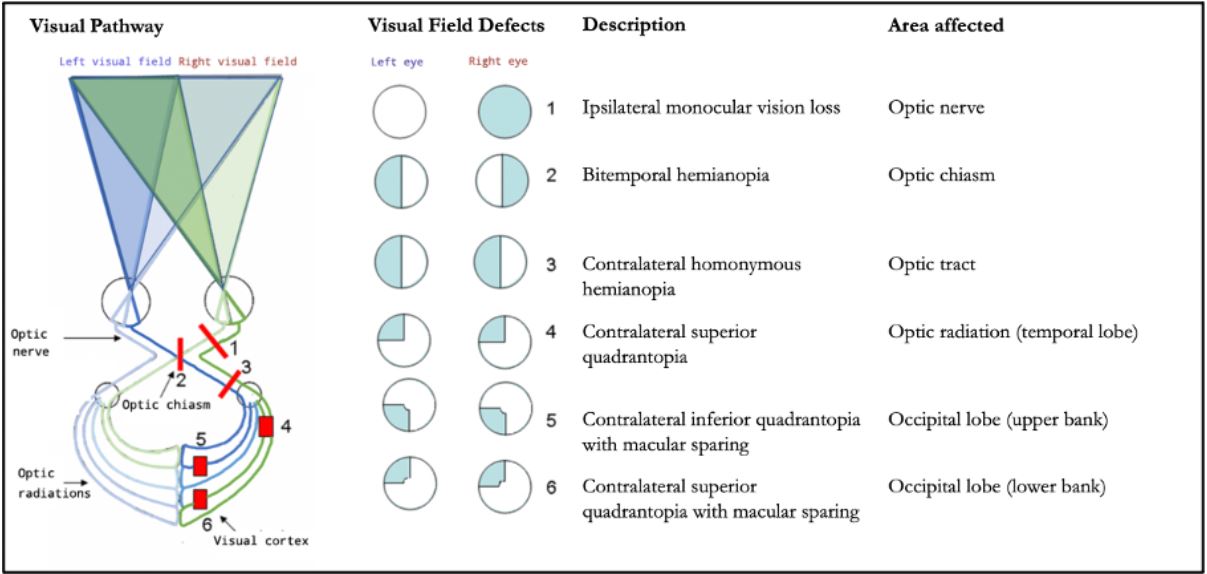
Image Source: Wikimedia Commons (2020). Hemianopsia en, Licensed under a creative commons licence. Available for free at: https://commons.wikimedia.org/wiki/File:Hemianopsia_en.jpg, via Wikimedia Commons (Accessed: 18 January 2024).
Oncological
Tumours are rare as a cause of vision loss but can result in severe and progressive visual field defects. The exact defect is according to the site of the lesion, but typical examples include:
- Cerebral tumours: contralateral homonymous hemianopia
- Pituitary tumours: bitemporal hemianopia. These tumours can be non functional or produce hormones, e.g. prolactin, ACTH or growth hormone. Be aware of idiosyncratic presentations, such as skin hyperpigmentation seen in ACTH-producing tumours due to its effect on melanocyte-stimulating hormone receptors.
References
- BMJ Best Practice. Assessment of vision loss [Internet]. BMJ Publishing Group. 2023 [cited July 22 2023]. Available from: https://bestpractice.bmj.com/topics/en-gb/960
- Baldwin A, Hjelde N, Goumalatsou C, Myers G. Oxford Handbook of Clinical Specialties. 10 edn. Oxford Medical Handbooks, UK: Oxford; 2016
- Rigi M, Almarzouqi SJ, Morgan ML, Lee AG. Papilledema: epidemiology, etiology, and clinical management. Eye Brain. 2015 Aug 17;7:47-57. doi: 10.2147/EB.S69174. PMID: 28539794; PMCID: PMC5398730
- Bennett JL. Optic neuritis. Continuum (Minneapolis, Minn.). 2019 Oct;25(5):1236
- Hayreh SS. Management of ischemic optic neuropathies. Indian J Ophthalmol. 2011 Mar-Apr;59(2):123-36. doi: 10.4103/0301-4738.77024. PMID: 21350282; PMCID: PMC3116541
- Tadi P, Najem K, Margolin E. Amaurosis Fugax. [Updated 2022 Aug 8]. In: StatPearls [Internet]. Treasure Island (FL): StatPearls Publishing; 2023 Jan-. Available from: https://www.ncbi.nlm.nih.gov/books/NBK470528/
- Lim J et al. Retinal Artery Occlusion, EyeWiki. [Internet]. July 2023 [cited July 23 2023]. Available from: https://eyewiki.aao.org/Retinal_Artery_Occlusion
Chronic vision loss
Overview of chronic vision loss
Causes of chronic vision loss can be categorised according to the presence or absence of pain. Painless chronic vision loss can be caused by:
- Refractive error – characterised by vision improvement with pinhole
- Cataracts
- Age-related macular degeneration (AMD)
- Diabetic retinopathy
- Open-angle glaucoma
- Optic nerve pathology
- Hereditary retinal dystrophies
- Drugs, toxins and deficiencies
Painful chronic vision loss is much less common. A serious underlying cause, such as an intracranial mass, choroidal melanoma or optic neuritis, should be investigated.
Age-related macular degeneration
AMD is the most common cause of irreversible blindness in developed countries and the leading cause for blindness in the United Kingdom. It is a potentially progressive disorder of the macula which affects people over 50 years. Risk factors include age, family history, Northern European ancestry and smoking (the latter in a dose-response relationship).
Symptoms include vision blurring or distortion, metamorphopsia and, later, a central scotoma (Figures 4 and 5). Note that as the macula is responsible for central vision, it is this part of the visual field that is affected in AMD.
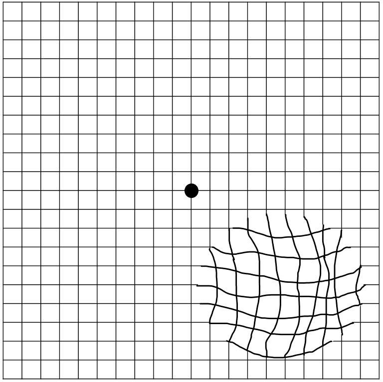
https://commons.wikimedia.org/wiki/File:Amsler_grid_-_distorted_vision_(metamorphopsia).png#/media/File:Amsler_grid_-_distorted_vision_(metamorphopsia).png (Accessed: 18 January 2024).

Image Source: Flickr (2020). 50337925921, Licensed under a creative commons licence. Available for free at: https://www.flickr.com/photos/nihgov/50337925921 (Accessed: 18 January 2024).
Classification of AMD
| NICE classification | Features | |
|---|---|---|
| Early AMD | Medium to large drusen with or without pigmentary abnormalities
No significant vision loss |
|
| Late AMD | Indeterminate | Degeneration of the retinal pigment epithelium (RPE) with presence of subretinal or intraretinal fluid |
| Dry | Geographic atrophy and significant vision loss with:
|
|
| Wet active | Choroidal neovascularisation, retinal angiomatous proliferation | |
| Wet inactive | Fibrous scar, sub-foveal atrophy or fibrosis, cystic degeneration | |
Table 2 NICE classification of age-related macular degeneration
Reminder of terminology:
- Drusen = collection of lipids below the retinal pigment epithelium (RPE) which appears as yellow deposits on fundoscopy. Drusen occur naturally with ageing and hence are a non-specific sign. Larger and more numerous drusen are associated with AMD.
- Geographic atrophy = atrophy of outer retinal tissue, RPE and choriocapillaris.
- Choroidal neovascularisation = new blood vessels in the choroid which breach the barrier membrane between the choroid and the retina.
- Retinal angiomatous proliferation = proliferation of retinal capillaries.
Management of AMD
There are currently no treatment options for dry AMD.
Anti-vascular endothelial growth factor (anti-VEGF) intravitreal injections are used to treat wet active AMD. While many options exist, there is currently no substantive evidence that one anti-VEGF medication is superior to others.
Patients should be offered certification of visual impairment when eligible and supported by low vision services.
Prognosis in AMD
Early and indeterminate AMD is not typically associated with loss of central vision.
Geographic atrophy (dry AMD) occurs more slowly than neovascularisation and causes less severe visual impairment.
In those with neovascularisation (wet AMD), vision loss can be rapid (over weeks to months), however timely treatment results in the vast majority maintaining their current vision level.
Diabetic retinopathy
Diabetes is the leading cause of vision loss in the working age population in the UK. Diabetic retinopathy (DR) is a chronic, progressive disease caused by prolonged hyperglycaemia affecting the retinal microvasculature.
It is potentially sight threatening through two mechanisms:
- Retinopathy = neovascularisation leading to intraocular haemorrhage causing global vision loss
- Maculopathy = localised damage to the macula causing loss of central vision
Patients with diabetes should be offered eye screening annually.
Classification of DR
Many classifications of DR exist but commonly include:
| Classification | Features | ||
|---|---|---|---|
| Retinopathy | Non-proliferative diabetic retinopathy | Mild | According to degree of ischaemia:
|
| Moderate | |||
| Severe | |||
| Proliferative diabetic retinopathy | Neovascularisation Vitreous haemorrhage |
||
| Maculopathy | Macular oedema | ||
Table 3 Classification of diabetic retinopathy
Management of DR
General management of DR includes adequate control of blood glucose, correcting hypertension and promoting smoking cessation. Proliferative DR can be managed by panretinal photocoagulation. Maculopathy can be managed by focal or grid photocoagulation, intravitreal steroids or anti-VEGF.
Complications of DR
Patients with proliferative DR are at risk of tractional retinal detachment, which is caused by contraction of the fibrous gel after haemorrhage and fibrosis.
Advanced retinal ischaemia can precipitate rubeosis iridis (neovascularisation affecting the iris), which can result in secondary glaucoma if the drainage channels are blocked.
Furthermore, patients with diabetes are more likely to have cataracts, retinal vein occlusion or ischaemic optic neuropathy.
Retinitis pigmentosa
Retinitis pigmentosa is the most common inherited cause for retinal degeneration. Mutations can be inherited in an autosomal dominant, autosomal recessive or X-linked manner and can be isolated or, less commonly, associated with systemic disease. Systemic associations include:
| Name | Inheritance | Other clinical features |
|---|---|---|
| Usher syndrome | Autosomal recessive | Hearing loss Most common cause of deaf-blindness |
| Bardet-Biedl syndrome | Autosomal recessive | Cognitive impairment Obesity Renal impairment Polydactyly |
| Kearns-Sayre syndrome | Mitochondrial | Progressive external ophthalmoplegia Muscle weakness with ‘ragged red fibres’ on muscle histology Cardiac conduction abnormalities (heart block) |
| Abetalipoproteinaemia | Autosomal recessive | Malabsorption of fats and fat-soluble vitamins Failure to thrive, diarrhoea, steatorrhoea |
Table 4 Systemic diseases associated with retinitis pigmentosa
These mutations result in rod photoreceptor dysfunction, presenting clinically as night blindness (nyctalopia) in the first instance, with subsequent degeneration of cone photoreceptors and the retinal pigment epithelium, which causes clinical deterioration and progressive peripheral vision loss.
Hydroxychloroquine retinopathy
Hydroxychloroquine is an anti-malarial medication also used in the management of autoimmune diseases such as rheumatoid arthritis and systemic lupus erythematosus. The incidence of hydroxychloroquine retinopathy is low but it can lead to irreversible vision loss.
Patients are screened through annual eye examinations if they are considered at high risk, including with treatment duration >5 years, high dose of hydroxychloroquine or co-existing significant renal or hepatic disease.
References
- Tidy, C. Gradual Loss of Vision [Internet]. Patient. Egton Medical Information Systems Limited. 2022 [cited 18 July 2023]. Available from: https://patient.info/doctor/gradual-loss-of-vision
- National Institute for Health and Care Excellence. Macular degeneration: age-related [Internet]. NICE. 2022 [cited May 17 2023]. Available from: https://cks.nice.org.uk/topics/macular-degeneration-age-related
- Baldwin A, Hjelde N, Goumalatsou C, Myers G. Oxford Handbook of Clinical Specialties. 10 edn. Oxford Medical Handbooks, UK: Oxford; 2016
- Royal College of Ophthalmologists. Diabetic Retinopathy Guidelines. 2012 [cited 18 July 2023]. Available from: https://www.rcophth.ac.uk/wp-content/uploads/2021/08/2012-SCI-267-Diabetic-Retinopathy-Guidelines-December-2012.pdf
- Public Health England. NHS Diabetic Eye Screening Programme: grading definitions for referable disease. 2021 [cited 13 July 2023] Available from: https://www.gov.uk/government/publications/diabetic-eye-screening-retinal-image-grading-criteria/nhs-diabetic-eye-screening-programme-grading-definitions-for-referable-disease
- Hsu, J. Retinitis Pigmentosa, EyeWiki. [Internet]. July 2023 [cited July 17 2023]. Available from: https://eyewiki.aao.org/Retinitis_Pigmentosa#History
- Ferrari S, Di Iorio E, Barbaro V, Ponzin D, Sorrentino FS, Parmeggiani F. Retinitis pigmentosa: genes and disease mechanisms. Curr Genomics. 2011 Jun;12(4):238-49. doi: 10.2174/138920211795860107. PMID: 22131869; PMCID: PMC3131731
- Pandya HK, Robinson M, Mandal N, Shah VA. Hydroxychloroquine retinopathy: A review of imaging. Indian J Ophthalmol. 2015 Jul;63(7):570-4. doi: 10.4103/0301-4738.167120. PMID: 26458473; PMCID: PMC4652246
Diplopia
Overview of diplopia
Diplopia, or double vision, is the separation of images horizontally, vertically or obliquely.
Diplopia can be monocular whereby the abnormality persists when the unaffected eye is closed. This is typically associated with local ocular causes such as refractive error or media abnormalities.
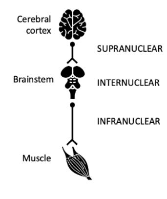
Binocular diplopia is the result of dysfunction of the extraocular muscles which leads to ocular misalignment, preventing fusion of the two images produced by each eye. The aetiology of binocular diplopia can be classified according to the segment of the efferent oculomotor pathway that is involved.
The following will be discussed:
| Location | Example | |||
|---|---|---|---|---|
 |
Supranuclear | Progressive supranuclear palsy | ||
| Nuclear and internuclear | Internuclear ophthalmoplegia | |||
| Infranuclear | Cranial nerves | Third nerve palsy
Fourth nerve palsy Sixth nerve palsy |
||
| Muscles and neuromuscular junction | Myasthenia gravis
Thyroid eye disease Miller Fisher syndrome |
|||
Table 5 Causes of binocular diplopia
Figure adapted from icons through The Noun Project, CC BY 3.0
Cerebral cortex – VectorsLab https://thenounproject.com/icon/brain-4224212/
Brainstem – Tezar tantular https://thenounproject.com/icon/brainstem-3656737/
Muscle – Amethyst Studio https://thenounproject.com/icon/muscle-4666756/
Progressive supranuclear palsy
Progressive supranuclear palsy (PSP) is an uncommon, atypical Parkinsonian disorder characterised by akinesia, postural instability, cognitive decline and ocular dysmotility. The latter typically consists of vertical gaze palsy or slow vertical saccades.
Internuclear ophthalmoplegia
Internuclear ophthalmoplegia (INO) is an ocular movement disorder caused by pathology of the medial longitudinal fasciculus (MLF) within the brainstem. It results in impaired adduction of the ipsilateral eye and nystagmus during abduction of the contralateral eye (Figure 6).
Neuroimaging in the form of an MRI brainstem is useful to identify a lesion. The underlying causes are:
- ⅓: infarction
- ⅓: demyelinating disorder, such as multiple sclerosis
- ⅓: other causes, e.g. trauma, infections, vasculitis
Treatment depends on the underlying cause. Approximately half of all cases of INO will resolve within 1 year.
Important differential diagnoses to consider include myasthenia gravis and sixth nerve palsy.
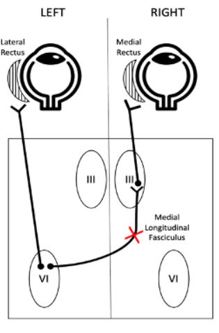
Image Source: Wikimedia Commons (2021). eyeball-4303894, Licensed under a creative commons licence. Available for free at: https://thenounproject.com/icon/eyeball-4303894/ (Accessed: 18 January 2024).
Cranial nerve palsy
| Cranial nerve | Third (oculomotor) | Fourth (trochlear) | Sixth (abducens) |
|---|---|---|---|
| Cause | Intracranial aneurysm: posterior communicating artery most common
Microvascular disease Trauma: significant injury Malignancy |
Congenital
Trauma: can occur following mild injury Microvascular disease: but less commonly affects CN IV compared to III and VI |
Microvascular disease
Malignancy Trauma: significant injury, e.g. basal skull fracture Increased intracranial pressure |
| Symptoms | Horizontal, vertical or oblique diplopia
Pain at the onset of symptoms is common |
Vertical diplopia and/or torsional diplopia which is worse at gaze towards the paretic superior oblique muscle
Head tilt towards the unaffected side reduces diplopia |
Horizontal diplopia which is worse at distance and at gaze towards paretic lateral rectus muscle
Head turned towards the affected side reduces diplopia |
| Examination | 
Primary position ipsilateral abduction, depression and intorsion. Fixed dilated pupil and ptosis may also be present if a complete palsy |

Primary position ipsilateral hypertropia and extorsion, more pronounced in ipsilateral head tilt |

Primary position esotropia with an ipsilateral abduction deficit |
| Prognosis | Most will recover over weeks to months
Lack of pupil involvement suggests a benign cause which can be observed |
Patients are often observed for several months before treatment is considered as symptoms may improve | Majority will recover spontaneously (3-6 months), more common if unilateral and non-traumatic |
Table 6 Summary of cranial nerve palsies
Figures adapted from icons through The Noun Project, CC BY 3.0
Eyes – Alone forever https://thenounproject.com/icon/eye-1436483/
Cranial nerve palsies are often investigated with neuroimaging, ideally MRI, to identify an underlying cause. This is essential in third nerve palsies with pupillary involvement to exclude an underlying intracranial aneurysm.
Management of cranial nerve palsies includes addressing the underlying cause as well as managing symptoms of diplopia with patching, prisms, strabismus surgery and/or botulinum toxin.
Muscular and neuromuscular junction
Myasthenia gravis
Myasthenia gravis (MG) is an autoimmune disease affecting the neuromuscular junctions of skeletal muscles. Pathophysiology includes the presence of autoantibodies to acetylcholine receptors (AChR).
MG typically presents with fatigable weakness and often affects the extraocular muscles. Patients can present with a spectrum of symptom severity from mild blurred vision to variable ptosis to severe diplopia. Examination characteristically reveals a fatigable muscle weakness not confined to a specific muscle or nerve and which is variable and asymmetrical in nature. Importantly, there is no associated eye pain and no pupil involvement. The tongue can also be affected, resulting in atrophy and a rare but distinctive ‘triple-groove’ appearance.
Occasionally, manifestations of MG are limited to the levator palpebrae superioris, orbicularis oculi and extraocular muscles, which is known as ‘ocular MG’. Unlike generalised MG where more females are affected, males are more likely to be affected by ocular MG. Furthermore, patients with ocular MG are more likely to be seronegative for AChR antibodies than those with generalised MG.
Diagnosis can be confirmed through various tests:
| Category | Test | Positive findings |
|---|---|---|
| Clinical | Lid fatigability test | Ptosis increases following prolonged upgaze. |
| Cogan’s lid twitch | Brief overshooting of upper lid retraction on returning to primary gaze after looking in downgaze. | |
| Ice test | Ptosis improves by ≥2 mm following an ice pack placed over the eyelid for 2-5 minutes. Cool temperature results in reduced acetylcholinesterase (AChE) activity. | |
| Pharmacological | Tensilon test (a.k.a. edrophonium test) | Improvement in clinical signs 1 minute after injection of IV edrophonium, a rapid-acting AChE.
Serious side effects include bradycardia and syncope, therefore atropine should be available during testing. Notably, up to 25% of patients with ocular MG may experience a paradoxical worsening of symptoms due to a depolarising block caused by excess ACh (acetylcholine). |
| Electrophysiological | Repetitive nerve stimulation | The number of individual muscle fibre action potentials and the amplitude of action potentials reduce with repeated electrical stimulation of the nerve. |
| Single fibre electromyography | Neuromuscular ‘jitter’ is recorded, which represents fluctuations in the time for end plate potentials at neuromuscular junctions to reach the threshold to produce an action potential.
More sensitive for MG than repetitive nerve stimulation. |
Table 7 Investigations to confirm a diagnosis of myasthenia gravis
Treatment with AChE inhibitors, such as pyridostigmine, improves symptoms but does not alter disease activity. Tapering courses of corticosteroids and immunosuppressants, such as azathioprine, are also used.
Thyroid eye disease
Thyroid eye disease is an autoimmune disorder. It is most commonly associated with thyrotoxicosis secondary to Graves’ disease and occurs due to shared autoantigens between the thyroid and the orbit. However, rarely, the patient can be euthyroid or hypothyroid. Common symptoms include altered periocular appearance, eye surface discomfort or lacrimation and diplopia at extremes of gaze. Diplopia occurs due to inflamed extraocular muscles and connective tissues and affects the inferior rectus, medial rectus and superior rectus in that order. The diplopia is worse in the direction opposite the affected muscle. Hence, hypertropia occurs if the inferior rectus is affected.
Diagnosis can be made clinically, but if required CT or MRI are the most valuable tests. Imaging demonstrates enlarged extraocular muscles with tendon sparing.
Management options include:
- Conservative: controlling exacerbating factors, e.g. smoking cessation, correction of thyroid dysfunction, lubricants, prisms
- Medical: steroids
- Surgical: orbital decompression
The natural course of thyroid eye disease is progression for 1-2 years, followed by a stable chronic phase. Complications include corneal ulceration precipitated by impaired eyelid closure and optic nerve compression.
Differential diagnoses include myasthenia gravis, allergic conjunctivitis and orbital tumours.
Miller Fisher syndrome
Guillain-Barré syndrome is an acute inflammatory demyelinating polyneuropathy which can affect extraocular muscles, when it is known as Miller Fisher syndrome (MFS). It is characterised by a triad of ophthalmoplegia, ataxia and areflexia. Approximately 85-90% of patients are positive for GQ1b antibodies. Pathophysiology includes a T-cell-mediated inflammatory response and destruction of myelin.
References
- Bienfang DC. Overview of diplopia. In: Brazis PW, Wilterdink JL, editors. UpToDate. [Internet]. Waltham (MA): UpToDate Inc; 2021 [updated September 9 2021; cited May 3 2023]. Available from: https://www.uptodate.com/contents/overview-of-diplopia
- Suleiman AO. Basic Approach to Diplopia. American Academy of Ophthalmology, EyeWiki. [Internet]. February 2023 [cited May 3 2023]. Available from: https://eyewiki.aao.org/w/index.php?title=Basic_Approach_to_Diplopia&oldid=89879
- Factor SA, Esper CD. Progressive Supranuclear Palsy (PSP). In: Hurtig HI, Eichler AF, editors. UpToDate. [Internet]. Waltham (MA): UpToDate Inc; 2022 [cited May 12 2023]. Available from:
- https://www.uptodate.com/contents/progressive-supranuclear-palsy-psp-clinical-features-and-diagnosis
- Feroze KB, Wang J. Internuclear Ophthalmoplegia. [Updated 2022 Jun 27]. In: StatPearls [Internet]. Treasure Island (FL): StatPearls Publishing; 2023 Jan. Available from: https://www.ncbi.nlm.nih.gov/books/NBK441970/
- Nair AG, Patil-Chhablani P, Venkatramani DV, Gandhi RA. Ocular myasthenia gravis: a review. Indian J Ophthalmol. 2014 Oct;62(10):985-91. doi: 10.4103/0301-4738.145987. PMID: 25449931; PMCID: PMC4278125.
- Perros P, Neoh C, Dickinson J. Thyroid eye disease. Bmj. 2009 Mar 6;338.
Complete Guide to Clinical Genetics ST3 Application and Recruitment (2027)
Written by Medibuddy. Last updated: October 2026. Clinical Genetics is

Complete Guide to Clinical Radiology ST3 Application and Recruitment (2027)
Recruitment paused: NHS England is not running Clinical Radiology ST3+ recru

Complete Guide to Paediatric Surgery ST3 Application and Recruitment in United Kingdom (2026)
Written by Medibuddy. Last updated: October 2026. If you’re planning


Take your subscriptions with you
Our mobile app allows you to access your interview and exam question banks wherever you are.

