


 Close
Close
 13th February 2025
Vascular
Retinal vein occlusion and retinal artery occlusion
Retinal vein occlusion is more common than retinal artery occlusion and less sudden. Both need to be managed as an emergency. Note that branch retinal vein or artery occlusions can occur and typically present with vision loss to a lesser extent, and the patient may not even be aware of the defect.
| Central retinal vein occlusion (CRVO) | Central retinal artery occlusion (CRAO) | |
|---|---|---|
| Cause | Atherosclerosis | Emboli (most common), thrombus or vasculitis |
| Risk factors | Hypertension
Diabetes mellitus Smoking Hyperlipidaemia Polycythaemia Renal failure Glaucoma |
Older age
Male Hypertension Diabetes mellitus Smoking Hyperlipidaemia Cardiovascular disease Periocular aesthetic injections (rare) |
| Symptoms | Painless monocular loss of vision | Painless monocular loss of vision over seconds |
| Fundoscopy appearance | Multiple retinal haemorrhages – the ‘stormy sunset’ appearance (Figure 1)
Cotton wool spots Optic disc swelling |
Pale retina with cherry red spot (Figure 2) |
| Management | Monitor for secondary complications including retinal neovascularisation and macular oedema | Patients should be reviewed by a Stroke team urgently. There is no evidence to demonstrate any treatment improves vision |
Table 1 Clinical features and management of CRVO and CRAO
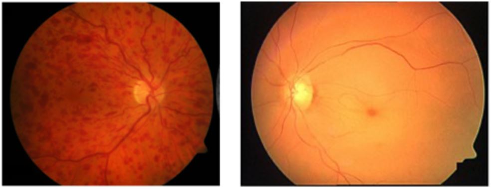
Image Source: Wikimedia Commons (2019). Fondo de ojo Ostrucción Vena central, Licensed under a creative commons licence. Available for free at: https://commons.wikimedia.org/wiki/File:Fondo_de_ojo_Ostrucción_Vena_central.png (Accessed: 18 January 2024).
Image Source: Wikimedia Commons (2014). Cherry Red Spot Fiess, Licensed under a creative commons licence. Available for free at: https://commons.wikimedia.org/wiki/File:Cherry_Red_Spot_Fiess.jpg, via Wikimedia Commons svg (Accessed: 18 January 2024).
Retinal vein occlusions can be clinically characterised into two types: ischaemic (25%) and non-ischaemic (75%) according to the area of non-perfusion in the posterior pole. Ischaemic retinal vein occlusions present with more severe vision loss, relative afferent pupillary defect and more marked fundoscopy findings. Prognosis is poorer and secondary complications are more common in ischaemic presentations.
Amaurosis fugax
Amaurosis fugax describes transient, painless monocular loss of vision caused by a carotid artery atheroma or embolus into the central retinal artery. It is considered as a retinal transient ischaemic attack (TIA) and hence the precursor to a stroke. Management should address any underlying cardiac risk factors and consider a carotid endarterectomy.
Vitreous haemorrhage
Vitreous haemorrhage can occur secondary to neovascularisation, retinal tears, retinal detachment or trauma. Neovascularisation is particularly significant in patients with diabetes because they can develop new retinal blood vessels (proliferative diabetic retinopathy) which are fragile and bleed easily. It is important the underlying cause is identified, and this may require an ultrasound if the blood obstructs the fundoscopy. Management may involve observation or a vitrectomy.
Anterior ischaemic optic neuropathy
Anterior ischaemic optic neuropathy (AION) is the most common cause of optic neuropathy in older people. It results from inflammation (arteritic) or atheroma (non-arteritic) causing blockage to the vascular supply of the optic nerve.
Arteritic AION is most commonly caused by giant cell (temporal) arteritis (GCA). GCA is classified as a medium-to-large-vessel vasculitis. Symptoms include monocular vision loss possibly preceded by episodes of amaurosis fugax, diplopia, new headache, jaw claudication and scalp tenderness. Examination reveals reduced visual acuity, optic disc oedema and thickened temporal arteries, which may be pulseless. Diagnosis can be confirmed with a raised ESR, raised CRP and a positive temporal artery biopsy. Note that due to skip lesions the biopsy may be falsely negative. Management with high-dose steroids should be started without delay (i.e. a positive biopsy result should not be awaited) to prevent vision loss in the other eye.
Other rarer causes of arteritic AION include other types of vasculitis, such as polyarteritis nodosa or systemic lupus erythematosus.
Non-arteritic AION is more common. It is typically painless and noticed on waking. Risk factors include hypertension, hypercholesterolaemia, diabetes mellitus and smoking. Management is to control these risk factors to prevent occurrence in the other eye.
Complete Guide to Clinical Genetics ST3 Application and Recruitment (2027)
Written by Medibuddy. Last updated: October 2026. Clinical Genetics is

Complete Guide to Clinical Radiology ST3 Application and Recruitment (2027)
Recruitment paused: NHS England is not running Clinical Radiology ST3+ recru

Complete Guide to Paediatric Surgery ST3 Application and Recruitment in United Kingdom (2026)
Written by Medibuddy. Last updated: October 2026. If you’re planning


Take your subscriptions with you
Our mobile app allows you to access your interview and exam question banks wherever you are.

