


 Close
Close
 13th February 2025
Chronic vision loss
Overview of chronic vision loss
Causes of chronic vision loss can be categorised according to the presence or absence of pain. Painless chronic vision loss can be caused by:
- Refractive error – characterised by vision improvement with pinhole
- Cataracts
- Age-related macular degeneration (AMD)
- Diabetic retinopathy
- Open-angle glaucoma
- Optic nerve pathology
- Hereditary retinal dystrophies
- Drugs, toxins and deficiencies
Painful chronic vision loss is much less common. A serious underlying cause, such as an intracranial mass, choroidal melanoma or optic neuritis, should be investigated.
Age-related macular degeneration
AMD is the most common cause of irreversible blindness in developed countries and the leading cause for blindness in the United Kingdom. It is a potentially progressive disorder of the macula which affects people over 50 years. Risk factors include age, family history, Northern European ancestry and smoking (the latter in a dose-response relationship).
Symptoms include vision blurring or distortion, metamorphopsia and, later, a central scotoma (Figures 4 and 5). Note that as the macula is responsible for central vision, it is this part of the visual field that is affected in AMD.
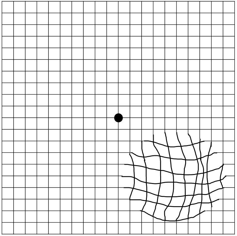
https://commons.wikimedia.org/wiki/File:Amsler_grid_-_distorted_vision_(metamorphopsia).png#/media/File:Amsler_grid_-_distorted_vision_(metamorphopsia).png (Accessed: 18 January 2024).

Image Source: Flickr (2020). 50337925921, Licensed under a creative commons licence. Available for free at: https://www.flickr.com/photos/nihgov/50337925921 (Accessed: 18 January 2024).
Classification of AMD
| NICE classification | Features | |
|---|---|---|
| Early AMD | Medium to large drusen with or without pigmentary abnormalities
No significant vision loss |
|
| Late AMD | Indeterminate | Degeneration of the retinal pigment epithelium (RPE) with presence of subretinal or intraretinal fluid |
| Dry | Geographic atrophy and significant vision loss with:
|
|
| Wet active | Choroidal neovascularisation, retinal angiomatous proliferation | |
| Wet inactive | Fibrous scar, sub-foveal atrophy or fibrosis, cystic degeneration | |
Table 2 NICE classification of age-related macular degeneration
Reminder of terminology:
- Drusen = collection of lipids below the retinal pigment epithelium (RPE) which appears as yellow deposits on fundoscopy. Drusen occur naturally with ageing and hence are a non-specific sign. Larger and more numerous drusen are associated with AMD.
- Geographic atrophy = atrophy of outer retinal tissue, RPE and choriocapillaris.
- Choroidal neovascularisation = new blood vessels in the choroid which breach the barrier membrane between the choroid and the retina.
- Retinal angiomatous proliferation = proliferation of retinal capillaries.
Management of AMD
There are currently no treatment options for dry AMD.
Anti-vascular endothelial growth factor (anti-VEGF) intravitreal injections are used to treat wet active AMD. While many options exist, there is currently no substantive evidence that one anti-VEGF medication is superior to others.
Patients should be offered certification of visual impairment when eligible and supported by low vision services.
Prognosis in AMD
Early and indeterminate AMD is not typically associated with loss of central vision.
Geographic atrophy (dry AMD) occurs more slowly than neovascularisation and causes less severe visual impairment.
In those with neovascularisation (wet AMD), vision loss can be rapid (over weeks to months), however timely treatment results in the vast majority maintaining their current vision level.
Diabetic retinopathy
Diabetes is the leading cause of vision loss in the working age population in the UK. Diabetic retinopathy (DR) is a chronic, progressive disease caused by prolonged hyperglycaemia affecting the retinal microvasculature.
It is potentially sight threatening through two mechanisms:
- Retinopathy = neovascularisation leading to intraocular haemorrhage causing global vision loss
- Maculopathy = localised damage to the macula causing loss of central vision
Patients with diabetes should be offered eye screening annually.
Classification of DR
Many classifications of DR exist but commonly include:
| Classification | Features | ||
|---|---|---|---|
| Retinopathy | Non-proliferative diabetic retinopathy | Mild | According to degree of ischaemia:
|
| Moderate | |||
| Severe | |||
| Proliferative diabetic retinopathy | Neovascularisation Vitreous haemorrhage |
||
| Maculopathy | Macular oedema | ||
Table 3 Classification of diabetic retinopathy
Management of DR
General management of DR includes adequate control of blood glucose, correcting hypertension and promoting smoking cessation. Proliferative DR can be managed by panretinal photocoagulation. Maculopathy can be managed by focal or grid photocoagulation, intravitreal steroids or anti-VEGF.
Complications of DR
Patients with proliferative DR are at risk of tractional retinal detachment, which is caused by contraction of the fibrous gel after haemorrhage and fibrosis.
Advanced retinal ischaemia can precipitate rubeosis iridis (neovascularisation affecting the iris), which can result in secondary glaucoma if the drainage channels are blocked.
Furthermore, patients with diabetes are more likely to have cataracts, retinal vein occlusion or ischaemic optic neuropathy.
Retinitis pigmentosa
Retinitis pigmentosa is the most common inherited cause for retinal degeneration. Mutations can be inherited in an autosomal dominant, autosomal recessive or X-linked manner and can be isolated or, less commonly, associated with systemic disease. Systemic associations include:
| Name | Inheritance | Other clinical features |
|---|---|---|
| Usher syndrome | Autosomal recessive | Hearing loss Most common cause of deaf-blindness |
| Bardet-Biedl syndrome | Autosomal recessive | Cognitive impairment Obesity Renal impairment Polydactyly |
| Kearns-Sayre syndrome | Mitochondrial | Progressive external ophthalmoplegia Muscle weakness with ‘ragged red fibres’ on muscle histology Cardiac conduction abnormalities (heart block) |
| Abetalipoproteinaemia | Autosomal recessive | Malabsorption of fats and fat-soluble vitamins Failure to thrive, diarrhoea, steatorrhoea |
Table 4 Systemic diseases associated with retinitis pigmentosa
These mutations result in rod photoreceptor dysfunction, presenting clinically as night blindness (nyctalopia) in the first instance, with subsequent degeneration of cone photoreceptors and the retinal pigment epithelium, which causes clinical deterioration and progressive peripheral vision loss.
Hydroxychloroquine retinopathy
Hydroxychloroquine is an anti-malarial medication also used in the management of autoimmune diseases such as rheumatoid arthritis and systemic lupus erythematosus. The incidence of hydroxychloroquine retinopathy is low but it can lead to irreversible vision loss.
Patients are screened through annual eye examinations if they are considered at high risk, including with treatment duration >5 years, high dose of hydroxychloroquine or co-existing significant renal or hepatic disease.
References
- Tidy, C. Gradual Loss of Vision [Internet]. Patient. Egton Medical Information Systems Limited. 2022 [cited 18 July 2023]. Available from: https://patient.info/doctor/gradual-loss-of-vision
- National Institute for Health and Care Excellence. Macular degeneration: age-related [Internet]. NICE. 2022 [cited May 17 2023]. Available from: https://cks.nice.org.uk/topics/macular-degeneration-age-related
- Baldwin A, Hjelde N, Goumalatsou C, Myers G. Oxford Handbook of Clinical Specialties. 10 edn. Oxford Medical Handbooks, UK: Oxford; 2016
- Royal College of Ophthalmologists. Diabetic Retinopathy Guidelines. 2012 [cited 18 July 2023]. Available from: https://www.rcophth.ac.uk/wp-content/uploads/2021/08/2012-SCI-267-Diabetic-Retinopathy-Guidelines-December-2012.pdf
- Public Health England. NHS Diabetic Eye Screening Programme: grading definitions for referable disease. 2021 [cited 13 July 2023] Available from: https://www.gov.uk/government/publications/diabetic-eye-screening-retinal-image-grading-criteria/nhs-diabetic-eye-screening-programme-grading-definitions-for-referable-disease
- Hsu, J. Retinitis Pigmentosa, EyeWiki. [Internet]. July 2023 [cited July 17 2023]. Available from: https://eyewiki.aao.org/Retinitis_Pigmentosa#History
- Ferrari S, Di Iorio E, Barbaro V, Ponzin D, Sorrentino FS, Parmeggiani F. Retinitis pigmentosa: genes and disease mechanisms. Curr Genomics. 2011 Jun;12(4):238-49. doi: 10.2174/138920211795860107. PMID: 22131869; PMCID: PMC3131731
- Pandya HK, Robinson M, Mandal N, Shah VA. Hydroxychloroquine retinopathy: A review of imaging. Indian J Ophthalmol. 2015 Jul;63(7):570-4. doi: 10.4103/0301-4738.167120. PMID: 26458473; PMCID: PMC4652246
Complete Guide to Clinical Genetics ST3 Application and Recruitment (2027)
Written by Medibuddy. Last updated: October 2026. Clinical Genetics is

Complete Guide to Clinical Radiology ST3 Application and Recruitment (2027)
Recruitment paused: NHS England is not running Clinical Radiology ST3+ recru

Complete Guide to Paediatric Surgery ST3 Application and Recruitment in United Kingdom (2026)
Written by Medibuddy. Last updated: October 2026. If you’re planning


Take your subscriptions with you
Our mobile app allows you to access your interview and exam question banks wherever you are.

