


 Close
Close
 13th February 2025
Age-related macular degeneration
AMD is the most common cause of irreversible blindness in developed countries and the leading cause for blindness in the United Kingdom. It is a potentially progressive disorder of the macula which affects people over 50 years. Risk factors include age, family history, Northern European ancestry and smoking (the latter in a dose-response relationship).
Symptoms include vision blurring or distortion, metamorphopsia and, later, a central scotoma (Figures 4 and 5). Note that as the macula is responsible for central vision, it is this part of the visual field that is affected in AMD.
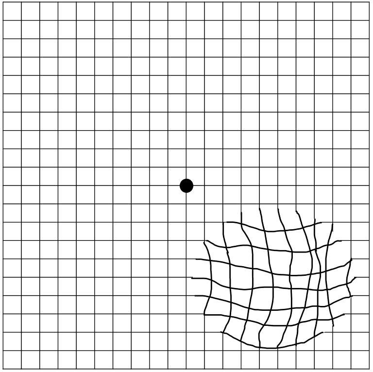
https://commons.wikimedia.org/wiki/File:Amsler_grid_-_distorted_vision_(metamorphopsia).png#/media/File:Amsler_grid_-_distorted_vision_(metamorphopsia).png (Accessed: 18 January 2024).

Image Source: Flickr (2020). 50337925921, Licensed under a creative commons licence. Available for free at: https://www.flickr.com/photos/nihgov/50337925921 (Accessed: 18 January 2024).
Classification of AMD
| NICE classification | Features | |
|---|---|---|
| Early AMD | Medium to large drusen with or without pigmentary abnormalities
No significant vision loss |
|
| Late AMD | Indeterminate | Degeneration of the retinal pigment epithelium (RPE) with presence of subretinal or intraretinal fluid |
| Dry | Geographic atrophy and significant vision loss with:
|
|
| Wet active | Choroidal neovascularisation, retinal angiomatous proliferation | |
| Wet inactive | Fibrous scar, sub-foveal atrophy or fibrosis, cystic degeneration | |
Table 2 NICE classification of age-related macular degeneration
Reminder of terminology:
- Drusen = collection of lipids below the retinal pigment epithelium (RPE) which appears as yellow deposits on fundoscopy. Drusen occur naturally with ageing and hence are a non-specific sign. Larger and more numerous drusen are associated with AMD.
- Geographic atrophy = atrophy of outer retinal tissue, RPE and choriocapillaris.
- Choroidal neovascularisation = new blood vessels in the choroid which breach the barrier membrane between the choroid and the retina.
- Retinal angiomatous proliferation = proliferation of retinal capillaries.
Management of AMD
There are currently no treatment options for dry AMD.
Anti-vascular endothelial growth factor (anti-VEGF) intravitreal injections are used to treat wet active AMD. While many options exist, there is currently no substantive evidence that one anti-VEGF medication is superior to others.
Patients should be offered certification of visual impairment when eligible and supported by low vision services.
Prognosis in AMD
Early and indeterminate AMD is not typically associated with loss of central vision.
Geographic atrophy (dry AMD) occurs more slowly than neovascularisation and causes less severe visual impairment.
In those with neovascularisation (wet AMD), vision loss can be rapid (over weeks to months), however timely treatment results in the vast majority maintaining their current vision level.
Complete Guide to Clinical Genetics ST3 Application and Recruitment (2027)
Written by Medibuddy. Last updated: October 2026. Clinical Genetics is

Complete Guide to Clinical Radiology ST3 Application and Recruitment (2027)
Recruitment paused: NHS England is not running Clinical Radiology ST3+ recru

Complete Guide to Paediatric Surgery ST3 Application and Recruitment in United Kingdom (2026)
Written by Medibuddy. Last updated: October 2026. If you’re planning


Take your subscriptions with you
Our mobile app allows you to access your interview and exam question banks wherever you are.

