


 Close
Close
 17th February 2025
Lower Respiratory Disease
Asthma
Diagnosis of asthma
Clinical features of asthma
Asthma is a clinical diagnosis; there is no singular definition but criteria normally include:
- Presence of episodic symptoms (cough, breathlessness, wheeze, chest tightness)
- Variable airflow obstruction
- Evidence of airway inflammation
| Features with high predictive value for asthma | Features with low predictive value for asthma |
|---|---|
| Episodic symptoms with identifiable triggers, e.g. viral, allergen, exercise, emotion
Variation in FEV1 or PEF (peak expiratory flow) during symptomatic episodes Wheeze on auscultation Diurnal variability Personal history of atopy (e.g. eczema, allergic rhinitis), family history of asthma Raised IgE or eosinophilia |
Constant symptoms
Signs and symptoms to suggest alternative diagnosis, e.g. COPD, obesity, dysfunctional breathing Repeatedly normal chest examination when symptomatic |
Table 1 Clinical features with a high and low predictive value for asthma
Investigations in chronic asthma
With a typical clinical picture of asthma, treatment can be initiated with inhaled corticosteroids (usually a 6-week trial). If there is a good objective response (shown using lung function tests or symptom scores), the diagnosis can be confirmed. Otherwise, investigations include the following:
- Spirometry with bronchodilator reversibility test:
- FEV1/FVC <70%, i.e. an obstructive picture
- Post-bronchodilator >12% improvement in FEV1 or >200 ml increase in volume
- Fractional exhaled nitrous oxide (FeNO): >40 parts per billion (a marker of airway inflammation)
Occupational asthma
Common triggers for occupational asthma include isocyanates (in spray paint, adhesives), resins and flour. To make the diagnosis, patients should record peak flow measurements at work and home. This diagnosis requires referral to a respiratory specialist.
Management of chronic asthma
Both the BTS (British Thoracic Society) 2019 guidelines and NICE 2017 guidelines cover the management of acute asthma. The following is a summary of the NICE stepwise management of asthma:
- Step 1 (new diagnosis): short-acting beta-2 agonist (SABA)
- Step 2 (not controlled with SABA or new diagnosis of asthma with moderate/severe symptoms, e.g. nighttime waking or frequent exacerbations): SABA + low-dose inhaled corticosteroid (ICS)
- Step 3: SABA + low-dose ICS + leukotriene receptor antagonist (LTRA)
- Step 4: SABA + low-dose ICS + long-acting beta agonist (LABA); continue LRTA if good response, otherwise stop
- Step 5: SABA ± LTRA; switch ICS + LABA for maintenance and reliever therapy (MART) including a low-dose ICS
- Step 6: SABA ± LTRA + medium-dose ICS MART
- Step 7: SABA ± LTRA + one of: high-dose ICS (fixed-dose regimen, not as a MART) or trial of long-acting muscarinic receptor antagonist (LAMA) or theophylline; referral to specialist asthma service
Guidelines by BTS are very similar, although they advise prescribing a leukotriene receptor antagonist in step 4 rather
| SABA: salbutamol, terbutaline
ICS: beclometasone, budesonide, fluticasone, mometasone LTRA: montelukast, zafirlukast LABA: salmeterol, formoterol MART (maintenance and reliever therapy – typically an ICS and fast-acting LABA in one inhaler): beclometasone/formoterol, budesonide/formoterol |
Box 1 Common medications used in asthma
| Low dose: less than 400 mcg of budesonide or equivalent
Intermediate dose: 400-800 mcg of budesonide or equivalent High dose: more than 800 mcg of budesonide or equivalent |
Box 2 ICS dosing
Acute asthma
Definitions in acute asthma
NICE guidance advises that acute asthma attacks be graded by severity using criteria such as peak flow, observations such as heart rate and respiratory rate, ABG (arterial blood gas) results and clinical symptoms.
Patients presenting with severe or life-threatening asthma attacks should be started on asthma treatment immediately and should be treated in secondary care facilities. Patients with moderate asthma attacks can be trialled on treatment at home or in primary care, in the first instance.
| Moderate | Acute severe | Life threatening | Near fatal |
|---|---|---|---|
| Increasing symptoms
PEF 50-75% best or predicted No features of acute severe asthma |
PEF 33-50% best or predicted
RR ≥25 /min HR ≥110 bpm Inability to complete sentences in one breath |
PEF <33% of predicted
PaO2 <8 kPA Normal PaCO2 (4.6-6.0) Exhaustion, altered consciousness Arrhythmia Hypotension Cyanosis Poor respiratory effort, silent chest |
Raised PaCO2
Requiring mechanical ventilation with raised inflation pressures |
Table 2 Grading of acute asthma
Investigations in acute asthma
These should include:
- ABG if saturations <92% or with other features of life-threatening asthma
- Chest X-ray if there is any suspicion of consolidation, pneumothorax, any features of life-threatening asthma or if the patient is not responding to initial treatment
Management of acute asthma
General management includes:
- Oxygen to keep saturations 94-98%
- Corticosteroids: prednisolone 40 mg for a minimum of 5 days; ICS should be continued during this time
- Salbutamol (SABA): the nebulised route (2.5-5 mg) is preferred if there are any features of acute severe asthma; IV is reserved for those in whom inhaled routes are unreliable, e.g. with altered consciousness
- Ipratropium bromide (short-acting muscarinic antagonist): nebulised; if no response to above treatments
- IV magnesium sulphate if features of acute severe asthma are present
Intensive care referral should be considered with any of the following:
- Ventilatory support is required
- Acute severe or life-threatening asthma failing to respond to therapy, as evidenced by:
- Investigations: deteriorating PEF, persistent or worsening hypoxia, hypercapnia, worsening acidosis
- Clinical features: exhaustion, drowsiness, altered conscious state, respiratory arrest
Criteria for discharge
Prior to discharge, a patient should be stable off nebulisers and oxygen for 12-24 hours, have a peak flow >75% of predicted or best, and have had their inhaler technique checked.
Asthma differential diagnosis
It is important to be aware of other differentials that may present with similar symptoms to asthma, such as wheezing, cough and shortness of breath:
- COPD: consider in older patients with a strong smoking history. Typically of chronic onset, with sputum production and more frequent respiratory tract infections. Be aware of asthma-COPD-overlap syndrome, where patients may have elements of both COPD and asthma.
- Heart failure: patients can present with wheeze, cough and breathlessness; assess for evidence of fluid overload.
- Angioedema: could be secondary to an allergic reaction, hereditary angioedema or ACE-inhibitor related. Swelling in the connective tissue around the airways can cause wheeze and breathlessness.
- Eosinophilic granulomatosis with polyangiitis (EGPA): a small-medium vessel vasculitis that can commonly affect the lungs, skin and nerves, and is associated with eosinophilia and p-ANCA (usually anti-MPO antibodies). Adult-onset asthma is one of the most common signs of EGPA and it typically does not respond well to standard asthma treatment.
- Inhalation of a foreign body: typically sudden-onset wheeze and breathlessness following a history of inhaling or aspirating a foreign body.
References
- British guideline on the management of asthma. A national clinical guideline [Internet]. 2003 [cited 2022 Jan 31] Available from: https://www.brit-thoracic.org.uk/document-library/guidelines/asthma/btssign-guideline-for-the-management-of-asthma-2019/
- National Institute for Health and Care Excellence. Asthma: diagnosis, monitoring and chronic asthma management [Internet]. 2017 [cited 2022 Jan 31] Available from: https://www.nice.org.uk/guidance/ng80/resources/asthma-diagnosis-monitoring-and-chronic-asthma-management-pdf-1837687975621
- Chapman S. Oxford handbook of respiratory medicine. Oxford, United Kingdom: Oxford University Press; 2014
Chronic obstructive pulmonary disease
Overview of COPD
COPD is an umbrella term used to encompass chronic bronchitis and emphysema. It is a chronic, life-limiting condition, with causes including:
- Smoking – the most important cause
- Alpha-1 antitrypsin (AAT) deficiency – should be considered in younger patients with no smoking history or a positive family history
- Occupational – secondary to exposure to cadmium, coal, cement or cotton
Prevalent features are productive cough, dyspnoea, reduced exercise tolerance, wheeze and recurrent ‘winter bronchitis’.
Diagnosis of COPD
Typical investigation findings include the following:
- Blood tests: FBC and CRP to identify anaemia and secondary polycythaemia, and rule out infection
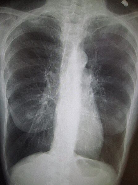
- Chest X-ray: flattened diaphragm, hyperexpanded lungs, bullae
- Lung function tests, especially post-bronchodilator spirometry, which should show an obstructive picture (FEV1/FVC <70%) to be in keeping with a diagnosis of COPD
- BMI: low BMI or weight loss is common in severe COPD and associated with a worse prognosis. It can also be associated with a co-existing lung cancer
Image Source: Wikimedia Commons (2010). COPD, Licensed under a creative commons licence. Available for free at: https://commons.wikimedia.org/wiki/File:COPD.JPG (Accessed: 19 January 2024).
FEV₁ is used to determine the severity of COPD:
| >80% of predicted | Stage 1: mild COPD – should have symptoms, e.g. cough or breathlessness, to make diagnosis |
| 50-80% of predicted | Stage 2: moderate COPD |
| 30-50% of predicted | Stage 3: severe COPD |
| <30% of predicted | Stage 4: very severe COPD |
Table 3 Staging of COPD using FEV₁
Management of COPD
Acute exacerbation of COPD
An acute exacerbation of COPD can be infective or non-infective. Key pathogens include:
- Bacterial: Haemophilus influenzae (most common bacterial pathogen in COPD), Streptococcus pneumoniae
- Viral: rhinovirus is the most common
Management is as follows:
- ABG to determine target SaO2: normally aim for 88-92%, but increase to 94-98% if normal pCO2
- Keep saturations 88-92% with a history of T2RF (type 2 respiratory failure), chronic hypercapnia or if baseline saturations are known to be <94%
- Bronchodilators: nebulised delivery and increased frequency are often required during an exacerbation
- Steroids: 30 mg prednisolone for 5 days
- Antibiotics are indicated only with signs of infection, e.g. purulent sputum, raised inflammatory markers or chest X-ray changes
Non-invasive ventilation (NIV) is typically indicated in the presence of T2RF with acidosis:
- Recommended with pH 7.25-7.35
- If pH <7.25, ITU involvement and possible intubation should be considered
- NIV delivers different air pressures for inspiration and expiration. The inspiratory positive airway pressure (IPAP) is set higher to improve ventilation and aid the patient’s own respiratory effort. The expiratory positive airway pressure (EPAP) recruits collapsed alveoli to increase gas exchange and allow removal of exhaled gases. Typical initial settings for NIV are IPAP 10-15 cmH₂O and EPAP 4-5 cmH₂O
Long-term management of COPD
The most important intervention is smoking cessation. Other facets to long-term management include vaccinations (once-off pneumococcal and annual influenza), pulmonary rehabilitation and inhaler therapy as per NICE guidelines:
- Step 1 (new diagnosis): SABA or short-acting muscarinic antagonist (SAMA) as needed
Beyond this stage, it is important to check for features of asthma or steroid responsiveness, e.g. history of asthma, high eosinophils, diurnal variation in PEF or FEV1 - Step 2a (no features of steroid responsiveness): SABA (PRN) + LABA + LAMA
- Step 2b (features of steroid responsiveness): SABA or SAMA (PRN) + LABA + ICS
- Step 3: SABA (PRN) + LABA + LAMA + ICS
| SABA: salbutamol, terbutaline
SAMA: ipratropium LABA: salmeterol, formoterol LAMA: tiotropium, umeclidinium, glycopyrrolate ICS: beclometasone, budesonide, fluticasone, mometasone |
Box 3 Common medications used in COPD
Other treatments may include:
- Theophylline if symptomatic with maximal inhaler therapy
- Mucolytic, e.g. carbocisteine, in cases of ongoing productive cough
- Prophylactic antibiotics in selected cases, e.g. azithromycin (usually 3 times a week) can be considered for non-smokers already receiving optimal medical management with frequent (>4 per year) or severe (prolonged or resulting in hospitalisation) exacerbations of COPD
- Surgical input (for selected patients), e.g. lung reduction surgery or bullectomy
- Diuretics if evidence of cor pulmonale (right-sided heart failure due to chronic raised pulmonary pressures)
Long-term oxygen therapy is indicated for non-smokers in selected cases:
- pO₂ on room air <7.3 kPa OR
- pO₂ on room air 7.3-8.0 kPa and one of secondary polycythaemia, pulmonary hypertension or fluid overload
End-stage COPD
COPD is a chronic progressive illness and the end stage of the disease will typically feature a progressive decline with increasing frequency and severity of acute exacerbations.
Factors that may indicate a patient is approaching end-stage COPD and end of life care are:
- Increasing frequency of hospitalisation and worsening severity of acute exacerbations
- Low body mass index
- Poor lung function on spirometry testing
- Other comorbidities such as cardiac disease or malignancy
Management of end-stage COPD should involve advance care planning and ensuring optimal medical management. MDT approach should involve respiratory specialist nurses, district nurses, palliative care team and social services/occupational therapy as appropriate.
It is important to ensure management of symptoms associated with COPD in conjunction with palliative care specialists:
- Breathlessness: simple breathing exercises, home oxygen, opiates and benzodiazepines
- Cough and secretions
- Pain
- Anxiety and depression
Alpha-1 antitrypsin deficiency
AAT deficiency is an inherited condition which causes reduced levels of alpha-1 antitrypsin, a protease inhibitor that inhibits elastase, an enzyme that causes inflammatory damage to the lungs. It should be considered in younger non-smokers who present similarly to COPD.
Genetics of AAT deficiency
The inheritance for alpha-1 antitrypsin deficiency is co-dominant.
The affected gene is on chromosome 14. Many different alleles have been identified but the most common are the M allele (normal), S allele (partially defective) and Z allele (nearly fully defective).
- MM – normal phenotype
- MS/MZ – reduced levels of AAT (50-75% of normal) but if non-smoker, risk of developing COPD is low
- SS/SZ – increased risk of developing COPD compared to the general population
- ZZ – worst prognosis, with an almost 100% chance of developing COPD
Features of AAT deficiency
Pulmonary manifestations include COPD and bronchiectasis, which is worse in smokers. Hepatic manifestations include cirrhosis, increased risk of hepatocellular carcinoma and cholestasis. There are also skin manifestations such as panniculitis and vasculitis (particularly ANCA-positive disease).
Management of AAT deficiency
Smoking cessation is crucial. Other treatments include bronchodilator therapy, alpha-1 antitrypsin augmentation therapy and surgical management options (lung transplant or lung volume reduction surgery).
References
- National Institute for Health and Care Excellence. Chronic obstructive pulmonary disease in over 16s: diagnosis and management [Internet]. 2018 [cited 2022 Jan 31] Available from: https://www.nice.org.uk/guidance/ng115/resources/chronic-obstructive-pulmonary-disease-in-over-16s-diagnosis-and-management-pdf-66141600098245
- The National Institute for Health and Care Excellence. Oxygen [Internet]. NICE; 2019 [cited 2022 March 28] Available from: https://bnf.nice.org.uk/treatment-summary/oxygen.html
- Sethi S. Bacteria in Exacerbations of Chronic Obstructive Pulmonary Disease: Phenomenon or Epiphenomenon? Proceedings of the American Thoracic Society. 2004 Apr 1;1(2):109–14
- Davidson AC, Banham S, Elliott M, Kennedy D, Gelder C, Glossop A, et al. BTS/ICS guideline for the ventilatory management of acute hypercapnic respiratory failure in adults. Thorax. 2016 Mar 14;71(Suppl 2):ii1–35
- Chapman S. Oxford handbook of respiratory medicine. Oxford; New York: Oxford University Press; 2005
Cystic fibrosis (CF)
Overview of CF
Cystic fibrosis is a genetic condition characterised by an abnormality in salt and water transport, resulting in thick, sticky mucus that can block the lungs and digestive system.
Genetics of CF
Cystic fibrosis has an autosomal recessive inheritance caused by a defect in the cystic fibrosis transmembrane conductance regulator (CFTR) (chromosome 7), which is a chloride channel in cells that line the lungs, pancreas, digestive tract, sweat glands and reproductive organs.
There are over 200 genetic mutations associated with CF, but the commonest is deletion of the 508th amino acid (∆508).
Diagnosis of CF
Cystic fibrosis is mostly now picked up at birth via heel-prick testing in the UK (blood spot immunoreactive trypsin test), although, rarely, milder disease may present later. This can be confirmed by a sweat test (people with cystic fibrosis tend to have high levels of chloride in their sweat) and genetic screening.
Clinical features of CF
Viscous secretions in the lungs can cause recurrent chest infections and subsequent bronchiectasis.
In the pancreas, secretions can block exocrine ducts, leading to pancreatic enzyme insufficiency (malabsorption, steatorrhoea) and pancreatic destruction, causing diabetes mellitus.
Thickened secretions in the reproductive organs can cause infertility, reduced fertility and delayed puberty. Fertility issues are often more severe in men than women, due to congenital bilateral absence of the vas deferens.
Patients with cystic fibrosis are also at high risk of intestinal obstruction; meconium ileus is seen in newborns.
Management of CF
The mainstay of management is respiratory physiotherapy, such as postural drainage techniques to help clear viscous sputum and avoid chest infections. Other aspect of management may include:
- Pancreatic enzyme supplementation – to treat pancreatic insufficiency
- Specialised diet – high in calories, fat and salt to counteract malabsorption
- Mucolytics – to help break down thickened sputum
- Antibiotics for recurrent infections – patients with CF generally need longer courses and higher doses. Pseudomonas aeruginosa cover should always be considered, especially if the patient has been previously infected. This is a common coloniser in patients with cystic fibrosis and is identified by Gram-negative rods on blood cultures.
- Lung transplantation – bilateral lung transplant is usually the procedure of choice and should be considered once a patient is on optimal medical therapy and their disease is impacting their quality of life
- Routine immunisations and yearly flu vaccine
References
- Chapman S. Oxford handbook of respiratory medicine. Oxford; New York: Oxford University Press; 2005
- National Institute for Health and Care Excellence. Cystic fibrosis: diagnosis and management [Internet]. 2017 [cited 2022 Jan 31] Available from: https://www.nice.org.uk/guidance/ng78/resources/cystic-fibrosis-diagnosis-and-management-pdf-1837640946373
- August 2 Pool JJ, 2019 [cited 2022 Jan 31] Diet and Nutrition Considerations with Cystic Fibrosis [Internet]. Cystic-Fibrosis.com. Available from: https://cystic-fibrosis.com/diet-nutrition
Bronchiectasis
Overview of bronchiectasis
Bronchiectasis is the chronic and irreversible dilatation of the airways due to recurrent inflammation or infection. It is associated with chronic sputum production, airflow obstruction and recurrent chest infections.
Bronchiectasis can be idiopathic or secondary to one of many causes, including:
- Previous infection: bacterial pneumonia, TB, viral illness, measles
- Genetic causes: cystic fibrosis, Kartagener’s syndrome, primary ciliary dyskinesia
- Allergic bronchopulmonary aspergillosis
- Immune deficiency:
- Primary: hypogammaglobulinaemia, common variable immunodeficiency
- Secondary: HIV, CLL, nephrotic syndrome
- Yellow nail syndrome
- Chronic aspiration: GORD, alcohol excess
- Bronchial obstruction: lung cancer
- Systemic conditions: rheumatoid arthritis, systemic lupus erythematosus, inflammatory bowel disease
Presentation of bronchiectasis
Bronchiectasis often presents as a chronic productive cough with recurrent chest infections or “bronchitis” over a number of years. Patients typically have daily mucopurulent sputum with intermittent haemoptysis. They may also have chronic progressive breathlessness.
On examination, patients may have coarse inspiratory and expiratory crackles or wheeze and finger clubbing.
Investigations in bronchiectasis
Key investigations and findings in bronchiectasis include:
- Blood tests:
- White cell count and CRP to rule in/out infection
- Raised eosinophils may be suggestive of allergic bronchopulmonary aspergillosis
- To investigate an underlying cause, e.g. rheumatoid factor, immunoglobulins, HIV test
- Sputum culture: pathogenic organisms are often found in sputum and it is advisable to send a sputum culture and sensitivity, as well as a viral and atypical screen. Pseudomonas aeruginosa is often found and can correlate with more severe lung damage. It is more likely to be antibiotic resistant than other common respiratory pathogens.
- CXR: classically shows the tram-track sign (thick-walled, dilated bronchi).
- HRCT (gold standard for diagnosis): classically shows dilated airways, with or without a thickened bronchial wall (tram-track sign) and signet-ring sign (airway appearing larger than accompanying blood vessel) (Figure 2).
- Pulmonary function tests: obstructive picture and reduced FEV1.
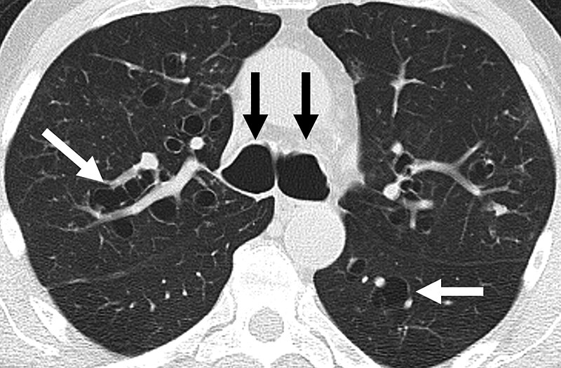
Image Source: Wikimedia Commons (2012). Bronquiectasia, Licensed under a creative commons licence. Available for free at: https://commons.wikimedia.org/wiki/File:Bronquiectasia.jpeg#file (Accessed: 19 January 2024).
Management of bronchiectasis
The mainstay of management is to treat any underlying cause and minimise the frequency and severity of exacerbations.
- Physiotherapy is key in minimising exacerbations and preventing progression of lung disease. Techniques such as postural drainage and active cycle breathing help to clear the airway
- Antibiotics are used for exacerbations and, in some cases, for long-term prophylaxis.
- Antibiotics can be nebulised, oral or intravenous
- Patients will normally need a higher dose and longer course (usually 2 weeks) of antibiotics
- Pseudomonas colonisation is a poor prognostic marker, associated with worse lung function, more frequent exacerbations and more severe disease
- Bronchodilators and inhaled steroids are used if there are symptoms or evidence of airflow obstruction
- Annual influenza and pneumococcal vaccinations
- Surgical input can be curative if the disease is localised
- Lung transplant can be considered in some specific, severe cases
Complications of bronchiectasis
Massive haemoptysis
Massive haemoptysis is defined as more than 250 mL/day. It is normally thought to occur from bronchial arteries around damaged areas of the lung.
Airway protection is important and patients may need intubation in extreme cases. If bleeding is not stopping, involvement of thoracic surgeons or interventional radiologists for further interventions such as bronchial artery embolisation, bronchoscopy-guided tamponade or surgical resection may be required.
Cor pulmonale
Untreated or progressive lung disease can cause high blood pressure in the pulmonary arteries (pulmonary hypertension) which causes right-sided heart failure.
Management of cor pulmonary involves optimising underlying lung disease but can also include supportive measures such as long-term oxygen therapy and diuretics. In extreme cases, a heart-lung transplant may be appropriate.
Primary ciliary dyskinesia (PCD)
This is a rare autosomal recessive condition characterised by abnormal movement of the cilia lining the respiratory tract (upper and lower), ear, fallopian tubes and sperm flagella. The abnormal ciliary movements reduce mucociliary clearance, increasing likelihood of infections.
Features include recurrent chest infection and bronchiectasis, sinusitis, hearing loss and subfertility or infertility.
There is no gold-standard diagnostic test for primary ciliary dyskinesia, however nasal nitric oxide testing, which is very low in people with PCD, is the most specific and sensitive screening test. If this is positive, a ciliary biopsy can also confirm the diagnosis by assessing the cilial beat pattern and frequency under a microscope. Genetic testing can also aid diagnosis.
Kartagener’s syndrome
Approximately 50% of people with PCD have associated dextrocardia or situs inversus, as cilia also helps to determine organ siting. This is known as Kartagener’s syndrome.
Yellow nail syndrome
Yellow nail syndrome is a rare cause of bronchiectasis and is a constellation of signs and symptoms that includes thickened yellow nails (slowed nail growth), lymphoedema, and respiratory signs such as pleural effusions, bronchiectasis and chronic sinusitis. The cause of this syndrome is unknown.
References
- Chapman S. Oxford handbook of respiratory medicine. Oxford; New York: Oxford University Press; 2005
- Lucas JS, Burgess A, Mitchison HM, Moya E, Williamson M, Hogg C. Diagnosis and management of primary ciliary dyskinesia. Archives of Disease in Childhood [Internet]. 2014 Apr 25 [cited 2022 Jan 31];99(9):850–6. Available from: https://www.ncbi.nlm.nih.gov/pmc/articles/PMC4145427/
- The National Institute for Health and Care Excellence. Bronchiectasis (non-cystic fibrosis), acute exacerbation: antimicrobial prescribing [Internet]. 2018 [cited 2022 Jan 31] Available from: https://www.nice.org.uk/guidance/ng117/resources/bronchiectasis-noncystic-fibrosis-acute-exacerbation-antimicrobial-prescribing-pdf-66141603457477
Complete Guide to Clinical Genetics ST3 Application and Recruitment (2027)
Written by Medibuddy. Last updated: October 2026. Clinical Genetics is

Complete Guide to Clinical Radiology ST3 Application and Recruitment (2027)
Recruitment paused: NHS England is not running Clinical Radiology ST3+ recru

Complete Guide to Paediatric Surgery ST3 Application and Recruitment in United Kingdom (2026)
Written by Medibuddy. Last updated: October 2026. If you’re planning


Take your subscriptions with you
Our mobile app allows you to access your interview and exam question banks wherever you are.

