


 Close
Close
 17th February 2025
Bronchiectasis
Overview of bronchiectasis
Bronchiectasis is the chronic and irreversible dilatation of the airways due to recurrent inflammation or infection. It is associated with chronic sputum production, airflow obstruction and recurrent chest infections.
Bronchiectasis can be idiopathic or secondary to one of many causes, including:
- Previous infection: bacterial pneumonia, TB, viral illness, measles
- Genetic causes: cystic fibrosis, Kartagener’s syndrome, primary ciliary dyskinesia
- Allergic bronchopulmonary aspergillosis
- Immune deficiency:
- Primary: hypogammaglobulinaemia, common variable immunodeficiency
- Secondary: HIV, CLL, nephrotic syndrome
- Yellow nail syndrome
- Chronic aspiration: GORD, alcohol excess
- Bronchial obstruction: lung cancer
- Systemic conditions: rheumatoid arthritis, systemic lupus erythematosus, inflammatory bowel disease
Presentation of bronchiectasis
Bronchiectasis often presents as a chronic productive cough with recurrent chest infections or “bronchitis” over a number of years. Patients typically have daily mucopurulent sputum with intermittent haemoptysis. They may also have chronic progressive breathlessness.
On examination, patients may have coarse inspiratory and expiratory crackles or wheeze and finger clubbing.
Investigations in bronchiectasis
Key investigations and findings in bronchiectasis include:
- Blood tests:
- White cell count and CRP to rule in/out infection
- Raised eosinophils may be suggestive of allergic bronchopulmonary aspergillosis
- To investigate an underlying cause, e.g. rheumatoid factor, immunoglobulins, HIV test
- Sputum culture: pathogenic organisms are often found in sputum and it is advisable to send a sputum culture and sensitivity, as well as a viral and atypical screen. Pseudomonas aeruginosa is often found and can correlate with more severe lung damage. It is more likely to be antibiotic resistant than other common respiratory pathogens.
- CXR: classically shows the tram-track sign (thick-walled, dilated bronchi).
- HRCT (gold standard for diagnosis): classically shows dilated airways, with or without a thickened bronchial wall (tram-track sign) and signet-ring sign (airway appearing larger than accompanying blood vessel) (Figure 2).
- Pulmonary function tests: obstructive picture and reduced FEV1.
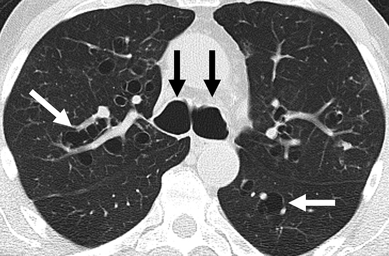
Image Source: Wikimedia Commons (2012). Bronquiectasia, Licensed under a creative commons licence. Available for free at: https://commons.wikimedia.org/wiki/File:Bronquiectasia.jpeg#file (Accessed: 19 January 2024).
Management of bronchiectasis
The mainstay of management is to treat any underlying cause and minimise the frequency and severity of exacerbations.
- Physiotherapy is key in minimising exacerbations and preventing progression of lung disease. Techniques such as postural drainage and active cycle breathing help to clear the airway
- Antibiotics are used for exacerbations and, in some cases, for long-term prophylaxis.
- Antibiotics can be nebulised, oral or intravenous
- Patients will normally need a higher dose and longer course (usually 2 weeks) of antibiotics
- Pseudomonas colonisation is a poor prognostic marker, associated with worse lung function, more frequent exacerbations and more severe disease
- Bronchodilators and inhaled steroids are used if there are symptoms or evidence of airflow obstruction
- Annual influenza and pneumococcal vaccinations
- Surgical input can be curative if the disease is localised
- Lung transplant can be considered in some specific, severe cases
Complications of bronchiectasis
Massive haemoptysis
Massive haemoptysis is defined as more than 250 mL/day. It is normally thought to occur from bronchial arteries around damaged areas of the lung.
Airway protection is important and patients may need intubation in extreme cases. If bleeding is not stopping, involvement of thoracic surgeons or interventional radiologists for further interventions such as bronchial artery embolisation, bronchoscopy-guided tamponade or surgical resection may be required.
Cor pulmonale
Untreated or progressive lung disease can cause high blood pressure in the pulmonary arteries (pulmonary hypertension) which causes right-sided heart failure.
Management of cor pulmonary involves optimising underlying lung disease but can also include supportive measures such as long-term oxygen therapy and diuretics. In extreme cases, a heart-lung transplant may be appropriate.
Primary ciliary dyskinesia (PCD)
This is a rare autosomal recessive condition characterised by abnormal movement of the cilia lining the respiratory tract (upper and lower), ear, fallopian tubes and sperm flagella. The abnormal ciliary movements reduce mucociliary clearance, increasing likelihood of infections.
Features include recurrent chest infection and bronchiectasis, sinusitis, hearing loss and subfertility or infertility.
There is no gold-standard diagnostic test for primary ciliary dyskinesia, however nasal nitric oxide testing, which is very low in people with PCD, is the most specific and sensitive screening test. If this is positive, a ciliary biopsy can also confirm the diagnosis by assessing the cilial beat pattern and frequency under a microscope. Genetic testing can also aid diagnosis.
Kartagener’s syndrome
Approximately 50% of people with PCD have associated dextrocardia or situs inversus, as cilia also helps to determine organ siting. This is known as Kartagener’s syndrome.
Yellow nail syndrome
Yellow nail syndrome is a rare cause of bronchiectasis and is a constellation of signs and symptoms that includes thickened yellow nails (slowed nail growth), lymphoedema, and respiratory signs such as pleural effusions, bronchiectasis and chronic sinusitis. The cause of this syndrome is unknown.
References
- Chapman S. Oxford handbook of respiratory medicine. Oxford; New York: Oxford University Press; 2005
- Lucas JS, Burgess A, Mitchison HM, Moya E, Williamson M, Hogg C. Diagnosis and management of primary ciliary dyskinesia. Archives of Disease in Childhood [Internet]. 2014 Apr 25 [cited 2022 Jan 31];99(9):850–6. Available from: https://www.ncbi.nlm.nih.gov/pmc/articles/PMC4145427/
- The National Institute for Health and Care Excellence. Bronchiectasis (non-cystic fibrosis), acute exacerbation: antimicrobial prescribing [Internet]. 2018 [cited 2022 Jan 31] Available from: https://www.nice.org.uk/guidance/ng117/resources/bronchiectasis-noncystic-fibrosis-acute-exacerbation-antimicrobial-prescribing-pdf-66141603457477
Complete Guide to Clinical Genetics ST3 Application and Recruitment (2027)
Written by Medibuddy. Last updated: October 2026. Clinical Genetics is

Complete Guide to Clinical Radiology ST3 Application and Recruitment (2027)
Recruitment paused: NHS England is not running Clinical Radiology ST3+ recru

Complete Guide to Paediatric Surgery ST3 Application and Recruitment in United Kingdom (2026)
Written by Medibuddy. Last updated: October 2026. If you’re planning


Take your subscriptions with you
Our mobile app allows you to access your interview and exam question banks wherever you are.

