


 Close
Close
 22nd July 2024
Malignant Haematology
Leukaemia
There are four major types of leukaemia:
| Cell type | Acute | Chronic |
|---|---|---|
| Lymphoid | Acute lymphoblastic leukaemia (ALL) | Chronic lymphocytic leukaemia (CLL) |
| Myeloid | Acute myeloid leukaemia (AML) | Chronic myeloid leukaemia (CML) |
There are also other types of leukaemia, some of which shall be discussed briefly.
Acute myeloid leukaemia
Background of AML
AML occurs due to clonal proliferation of haematopoietic precursor cells of myeloid lineage in the bone marrow.
In adults, it is the most common acute leukaemia and the second most common leukaemia. The median age of diagnosis is 65 years, with a male predominance.
The exact aetiology is unknown, however there are numerous associated risk factors:
- Environmental exposure to radiation and benzene
- Pre-existing haematological pathology such as myelodysplastic syndrome or myeloproliferative neoplasm
- Previous treatment with chemotherapy, particularly alkylating agents and topoisomerase II inhibitors
Presentation and diagnosis of AML
The proliferation of precursor myeloid cells (blast cells) in the BM leads to BM failure, causing:
- Anaemia: pallor, palpitations, dyspnoea, fatigue
- Thrombocytopenia: bruising, excess bleeding
- Neutropenia: infections
Gum hypertrophy can also be a presenting feature, seen most commonly in the monocytic subtypes of AML.
Diagnosis is multi-faceted:
- Blood tests: FBC (cytopenias); can include a metabolic panel to screen for tumour lysis syndrome (high potassium, low calcium, high phosphate, high uric acid)
- Blood film: immature myeloid blasts with pathognomonic Auer rods (Figure 1), ≥20% blasts
- BM biopsy: diagnosis if ≥20% blasts; carry out immunophenotyping to differentiate AML from ALL (see acute lymphocytic leukaemia); cytogenetic and molecular analysis for prognostication and classification
Tumour lysis syndrome occurs due to the rapid breakdown of a large number of cancer cells. This occurs most commonly a few weeks after treatment initiation but can also occur spontaneously. A high phosphate is not seen as commonly in spontaneous tumour lysis syndrome, compared to post-treatment.

Image Source: Wikimedia Commons (2010). Myeloblast with Auer rod smear 2010-01-27, Licensed under a creative commons licence. Available for free at: https://en.wikipedia.org/wiki/Auer_rod#/media/File:Myeloblast_with_Auer_rod_smear_2010-01-27.JPG (Accessed: 16 January 2024)
Management and prognosis of AML
Full treatment for AML in medically fit and willing patients consists of two stages:
- Remission induction: generally carried out with cytarabine and an anthracycline (e.g. daunorubicin)
- Consolidation therapy: often carried out with high dose cytarabine
Allogeneic haematopoietic stem cell transplantation (SCT) can be used in patients with an intermediate or poor prognosis in addition to consolidation therapy, especially in patients <60 years old.
Less intensive chemotherapy options are available for patients not likely to tolerate high intensity regimes. These are less likely to induce complete remission and allow long-term disease control.
Maintenance chemotherapy is generally not used.
Various parameters are used for prognosis:
| Favourable prognostic factors | Age <60 Cytogenetics, e.g. t(8;21)(q22;q22) or inv16(p13q22) |
| Poor prognostic factors | Age >60
Pre-morbid myelodysplastic syndrome or myeloproliferative neoplasm Cytogenetics, e.g. abnormal 3q, del(5q), or t(9;22)(q34;q11) >15% blast cells in BM after remission induction |
Acute promyelocytic leukaemia
Acute promyelocytic leukaemia (APL) is a subtype of AML. Peak incidence is in early adulthood. The vast majority of cases are caused by t(15;17) causing formation of the PML-RARα fusion gene.
APL is a medical emergency that warrants quick diagnosis and treatment as it can result in (and often presents with) disseminated intravascular coagulation or haemorrhage, which has high mortality.
Treatment is with all-trans-retinoic acid with an anthracycline for induction remission. Consolidation therapy uses the same combination of chemotherapy.
APL has a good prognosis with >80% cure rates.
Acute lymphoblastic leukaemia
Background of ALL
ALL occurs due to clonal proliferation of haematopoietic precursor cells of lymphoid lineage in the BM.
ALL is the most common malignancy in childhood. Peak incidence is between the ages of two and five, with further smaller peaks in the mid-30s and mid-80s. Incidence is higher in males than females.
Approximately 85% of ALL is of B-cell lineage and 15% is of T-cell lineage.
The exact cause is not known, but there are known associated contributing factors. These include genetic factors (higher likelihood of development between monozygotic twins than singleton siblings) and pre-existing genetic syndromes (e.g. Down syndrome, Klinefelter’s syndrome, ataxia-telangiectasia). Contributing environmental factors include radiation and smoking exposure.
Presentation and diagnosis of ALL
The presenting signs and symptoms are from BM failure (see acute myeloid leukaemia).
In addition, children can often present with hepatomegaly and/or splenomegaly and lymphadenopathy.
As with AML, diagnosis requires blood tests, blood film and BM biopsy (for immunophenotyping, cytogenetics and molecular analysis).

Image Source: Wikimedia Commons (2007). Acute leukemia-ALL, Licensed under a creative commons licence. Available for free at: https://en.wikipedia.org/wiki/Acute_lymphoblastic_leukemia#/media/File:Acute_leukemia-ALL.jpg (Accessed: 16 January 2024).
Differentiating between AML and ALL
| AML | ALL | |
|---|---|---|
| Epidemiology | Older patients (generally >60) | Children (<10) |
| Presentation | Hepatosplenomegaly and lymphadenopathy not a prominent feature | Generally present with hepatosplenomegaly or lymphadenopathy |
| Blood film morphology | Large cells
Variable cytoplasm (often larger amounts) Large, regular nuclei (often round) Auer rods (pathognomonic) |
Large cells
Scant to moderate cytoplasm Irregular/convoluted nuclei No Auer rods |
| Immunophenotyping | MPO, CD33, CD34 | B-cell lineage: CD19, HLA-DR
T-cell lineage: CD3, CD7, CD4 Both lineages: TdT |
Management and prognosis of ALL
Treatment of ALL consists of four main stages:
- Remission induction: generally a four drug regimen.
- Consolidation therapy: several combinations, aiming to use drugs that differ in mechanism of action to those used for induction.
- CNS prophylaxis (or treatment if there is CNS involvement): intrathecal CNS prophylaxis is given alongside all stages of treatment to reduce risk of relapse with leukaemic meningitis.
- Stem cell transplant/maintenance therapy: SCT is preferred in patients after consolidation therapy who are at high risk of relapse. Maintenance therapy is recommended in patients who are high risk, but ineligible for SCT, or who are at standard risk for relapse.
Prognosis is significantly worse in adults than in children. Cure rates in children are high at around 80%, whereas 35-40% of adults treated with intensive chemotherapy survive two years. Prognostic factors include:
| Favourable prognostic factors | Age 1-9 years
Cytogenetics, e.g. t(12;21) High hyperdiploidy (>50 chromosomes) |
| Poor prognostic factors | In children: age <1 or >10 years; in adults: age >35 years
WCC >30 x109/L (for B-cell ALL), >100 x109/L (for T-cell ALL) WCC >50 x109/L for children Pre-morbid myelodysplastic syndrome or myeloproliferative neoplasm Cytogenetics, e.g. t(9;22), MLL translocations or abnormal 17p |
Chronic myeloid leukaemia
Background of CML
CML is a myeloproliferative disorder caused by clonal proliferation of mature and maturing granulocytes, predominantly neutrophils. Cell differentiation is relatively preserved, in contrast to AML. CML accounts for 15-20% of adult leukaemias. Median age of presentation is 50 years and it has a slight male predominance.
Ionising radiation is the only known risk factor.
Translocation between chromosomes 9 and 22, t(9;22)(q34;q11), causes formation of an abnormal chromosome 22, called the Philadelphia chromosome. This is pathognomonic for CML. The translocation results in formation of the BCR-ABL1 fusion gene, an oncogene that produces the BCR-ABL1 protein, a dysregulated and constitutively active tyrosine kinase.
Presentation and diagnosis of CML
CML is a triphasic or biphasic disease:
- Chronic phase: <10% blasts in BM
- Accelerated phase: 10-19% blasts in BM (some patients go straight from the chronic phase to a blast crisis)
- Blast crisis: ≥20% blasts in BM, resembles an acute leukaemia
Signs and symptoms depend on which phase patients present in. Around 85% of patients present in the chronic phase. As a result, up to half of patients are asymptomatic on presentation, being picked up incidentally on an FBC.
Splenomegaly, present in ~75% of patients, can cause abdominal pain or early satiety. Patients can also present with constitutional symptoms of fatigue, weight loss and sweats. Patients in a blast crisis can present with BM failure.
Diagnostic workup includes:
- Blood tests: FBC (raised WCC)
- Blood film: mature/maturing myeloid cells
- BM biopsy: to confirm disease phase
Definitive diagnosis is made by cytogenetic confirmation of the presence of the Philadelphia chromosome, and hence the BCR-ABL1 fusion gene, by fluorescent in situ hybridisation or quantitative reverse transcription polymerase chain reaction.

Image Source: Wikimedia Commons (2009). Chronic Myeloid Leukemia smear 2009-04-09, Licensed under a creative commons licence. Available for free at: https://en.wikipedia.org/wiki/Chronic_myelogenous_leukemia#/media/File:Chronic_Myeloid_Leukemia_smear_2009-04-09.JPG (Accessed: 16 January 2024).
Management and prognosis of CML
Management options for CML include:
- Tyrosine kinase inhibitors (TKI): first line TKIs include the first-generation imatinib and second-generation medications dasatinib and nilotinib. TKIs are generally continued indefinitely as long as they are working, and they provide good disease control long-term.
- Allogeneic SCT: can be considered in any patient not responding to initial therapy or with relapse.
- Hydroxycarbamide: can be used for symptom control while awaiting a diagnosis for patients with significant leucocytosis, systemic symptoms or symptomatic splenomegaly. It has no bearing on survival.
Patients in the chronic phase are treated with a TKI with a generally very good response.
Patients in the accelerated phase or blast crisis are treated with the same TKIs but have a lower likelihood of response and higher risk of relapse. As such, allogeneic SCT is more likely to be required and considered earlier.
TKIs allow patients with CML to have a near normal life expectancy. Stage of disease at presentation has the strongest impact on outcome.
Chronic lymphocytic leukaemia
Background of CLL
CLL (known as small lymphocytic lymphoma, SLL, if the disease is predominantly in the lymph nodes) is an indolent lymphoproliferative disorder of mature monoclonal B-cells. CLL is the most common leukaemia in Western adults. Median age of diagnosis is 70 years with a male predominance.
There appears to be a genetic predisposition to CLL. No environmental risk factors have been identified.
Presentation and diagnosis of CLL
The majority of patients are asymptomatic at diagnosis, usually identified on a routine FBC.
If symptomatic, patients may present with lymphadenopathy, splenomegaly, hepatomegaly, signs of BM failure (as discussed in the section on AML) or with ‘B’ symptoms of lymphoma (fever, fatigue, weight loss, night sweats).
Cytopenias can also occur as a result of autoimmune haemolytic anaemia (AIHA; in ~10% of patients) and immune thrombocytopenia (ITP; in ~1-2%).
Diagnosis usually involves:
- Blood tests: FBC with absolute lymphocytosis (>5 x10⁹/L); there may be anaemia or thrombocytopenia
- Blood film: lymphocytosis with ‘smudge’ or ‘smear’ cells
- Immunophenotyping with flow cytometry: positive for CD5, CD19, CD20, CD23, kappa or lambda immunoglobulin light chain
BM biopsy is usually not required.

Image Source: Wikimedia Commons (2014). Smear cells, Licensed under a creative commons licence. Available for free at: https://en.wikipedia.org/wiki/Chronic_lymphocytic_leukemia#/media/File:Smear_cells.jpg (Accessed: 16 January 2024).

Management and prognosis of CLL
CLL is staged using cell counts, lymphadenopathy and hepatosplenomegaly.
Early stage asymptomatic disease is managed with observation. Indications to initiate treatment include:
- Progressive bone marrow failure
- AIHA/ITP unresponsive to steroids
- Massive (≥6 cm below costal margin) or progressive splenomegaly
- Massive (≥10 cm) or progressive lymphadenopathy
- Progressive lymphocytosis with >50% increase in 2 months or doubling time <6 months
- Significant constitutional symptoms (weight loss ≥10% in 6 months, fevers >2 weeks without infection, night sweats without infection, significant fatigue)
If the decision is made to begin chemotherapy, factors such as age, performance status, comorbidities and severity of symptoms can be used to decide between a palliative approach, minimising toxicity, and an approach aiming for complete remission. Chemotherapy choice is guided by cytogenetic analysis.
If treatment is required, first-line chemotherapy can achieve 25-50% remission rates. However, there is a high risk of relapse, with most patients requiring further treatment after 2-3 years.
Hairy cell leukaemia
Hairy cell leukaemia (HCL) is an uncommon B-cell lymphoproliferative malignancy, which is distinct from CLL.
Median age of presentation of 50-55 years and there is a strong male predominance.
Patients generally present with symptoms related to BM failure or splenomegaly.
Diagnosis usually requires blood tests, blood film and immunophenotyping (CD19, CD20 and CD22 positive). BM biopsy is usually not possible due to increased BM fibrosis from HCL invasion.

Image Source: Wikimedia Commons (2015). Hairy Cell Leukaemia, Licensed under a creative commons licence. Available for free at: https://commons.wikimedia.org/wiki/File:Hairy_Cell_Leukaemia.jpg (Accessed: 16 January 2024).
As with CLL, asymptomatic patients are managed with observation and can be observed for months or years. Symptomatic patients (significant cytopenia, symptomatic splenomegaly or lymphadenopathy or significant constitutional symptoms) are treated with purine analogues first line. These have a very high response rate and most patients with HCL survive >10 years after diagnosis.
References
- Kolitz JE. Overview of acute myeloid leukaemia in adults [Internet]. Larson RA, Rosmarin AG, editors. UpToDate. 2020 [cited 2021 Dec 19]. Available from: https://www.uptodate.com/contents/overview-of-acute-myeloid-leukemia-in-adults
- Raj K, Mehta P. Acute myelogenous leukaemia [Internet]. BMJ Best Practice. 2017 [cited 2021 Dec 19]. Available from: https://bestpractice.bmj.com/topics/en-gb/274
- Provan D, Baglin T, Dokal I, de Vos J. Oxford Handbook of Clinical Haematology. 4th ed. Oxford: Oxford University Press; 2015
- Milligan DW, Grimwade D, Cullis JO, Bond L, Swirsky D, Craddock C, et al. Guidelines on the management of acute myeloid leukaemia in adults. British Journal of Haematology. 2006;135(4);450-474. Available from: doi: 10.1111/j.1365-2141.2006.06314.x
- Horton TM, Steuber CP, Aster JC. Overview of the clinical presentation and diagnosis of acute lymphoblastic leukemia/lymphoma in children [Internet]. Park JR, Rosmarin AG, editors. UpToDate. 2021 [cited 2021 Dec 20]. Available from: https://www.uptodate.com/contents/overview-of-the-clinical-presentation-and-diagnosis-of-acute-lymphoblastic-leukemia-lymphoma-in-children
- Ooi M, Poon M, Chan E, Ng CH. Acute lymphocytic leukaemia [Internet]. BMJ Best Practice. 2021 [cited 2021 Dec 20]. Available from: https://bestpractice.bmj.com/topics/en-gb/273
- Clarke RT, van den Bruel A, Bankhead C, Mitchell CD, Phillips B, Thompson MJ. Clinical presentation of childhood leukaemia: a systematic review and meta-analysis. Archives of Disease in Childhood. 2016;101(10);894-901. Available from: doi: 10.1136/archdischild-2016-311251
- Schiffer CA, Gurbuxani S. Clinical manifestations, pathologic features, and diagnosis of acute myeloid leukemia [Internet]. Larson RA, Rosmarin AG, editors. UpToDate. 2021 [cited 2021 Dec 20]. Available from: https://www.uptodate.com/contents/clinical-manifestations-pathologic-features-and-diagnosis-of-acute-myeloid-leukemia
- Moorman AV, Ensor HM, Richards SM, Chilton L, Schwab C, Kinsey SE et al. Prognostic effect of chromosomal abnormalities in childhood B-cell precursor acute lymphoblastic leukaemia: results from the UK Medical Research Council ALL97/99 randomised trial. The Lancet Oncology. 2010;11(5):429-438. Available from: doi: 10.1016/S1470-2045(10)70066-8
- van Etten RA. Clinical manifestations and diagnosis of chronic myeloid leukamia [Internet]. Larson RA, Rosmarin AG, editors. UpToDate. 2020 [cited 2021 Dec 20]. Available from: https://www.uptodate.com/contents/clinical-manifestations-and-diagnosis-of-chronic-myeloid-leukemia
- Somervaille T. Chronic myelogenous leukaemia [Internet]. BMJ Best Practice. 2018 [cited 2021 Dec 20]. Available from: https://bestpractice.bmj.com/topics/en-gb/276
- Schiffer CA, Atallah E. Overview of the treatment of chronic myeloid leukamia [Internet]. Larson RA, Rosmarin AG, editors. UpToDate. 2020 [cited 2021 Dec 20]. Available from: https://www.uptodate.com/contents/overview-of-the-treatment-of-chronic-myeloid-leukemia
- Rai KR, Stilgenbauer S, Aster JC. Clinical features and diagnosis of chronic lymphocytic leukemia/small lymphocytic lymphoma [Internet]. Larson RA, Connor RF, editors. UpToDate. 2021 [cited 2021 Dec 20]. Available from: https://www.uptodate.com/contents/clinical-features-and-diagnosis-of-chronic-lymphocytic-leukemia-small-lymphocytic-lymphoma
- Murthy V, Paneesha S. Chronic lymphocytic leukaemia [Internet]. BMJ Best Practice. 2021 [cited 2021 Dec 20]. Available from: https://bestpractice.bmj.com/topics/en-gb/275
- Rai KR, Stilgenbauer S. Overview of the treatment of chronic lymphocytic leukaemia [Internet]. Larson RA, Connor RF, editors. UpToDate. 2021 [cited 2021 Dec 20]. Available from: https://www.uptodate.com/contents/overview-of-the-treatment-of-chronic-lymphocytic-leukemia
- Tallman MS, Aster JC. Clinical features and diagnosis of hairy cell leukemia [Internet]. Larson RA, Connor RF, editors. UpToDate. 2021 [cited 2021 Dec 20]. Available from: https://www.uptodate.com/contents/clinical-features-and-diagnosis-of-hairy-cell-leukemia
- Tallman MS. Treatment of hairy cell leukemia [Internet]. Larson RA, Connor RF, editors. UpToDate. 2021 [cited 2021 Dec 20]. Available from: https://www.uptodate.com/contents/treatment-of-hairy-cell-leukemia
- Kumar A, Kharfan-Dabaja MA. Hairy cell leukaemia [Internet]. BMJ Best Practice. 2018 [cited 2021 Dec 20]. Available from: https://bestpractice.bmj.com/topics/en-gb/890
Lymphoma
Overview of lymphoma
Lymphomas are a heterogenous group of malignancies resulting from clonal proliferation of lymphocytes in lymphoid tissue. They are the most common haematological malignancy.
Lymphomas are subdivided into two broad groups:
- Hodgkin lymphoma (HL): around 10% of lymphomas are characterised by the presence of Reed-Sternberg cells on blood film. This is a B-cell lymphoma.
- Non-Hodgkin lymphoma (NHL): around 90% of lymphomas, no Reed-Sternberg cells on blood film. Around 85% originate from B-cells and 15% from T-cells/NK cells.
Diagnosis and staging of lymphoma
There are three main aspects to definitive diagnosis of HL and NHL:
- Tissue biopsy: excisional biopsy of a peripheral lymph node (LN). Fine needle aspiration does not provide sufficient material for a histological diagnosis and classification.
- Imaging: chest X-ray, CT of neck, chest, abdomen and pelvis with contrast and FDG-PET scan. Used to stage disease and guide tissue biopsy.
- Immunohistochemistry
Staging of HL and NHL is via the Lugano classification, a modification of the classic Ann Arbor staging system. This takes into account the number and site of LNs and/or extra-lymphatic organs affected, as well as the presence or absence of ‘B’ symptoms, defined as:
- Unexplained fever >38°C in the last month
- Recurrent drenching night sweats in the last month
- Weight loss >10% within six months of diagnosis
A sub-classification of ‘A’ denotes the absence of ‘B’ symptoms, whereas ‘B’ denotes their presence, e.g. IIA or IIIB.
| Stage | Description |
|---|---|
| I (early) | Involvement of a single LN region/structure or of a single extra-lymphatic organ/site |
| II (early) | Involvement of 2+ LN regions/structures on the same side of the diaphragm or with localised involvement of an extra-lymphatic organ/site |
| III (advanced) | Involvement of LNs or lymphoid organs on both sides of the diaphragm |
| IV (advanced) | Diffuse involvement of one or more extra-lymphatic organs, with or without LN involvement |
Hodgkin lymphoma
Categories of HL
HL can be further sub-categorised:
- Classical HL (~90% of HL)
- Nodular sclerosis classical HL (NSCHL)
- Mixed cellularity classical HL (MCCHL)
- Lymphocyte rich classical HL (LRCHL)
- Lymphocyte depleted classical HL (LDCHL)
- Nodular lymphocyte predominant HL (NLPHL) (~10% of HL)
Hodgkin lymphoma is characterised by the presence of Reed-Sternberg cells. They often have bilobed nuclei with prominent nucleoli (described as having an ‘owl’s eyes’ appearance) but can also be multinucleated.
Lymphocytic and histiocytic (‘popcorn’) cells are a variant of Reed-Sternberg cells characterised by a single multilobulated nucleus and are required to make a diagnosis of NLPHL.

Image Source: Ncbi (2022). Reed Sternberg Cells, Licensed under a creative commons licence. Available for free at: https://www.ncbi.nlm.nih.gov/books/NBK542333/ (Accessed: 16 January 2024)
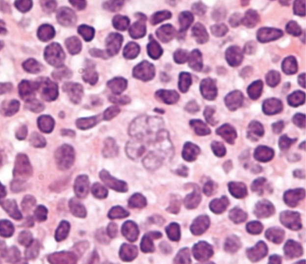
Image Source: Wikimedia Commons (2010). Popcorn cell in nodular lymphocyte predominant Hodgkin lymphoma – very high mag cropped, Licensed under a creative commons licence. Available for free at: https://en.wikipedia.org/wiki/Nodular_lymphocyte_predominant_Hodgkin_lymphoma#/media/File:Popcorn_cell_in_nodular_lymphocyte_predominant_Hodgkin_lymphoma_-_very_high_mag_cropped.jpg (Accessed: 16 January 2024)
Background of HL
HL incidence demonstrates a bimodal age distribution, with the first peak between 15 and 34 years and the second peak over 60 years. There is a higher incidence in men.
The precise aetiology of HL remains unclear, but there are numerous associated risk factors:
- Epstein-Barr virus (EBV): this is detected in Reed-Sternberg cells in many cases of HL. It has been linked to the pathogenesis of HL, but the absolute risk of developing HL after EBV infection remains very low. Its presence varies by subtype, location and immune status:
- EBV is hardly found in NLPHL but nearly always found in LDCHL
- EBV is found in 20-50% of classical HL in developed countries but found in nearly all cases in developing countries
- Almost all cases of HL in HIV positive patients are EBV positive
- Immunodeficiency (congenital and acquired)
- Autoimmune conditions
- Familial risk
- Environmental risk (obesity and smoking)
Presentation and diagnosis of HL
Common presenting symptoms include:
- Asymptomatic lymphadenopathy: most common presentation; painless, commonly cervical or supraclavicular
- Mediastinal mass on chest X-ray: may be incidental or done for symptoms such as cough, dyspnoea, chest pain or superior vena cava obstruction (classic signs of this are facial swelling that improves through the day and enlarged veins in the neck, upper chest and arms)
- Constitutional symptoms: ‘B’ symptoms of fever, night sweats, weight loss
- Fatigue
- Pruritus
- Pain on alcohol ingestion: at affected LNs or sites of bony involvement
Diagnosis relies on tissue biopsy, imaging and immunohistochemistry (see diagnosis and staging of lymphoma). Classical HL cells are always CD30 positive, mostly CD15 positive, occasionally CD20 positive and CD45 negative. NLPHL cells are CD30 and CD15 negative and CD20 and CD45 positive.
Management and prognosis of HL
Management of HL is complicated. It varies between paediatric and adult populations, disease stage and HL subtype. A summary of the treatment of adults is as follows:
| Early-stage classical HL | Combination chemotherapy¹ then involved-field radiotherapy (IFRT) |
| Late-stage classical HL | Combination chemotherapy¹ |
| Early-stage NLPHL | IFRT (or active surveillance) |
| Late-stage NLPHL | Chemoimmunotherapy² |
¹ A regime used commonly for classical HL in adults is ABVD: doxorubicin, bleomycin, vinblastine, dacarbazine
² Most often R-CHOP: rituximab, cyclophosphamide, doxorubicin, vincristine, prednisolone
Prognosis is generally very good. Patients treated in the early stage have an 85-90% chance of long-term complete remission. In children, even advanced HL has cure rates >85%.
Prognosis in advanced HL in adults is determined by the International Prognostic Score, which consists of seven adverse variables with independent prognostic value. It predicts freedom from disease progression at five years.
Due to the success of treatment, patients can develop complications of treatment many years after completing it. For example, radiation-induced hypothyroidism, chemotherapy side effects such as cardiomyopathy (doxorubicin) or pulmonary fibrosis (bleomycin), and secondary malignancies (e.g. AML).
Non-Hodgkin lymphoma
Background of NHL
NHL refers to any lymphoma that is not HL. As such, it encompasses a large and heterogenous group of conditions, only a few of which will be discussed here. NHL can be split into indolent and aggressive lymphomas.
NHL incidence increases with age and there is a male predominance.
There are numerous risk factors including congenital and acquired immunodeficiencies, autoimmune conditions, familial risk and environmental factors (e.g. pesticide exposure). There are also subtype-specific risk factors.
Prognosis is very dependent on subtype. In general, markers of poor prognosis include age >60, raised serum LDH, disease stage III or IV, ≤2 extranodal disease sites and poor performance status.
Presentation and diagnosis of NHL
Indolent (low-grade) lymphomas present insidiously, sometimes with symptoms over months to years. They are often disseminated at presentation and less likely to be curable. Aggressive (high-grade) lymphomas present sub-acutely or acutely with localised rapidly growing lymphadenopathy with or without ‘B’ symptoms but are more likely to be curable.
Painless lymphadenopathy is present in >2/3 of patients at presentation. ‘B’ symptoms are more likely to present in aggressive lymphomas. Hepatosplenomegaly is more common in indolent lymphomas.
As indolent lymphomas may be disseminated at presentation, they may present with extranodal symptoms. Gastrointestinal involvement may cause anorexia, early satiety and vomiting. Central nervous system involvement may cause headaches, focal neurological deficits or seizures.
Definitive diagnosis relies on tissue biopsy, imaging and immunohistochemistry (see above).
Follicular lymphoma
This is an indolent B-cell lymphoma. It is the most common indolent NHL and the second most common NHL. Median age at diagnosis is 65 years. The majority exhibit t(14;18), causing overexpression of the BCL2 oncogene. Lymphadenopathy is the most common presenting complaint; around 70% present with stage IV disease with BM involvement. Early-stage disease may be treated with IFRT. Advanced-stage disease may be treated with active surveillance if asymptomatic or if symptomatic, chemoimmunotherapy (e.g. R-CHOP). Median survival is 10 years.
Extranodal marginal zone lymphoma of mucosa-associated lymphoid tissue (MALT)
This is more commonly known as a MALT lymphoma. It is an indolent B-cell lymphoma. Median age of diagnosis is 66 years. MALT lymphomas can occur in any tissue with MALT e.g. stomach, small bowel, lung. They often arise on a background of chronic immune stimulation.
Gastric MALT lymphomas frequently develop following chronic gastritis caused by Helicobacter pylori. Over 90% of patients are H. pylori positive. They can present with dyspepsia, epigastric pain or gastrointestinal bleeding. Diagnosis is made with endoscopy and biopsy. Most patients present with localised disease. First line treatment for local disease in H. pylori positive patients is eradication therapy. In H. pylori negative patients or in H. pylori positive patients not responding to first line therapy, IFRT is used. 5-year survival is >80%.
H. pylori eradication therapy consists of a proton pump inhibitor (PPI) and antibiotics (first line: amoxicillin and clarithromycin/metronidazole; second line: amoxicillin and a tetracycline/quinolone, e.g. levofloxacin).
Diffuse large B-cell lymphoma (DLBCL)
This is an aggressive lymphoma and the most common NHL. Median age of diagnosis is 64 with a slight male predominance. DLBCL can occur de novo or from transformation of a low-grade lymphoma or of CLL (Richter’s transformation). It presents as rapidly progressing stage I or II disease in 50% of cases; extranodal disease is common. Early-stage disease is treated with R-CHOP chemoimmunotherapy +/- IFRT. Advanced-stage disease is treated with R-CHOP.
Mantle cell lymphoma
This is an uncommon, aggressive B-cell lymphoma, although there is a subset of patients who experience a more indolent disease course. Median age of diagnosis is 63 years, and it has a strong male predominance. It is highly associated with t(11;14). Around 70% of patients present with stage IV disease. There is no standardised first-line therapy; conventional chemoimmunotherapy with R-CHOP can be used. Median survival is 3-4 years.
Burkitt lymphoma
This is a highly aggressive B-cell lymphoma. There are three forms of the disease: endemic, sporadic and immunodeficiency associated. All three are clinically similar and dependent on dysregulation of the MYC gene on chromosome 8 through translocations.
- Endemic disease is seen primarily in Africa, presenting with extranodal disease of the jaw/facial bones (most common) or abdominal organs. Peak incidence is 4-7 years; 90% are associated with EBV infection.
- Sporadic disease is seen more in developed nations, usually presenting with abdominal symptoms, but a smaller percentage can present with jaw/facial bone disease. Median age of presentation is 31 years; 30% are associated with EBV infection.
- Immunodeficiency-associated disease is primarily seen in patients with HIV infection but also in other causes of acquired or congenital immunodeficiency.
Adult T-cell leukaemia-lymphoma (ATLL)
This is an aggressive lymphoma caused by chronic infection with human T-lymphotropic virus type 1 (HTLV-1). Incidence is highest where HTLV-1 infection is endemic: islands in southwest Japan, the Caribbean, parts of West Africa and Central and South America. There is often a long latency period of several decades between infection and development of ATLL; median age of diagnosis is 58 years. Median survival is very poor, ranging from months to a year, depending on subtype. Treatment is with combination chemotherapy.
Enteropathy-associated T-cell lymphoma (EATL)
EATL is an aggressive lymphoma highly associated with untreated coeliac disease. Management of coeliac disease with a gluten-free diet prevents development of EATL. Patients present with abdominal pain, often associated with gastrointestinal bleeding, bowel obstruction or perforation. Treatment is with combination chemotherapy with or without autologous SCT. Overall 5-year survival is 10-20%.
| Subtype | Cell type | Aggressiveness | Key features |
|---|---|---|---|
| Follicular | B-cell | Indolent | ~85% exhibit t(14;18)
Lymphadenopathy is the most common presenting complaint |
| MALT | B-cell | Indolent | >90% with gastric MALT are H. pylori positive |
| DLBCL | B-cell | Aggressive | Can occur de novo or from transformation of a low-grade lymphoma or CLL (Richter’s transformation) |
| Mantle cell | B-cell | Aggressive | Highly associated with t(11;14) |
| Burkitt | B-cell | Aggressive | Endemic: primarily in Africa, jaw/facial bone disease, 90% associated with EBV; sporadic: more in developed nations, primarily abdominal symptoms; immunodeficiency associated: mainly seen in HIV |
| ATLL | T-cell | Aggressive | Caused by chronic infection with HTLV-1 |
| EATL | T-cell | Aggressive | Highly associated with untreated coeliac disease |
References
- Provan D, Baglin T, Dokal I, de Vos J. Oxford Handbook of Clinical Haematology. 4th ed. Oxford: Oxford University Press; 2015.
- LaCasce AS, Ng AK. Hodgkin lymphoma: Epidemiology and risk factors [Internet]. Freedman AS, Rosmarin AG, editors. UpToDate. 2020 [cited 2021 Dec 20]. Available from: https://www.uptodate.com/contents/hodgkin-lymphoma-epidemiology-and-risk-factors
- LaCasce AS, Ng AK. Pretreatment evaluation, staging, and treatment stratification of classic Hodgkin lymphoma [Internet]. Freedman AS, Rosmarin AG, editors. UpToDate. 2020 [cited 2021 Dec 20]. Available from: https://www.uptodate.com/contents/pretreatment-evaluation-staging-and-treatment-stratification-of-classic-hodgkin-lymphoma
- Freedman AS, Friedberg JW. Pretreatment evaluation, staging, and response assessment of non-Hodgkin lymphoma [Internet]. Lister A, Rosmarin AG, editors. UpToDate. 2020 [cited 2021 Dec 20]. Available from: https://www.uptodate.com/contents/pretreatment-evaluation-staging-and-response-assessment-of-non-hodgkin-lymphoma
- Aggarwal P, Limaiem F. Reed Sternberg Cells [Internet]. NCBI. 2021 [cited 2021 Dec 20]. Available from: https://www.ncbi.nlm.nih.gov/books/NBK542333/
- Moskowitz A. Hodgkin’s lymphoma [Internet]. BMJ Best Practice. 2019 [cited 2021 Dec 21]. Available from: https://bestpractice.bmj.com/topics/en-gb/311
- McClain KL, Kamdar K. Overview of Hodgkin lymphoma in children and adolescents [Internet]. Park JR, Rosmarin AG, editors. UpToDate. 2021 [cited 2021 Dec 21]. Available from: https://www.uptodate.com/contents/overview-of-hodgkin-lymphoma-in-children-and-adolescents
- LaCasce AS, Ng AK, Aster JC. Clinical presentation and diagnosis of classic Hodgkin lymphoma in adults [Internet]. Freedman AS, Rosmarin AG, editors. UpToDate. 2020 [cited 2021 Dec 21]. Available from: https://www.uptodate.com/contents/clinical-presentation-and-diagnosis-of-classic-hodgkin-lymphoma-in-adults
- Canellos GP, Ng AK. Overview of the treatment of classic Hodgkin lymphoma in adults [Internet]. Freedman AS, Rosmarin AG, editors. UpToDate. 2020 [cited 2021 Dec 21]. Available from: https://www.uptodate.com/contents/overview-of-the-treatment-of-classic-hodgkin-lymphoma-in-adults
- LaCasce AS, Ng AK. Treatment of nodular lymphocyte-predominant Hodgkin lymphoma [Internet]. Freedman AS, Rosmarin AG, editors. UpToDate. 2021 [cited 2021 Dec 21]. Available from: https://www.uptodate.com/contents/treatment-of-nodular-lymphocyte-predominant-hodgkin-lymphoma
- Chan E, Ng CH, Ooi M, Poon M. Non-Hodgkin’s lymphoma [Internet]. BMJ Best Practice. 2021 [cited 2021 Dec 21]. Available from: https://bestpractice.bmj.com/topics/en-gb/312
- Freedman AS, Friedberg JW, Aster JC. Clinical presentation and initial evaluation of non-Hodgkin lymphoma [Internet]. Lister A, Rosmarin AG, editors. UpToDate. 2020 [cited 2021 Dec 21]. Available from: https://www.uptodate.com/contents/clinical-presentation-and-initial-evaluation-of-non-hodgkin-lymphoma
- Freedman AS, Aster JC. Clinical manifestations, pathologic features, diagnosis, and prognosis of follicular lymphoma [Internet]. Lister A, Connor RF, editors. UpToDate. 2020 [cited 2021 Dec 21]. Available from: https://www.uptodate.com/contents/clinical-manifestations-pathologic-features-diagnosis-and-prognosis-of-follicular-lymphoma
- Freedman AS, Aster JC. Clinical manifestations, pathologic features, and diagnosis of extranodal marginal zone lymphoma of mucosa associated lymphoid tissue (MALT) [Internet]. Lister A, Connor RF, editors. UpToDate. 2021 [cited 2021 Dec 21]. Available from: https://www.uptodate.com/contents/clinical-manifestations-pathologic-features-and-diagnosis-of-extranodal-marginal-zone-lymphoma-of-mucosa-associated-lymphoid-tissue-malt
- Freedman AS, Friedberg JW, Ng AK. Treatment of extranodal marginal zone lymphoma of mucosa associated lymphoid tissue (MALT lymphoma) [Internet]. Lister A, Connor RF, editors. UpToDate. 2021 [cited 2021 Dec 21]. Available from: https://www.uptodate.com/contents/treatment-of-extranodal-marginal-zone-lymphoma-of-mucosa-associated-lymphoid-tissue-malt-lymphoma
- Freedman AS, Aster JC. Epidemiology, clinical manifestations, pathologic features, and diagnosis of diffuse large B cell lymphoma [Internet]. Lister A, Rosmarin AG, editors. UpToDate. 2021 [cited 2021 Dec 21]. Available from: https://www.uptodate.com/contents/epidemiology-clinical-manifestations-pathologic-features-and-diagnosis-of-diffuse-large-b-cell-lymphoma
- Freedman AS, Aster JC. Clinical manifestations, pathologic features, and diagnosis of mantle cell lymphoma [Internet]. Lister A, Rosmarin AG, editors. UpToDate. 2021 [cited 2021 Dec 21]. Available from: https://www.uptodate.com/contents/clinical-manifestations-pathologic-features-and-diagnosis-of-mantle-cell-lymphoma
- Freedman AS, Aster JC. Epidemiology, clinical manifestations, pathologic features, and diagnosis of Burkitt lymphoma [Internet]. Lister A, Rosmarin AG, editors. UpToDate. 2021 [cited 2021 Dec 21]. Available from: https://www.uptodate.com/contents/epidemiology-clinical-manifestations-pathologic-features-and-diagnosis-of-burkitt-lymphoma
- Freedman AS, Aster JC, Suzuki R. Clinical manifestations, pathologic features, and diagnosis of adult T cell leukemia-lymphoma [Internet]. Lister A, Rosmarin AG, editors. UpToDate. 2020 [cited 2021 Dec 22]. Available from: https://www.uptodate.com/contents/clinical-manifestations-pathologic-features-and-diagnosis-of-adult-t-cell-leukemia-lymphoma
- Matsuoka M, Suzuki R. Treatment and prognosis of adult T cell leukemia-lymphoma [Internet]. Freedman AS, Rosmarin AG, editors. UpToDate. 2020 [cited 2021 Dec 22]. Available from: https://www.uptodate.com/contents/treatment-and-prognosis-of-adult-t-cell-leukemia-lymphoma
- Freedman AS, Aster JC. Clinical manifestations, pathologic features, and diagnosis of enteropathy-associated T cell lymphoma [Internet]. Lister A, Rosmarin AG, editors. UpToDate. 2021 [cited 2021 Dec 22]. Available from: https://www.uptodate.com/contents/clinical-manifestations-pathologic-features-and-diagnosis-of-enteropathy-associated-t-cell-lymphoma
Paraproteinaemias
A heterogenous group of conditions caused by clonal proliferation of plasma cells or B-cells associated with production of a monoclonal immunoglobulin (also called a paraprotein or M-protein), found in serum or urine.
Multiple myeloma (MM)
Background of MM
Multiple myeloma is the second most common haematological malignancy and accounts for 1-2% of all diagnosed malignancies. Diagnosis is most frequent between the ages of 65 and 74. It has a slight male predominance. MM is more common in Afro-Caribbeans and less common in Asians.
No clear aetiological factors have been identified. There may be a genetic component.
Common chromosomal aberrations are seen across MM. Hyperdiploidy or translocations involving the immunoglobulin heavy chain (IgH) region at 14q32 are seen in the majority of cases.
Myeloma is classified according to the combination of immunoglobulin (Ig) heavy chain (G, A, D, E or M) and light chain (kappa or lambda) secreted by the malignant cells.
IgG kappa myeloma is the most common subtype. Around 15% of patients exhibit light chain myeloma (Bence-Jones myeloma) where only light chains are secreted. IgM myeloma can occur in MM but is very rare; IgM production is more commonly seen in Waldenström’s macroglobulinaemia.
Diagnostic criteria for MM
Diagnosis is made using the International Myeloma Working Group diagnostic criteria. These criteria serve to differentiate active MM from monoclonal gammopathy of undetermined/unknown significance (MGUS) and smouldering myeloma.
MGUS is an asymptomatic premalignant stage that almost always precedes the development of MM. IgM MGUS is more likely to progress to Waldenström’s macroglobulinaemia, but all other types, if they progress, are likely to progress to MM.
Smouldering myeloma is an intermediate, asymptomatic stage between MGUS and MM, which has a higher chance of progression to MM than MGUS.
Myeloma-defining events (MDE) form part of the diagnostic criteria, and their presence differentiates smouldering myeloma from MM. MDEs include the well-known CRAB criteria, which are evidence of end-organ damage that can be attributed to a plasma cell proliferative disorder:
- C – hyperCalcaemia: 0.25 mmol/L above the normal upper limit or serum calcium >2.75 mmol/L
- R – Renal impairment: serum creatinine >177 µmol/L or creatinine clearance < 40 mL/min
- A – Anaemia: Hb 20 g/L below the lower limit of normal or Hb < 100 g/L
- B – Bony lesions: ≥1 osteolytic lesions on imaging
These are the diagnostic criteria:
| MGUS | Smouldering myeloma | MM | |
|---|---|---|---|
| Serum monoclonal protein (g/L) | <30 | ≥30 (or urine monoclonal protein ≥500mg/24hr) | ≥30 |
| Clonal BM plasma cells | <10% | 10-60% | ≥10% (or biopsy proven plasmacytoma*) |
| MDEs | Absent | Absent (no amyloidosis) | ≥1 MDE |
* A plasmacytoma is a mass of malignant plasma cells that can occur in bone or soft tissue
Presentation and investigations in MM
MM may be picked up incidentally. It may also present with signs and symptoms from the CRAB criteria.
Symptoms of anaemia are the most common presenting complaint. Anaemia can be from bone marrow failure or from erythropoietin deficiency. Bony pain is also very common. In MM there is osteoclast activation and osteoblast inhibition, leading to osteolytic bone lesions and hypercalcaemia. The lytic lesions may also result in pathological fractures. Renal failure may cause fatigue and generalised weakness. Renal failure can occur in MM due to light chain cast nephropathy (‘myeloma kidney’) or hypercalcaemia. If patients do not secrete light chains, then they are not at risk of developing myeloma kidney. Other signs of renal failure and hypercalcaemia may be present.
Investigations for diagnosis may include:
- Bloods: full blood count, urea and electrolytes, bone profile, serum protein electrophoresis, serum immunoglobulins, serum free light chains
- Blood film: may show rouleaux formation (stacking of red blood cells)
- Urine: urinalysis, urine Bence-Jones protein
- Bone marrow aspirate
- Imaging: whole-body low-dose CT (preferred over skeletal survey to identify lytic lesions), FDG-PET, MRI

Image Source: Wikimedia Commons (2012). Rouleaux, Licensed under a creative commons licence. Available for free at: https://commons.wikimedia.org/wiki/File:Rouleaux.jpg#/media/File:Rouleaux.jpg (Accessed: 16 January 2024)

Image Source: Wikimedia Commons (2023). Raindrop skull, Licensed under a creative commons licence. Available for free at: https://radiopaedia.org/articles/raindrop-skull?lang=gb (Accessed: 16 January 2024)
Management and prognosis of MM
MGUS and smouldering myeloma do not require treatment; they are managed with surveillance.
Treatment of MM depends on eligibility for autologous stem cell transplant (SCT). Generally, patients <65 years old with no significant comorbidities are eligible for SCT in MM.
- If SCT eligible, treatment is with induction therapy, which can be chemotherapy based (e.g. vincristine, doxorubicin, dexamethasone) or non-chemotherapy based (e.g. thalidomide and dexamethasone), followed by autologous SCT and maintenance therapy.
- If SCT ineligible, treatment is with induction therapy followed by maintenance therapy.
MM is incurable. Even following remission, relapse normally occurs after 2-5 years.
Prognosis is predicted using a combination of serum beta-2 microglobulin level, serum LDH and presence of high-risk chromosomal abnormalities.
Waldenström’s macroglobulinaemia (WM)
Background of WM
WM is an indolent B-cell lymphoma characterised by bone marrow infiltration of lymphoplasmacytic cells which secrete a monoclonal IgM into blood.
The median age of diagnosis is 70 years, with a higher incidence in males and Caucasians.
The specific aetiology of WM is unknown. A history of IgM MGUS or a first-degree relative with a B-cell lymphoproliferative disorder are the only known risk factors.
Presentation and diagnosis of WM
Some patients may be asymptomatic at presentation. Others may present with the following symptoms:
- Constitutional symptoms: fatigue, weakness, weight loss
- IgM paraproteinaemia: hyperviscosity syndrome (visual disturbance, headache, vertigo, tinnitus, bleeding), peripheral neuropathy (usually sensorimotor, distal, symmetric, slowly progressive, legs>arms), infections (reduced production of other Ig types)
- Tissue infiltration: anaemia, lymphadenopathy, hepatosplenomegaly
- Cryoglobulinaemia (discussed later)
Bone pain and bone lesions are rare in WM compared to MM.
Diagnosis requires blood tests, urine electrophoresis, bone marrow biopsy (with immunophenotyping) and imaging for disease staging. Blood tests include full blood count (anaemia), erythrocyte sedimentation rate (usually very high), serum immunoglobulins, serum protein electrophoresis and serum free light chains.
Management and prognosis of WM
Asymptomatic patients are considered to have smouldering WM and do not require treatment; they should be followed up every 3-6 months.
Symptomatic patients should be treated. WM is incurable, so treatment aims are to reduce symptoms and increase quality of life and survival. There is no standardised first-line therapy. Chemoimmunotherapy is normally used (e.g. bendamustine and rituximab).
Symptomatic hyperviscosity is a medical emergency that requires treatment with plasmapheresis.
Median survival is >10 years with many patients dying of causes unrelated to WM.
Cryoglobulinaemia
Cryoglobulins are Igs that precipitate out at temperatures below 37°C. Cryoglobulinaemia refers to the presence of cryoglobulins in serum.
There are three types:
- Type I: monoclonal Ig production (usually IgM, sometimes IgG). Associated with monoclonal gammopathies.
- Type II: mixed cryoglobulinaemia; monoclonal Ig (usually IgM) forms a complex with polyclonal Igs (usually IgG). Associated with chronic viral infections, especially hepatitis C, autoimmune disorders and lymphoproliferative disorders.
- Type III: mixed cryoglobulinaemia; polyclonal IgM forms a complex with polyclonal IgG. Associated with autoimmune conditions and sometimes chronic viral infections, mainly hepatitis C.
Type I presents with hyperviscosity syndrome and signs of small vessel obstruction (e.g. acrocyanosis, digital ischaemia, skin necrosis).
Mixed cryoglobulinaemia often presents with Meltzer’s triad of purpura, arthralgia and weakness. It can also cause glomerulonephritis and peripheral neuropathy.
Diagnosis is by identification of cryoglobulins in the blood with investigation for underlying precipitants.
Treatment requires management of any underlying conditions. Symptomatic mixed cryoglobulinaemia may be treated with steroids ± cyclophosphamide, depending on severity. Hyperviscosity syndrome requires urgent plasmapheresis.
References
- Laubach JP. Multiple myeloma: Clinical features, laboratory manifestations, and diagnosis [Internet]. Rajkumar SV, Connor RF, editors. UpToDate. 2021 [cited 2021 Dec 22]. Available from: https://www.uptodate.com/contents/multiple-myeloma-clinical-features-laboratory-manifestations-and-diagnosis
- Santo L, Lei MM, Raje N. Multiple myeloma [Internet]. BMJ Best Practice. 2021 [cited 2021 Dec 22]. Available from: https://bestpractice.bmj.com/topics/en-gb/179
- Provan D, Baglin T, Dokal I, de Vos J. Oxford Handbook of Clinical Haematology. 4th ed. Oxford: Oxford University Press; 2015
- International Myeloma Foundation. Types of Myeloma [Internet]. International Myeloma Foundation. 2021 [cited 2021 Dec 23]. Available from: https://www.myeloma.org/types-of-myeloma
- Rajkumar SV, Dimopoulos MA, Palumbo A, Blade J, Merlini G, Mateos MV et al. International Myeloma Working Group updated the criteria for the diagnosis of multiple myeloma. The Lancet Oncology. 2014;15(12):e538-548. Available from: doi: 10.1016/S1470-2045(14)70442-5
- Sive J, Cuthill K, Hunter H, Kazmi M, Pratt G, Smith D. Guidelines on the diagnosis, investigation and initial treatment of myeloma: a British Society for Haematology/UK Myeloma Forum Guideline. British Journal of Haematology. 2021;193(2):245-268. Available from: doi: 10.1111/bjh.17410
- Rajkumar SV. Multiple myeloma: Overview of management [Internet]. Kyle RA, Connor RF, editors. UpToDate. 2021 [cited 2021 Dec 22]. Available from: https://www.uptodate.com/contents/multiple-myeloma-overview-of-management
- Palumbo A, Avet-Loiseau H, Olivia S, Lokhorst HM, Goldschmidt H, Rosinol L et al. Revised International Staging System for Multiple Myeloma: A Report From International Myeloma Working Group. Journal of Clinical Oncology. 2015;33(26):2863-2869. Available from: doi: 10.1200/JCO.2015.61.2267
- Ansell SM. Epidemiology, pathogenesis, clinical manifestations, and diagnosis of Waldenström macroglobulinemia [Internet]. Rajkumar SV, Connor RF, editors. UpToDate. 2020 [cited 2021 Dec 27]. Available from: https://www.uptodate.com/contents/epidemiology-pathogenesis-clinical-manifestations-and-diagnosis-of-waldenstrom-macroglobulinemia
- Pratt G. Waldenström’s macroglobulinemia [Internet]. BMJ Best Practice. 2021 [cited 2021 Dec 27]. Available from: https://bestpractice.bmj.com/topics/en-gb/897
- Ansell SM. Treatment and prognosis of Waldenström macroglobulinemia [Internet]. Rajkumar SV, Connor RF, editors. UpToDate. 2021 [cited 2021 Dec 27]. Available from: https://www.uptodate.com/contents/treatment-and-prognosis-of-waldenstrom-macroglobulinemia
- Cacoub P. Overview of cryoglobulins and cryoglobulinemia [Internet]. Wener MH, Curtis MR, editors. UpToDate. 2021 [cited 2021 Dec 27]. Available from: https://www.uptodate.com/contents/overview-of-cryoglobulins-and-cryoglobulinemia
- Kaeley GS. Cryoglobulinaemia [Internet]. BMJ Best Practice. 2019 [cited 2021 Dec 27]. Available from: https://bestpractice.bmj.com/topics/en-gb/870
Myeloproliferative neoplasms and myelodysplastic syndromes
Overview of myeloproliferative neoplasms (MPN)
MPNs can be split according to Philadelphia chromosome presence:
| Philadelphia chromosome-negative MPNs | Polycythaemia vera (PV) |
| Essential thrombocythaemia (ET) | |
| Primary myelofibrosis (PMF) | |
| Philadelphia chromosome-positive MPN | Chronic myeloid leukaemia |
The differences between MPN, myelodysplastic syndromes (MDS) and acute leukaemias can be grossly summarised as below; there is a degree of overlap between them:

Polycythaemia vera (PV)
Background of PV
PV is a true primary erythrocytosis from clonal proliferation of haematopoietic stem cells, as opposed to a secondary erythrocytosis such as from hypoxia or a relative erythrocytosis from reduced plasma volume.
Median age at diagnosis is ~60 years with a slight male predominance.
JAK2 mutations are associated with almost all cases of PV. The majority of these exhibit the V617F mutation on exon 14 of the JAK2 gene; more rarely there are mutations on exon 12.
Presentation and diagnosis of PV
Most patients are identified incidentally with an elevated haemoglobin or haematocrit.
Common presenting complaints include aquagenic pruritus (pruritus after a warm bath or shower) and erythromelalgia (red, painful extremities). Arterial or venous thrombosis (stroke, myocardial infarction, deep vein thrombosis) and haemorrhage (intracranial or gastrointestinal) can occur. Hyperviscosity symptoms may be present (headache, visual disturbance, fatigue).
Signs include facial plethora (ruddy complexion), gouty arthritis, splenomegaly and hypertension.
Diagnosis:
- Bloods: full blood count (erythrocytosis, raised haematocrit and red cell mass, thrombocytosis and neutrophilia are common)
- JAK2 mutation analysis
If haematocrit (or red cell mass) is raised and JAK2 mutations are present, this is sufficient for diagnosis. If the patient is JAK2 mutation negative, then further investigations including serum erythropoietin (normal or low in PV, raised in patients with secondary polycythaemia), bone marrow biopsy and ultrasound imaging may be necessary.
Management and prognosis of PV
Treatment is not curative; aims are to reduce plasma viscosity, aiming for a haematocrit <0.45, and reduce thrombosis risk. All patients should be given advice on managing other cardiovascular risk factors.
Plasma viscosity is reduced with regular venesection and cytoreductive therapy (e.g. hydroxycarbamide).
Thrombosis risk is reduced with regular low-dose aspirin.
PV may progress to PMF (spent-phase PV, 10-15% after 10 years) or transform to MDS or AML. Occurrence of these confers a poor prognosis.
Median survival in untreated symptomatic patients is ~18 months. Treatment extends median survival to >13 years.
Essential thrombocythaemia (ET)
Background of ET
ET is a true primary thrombocytosis that is not a reactive erythrocytosis or due to another MPN or MDS.
Median age at diagnosis is ~60 years with a female predominance.
The precise aetiology is unknown. JAK2 V617F mutations are found in ~50% of ET. Mutually exclusive JAK2, CALR or MPL mutations are found in ~80-90% of ET.
Presentation and diagnosis of ET
Patients are asymptomatic and diagnosis is incidental in ~50% of cases.
Vasomotor symptoms may be the presenting complaint, including headaches, lightheadedness, atypical chest pain, paraesthesia, livedo reticularis and erythromelalgia. Arterial or venous thrombosis and bleeding (mucosal or gastrointestinal) can occur. Aquagenic pruritus is very uncommon.
Splenomegaly may be present on examination.
Diagnostic criteria for ET include:
- Platelets >450 x10⁹/L (with blood film showing marked thrombocytosis with platelet anisocytosis)
- Bone marrow biopsy showing proliferation mainly of the megakaryocytic lineage
- Exclusion of reactive thrombocytosis and other MPNs and MDSs
- Demonstration of JAK2, CALR or MPL mutations
Management and prognosis of ET
Treatment is not curative; aims are to reduce symptoms and risk of complications. Treatment is determined by risk of thrombosis. All patients should be given advice on managing other cardiovascular risk factors.
High risk patients (≥60 years old or history of arterial or venous thrombosis) receive cytoreductive therapy with hydroxycarbamide. Patients with previous venous thrombosis receive anticoagulation and patients with previous arterial thrombosis receive low-dose aspirin.
Intermediate risk patients receive low-dose aspirin ± hydroxycarbamide.
Low risk patients can be monitored with no therapy or receive low-dose aspirin.
Generally, ET is an indolent disease with most patients having a normal life expectancy. Transformation to AML or primary myelofibrosis can occur but is uncommon.
Primary myelofibrosis (PMF)
Background of PMF
PMF is an MPN characterised by bone marrow fibrosis, extramedullary haematopoiesis (causing splenomegaly) and leucoerythroblastic blood films. It primarily causes proliferation of cells of megakaryocyte and granulocyte lineage. PMF refers to only de novo myelofibrosis. Myelofibrosis from another cause is called secondary or reactive myelofibrosis. Myelofibrosis occurring from PV or ET are termed post-PV and post-ET myelofibrosis respectively.
PMF is the least common MPN. Median age of diagnosis is ~65 years with a slight male predominance.
Precise aetiology is unknown. The JAK2 V617F mutation is found in up to 58% of patients. Mutually exclusive JAK2, CALR or MPL mutations are found in ~90% of PMF.
Presentation and diagnosis of PMF
Fatigue is the most common presenting complaint. Weight loss, fevers and night sweats are also common and are due to an overexpression of inflammatory cytokines, resulting in a hypercatabolic state.
Marked splenomegaly is a hallmark of PMF. It is present in almost all patients, and they may present with symptoms from this such as left upper quadrant discomfort and early satiety.
A significant minority are asymptomatic at diagnosis, incidentally being identified with abnormal bloods or splenomegaly.
Diagnosis is multimodal:
- Bloods: usually show anaemia; platelets and white cells may be low, normal or high (thrombocytosis>thrombocytopenia)
- Blood film: teardrop-shaped red blood cells and a leucoerythroblastic picture (nucleated red cells, immature white cells) are disease hallmarks
- Bone marrow aspirate: usually unsuccessful, ‘dry tap’
- Bone marrow biopsy: extensive fibrosis, may be hypercellular
Further immunophenotyping and mutation analysis are required to ensure secondary causes of myelofibrosis are ruled out.
Management and prognosis of PMF
If eligible, patients should undergo allogeneic SCT. This can prolong survival and has curative potential.
If ineligible for transplant, treatment is for symptomatic relief. Ruxolitinib and fedratinib (JAK2 inhibitors) are effective at improving symptoms (reducing organomegaly and improving blood counts). Hydroxycarbamide can be used if JAK2 inhibitors are ineffective but it is less efficacious. Splenectomy and red cell transfusion are also used to manage symptoms. Symptomatic treatments do not affect prognosis.
Median survival is ~6 years, ranging from <1 to >30 years.
Death is usually from leukaemic transformation or from bone marrow failure complications (infection, haemorrhage).
Myelodysplastic syndromes (MDS)
Background of MDS
MDSs are a heterogenous group of conditions characterised by ineffective haematopoiesis, causing impaired cell maturation and proliferation.
Median age of diagnosis is 70 years with a male predominance.
Around 80% of cases are primary. Common chromosomal abnormalities include deletions of chromosomes 5, 7, 11, 12, 13 and 20.
Secondary MDSs can arise from risk factors including previous exposure to chemotherapy (alkylating agents and topoisomerase II inhibitors), previous SCT and environmental factors (benzene and tobacco).
Presentation and diagnosis of MDS
Patients may be asymptomatic at diagnosis, being identified with abnormal blood tests.
They may present with signs and symptoms of anaemia, neutropenia and thrombocytopenia. Autoimmune conditions are more common in patients with MDS, most commonly rheumatic heart disease, rheumatoid arthritis and pernicious anaemia.
Diagnosis relies upon:
- Bloods: anaemia with variable thrombocytopenia and neutropenia
- Blood film: red cell dysplasia (e.g. oval macrocytic cells, basophilic stippling) and white cell dysplasia (e.g. pseudo-Pelger-Huet abnormality) (Figures 11 and 12)
- Bone marrow: hypercellular with single or multilineage dysplasia; further investigation of the sample with cytochemistry, immunocytochemistry, flow cytometry and cytogenetic analysis for diagnosis and prognosis
MDSs are classified based on the number of cell lineages demonstrating dysplasia, percentage of blast cells, presence of Auer rods, percentage of ringed sideroblasts and cytogenetics. Classification is important for treatment and prognosis.
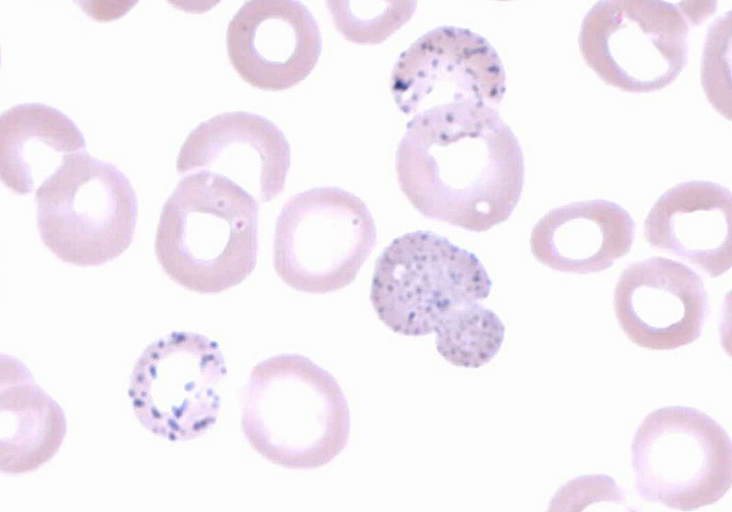
Image Source: Wikimedia Commons (2014). Basophilic stippling 2, Licensed under a creative commons licence. Available for free at: https://en.wikipedia.org/wiki/Basophilic_stippling#/media/File:Basophilic_stippling_2.jpg (Accessed: 16 January 2024)

Image Source: Wikimedia Commons (2008). Hypogranular neutrophil with a pseudo-Pelger-Huet nucleus in MDS, Licensed under a creative commons licence. Available for free at: https://commons.wikimedia.org/wiki/File:Hypogranular_neutrophil_with_a_pseudo-Pelger-Huet_nucleus_in_MDS.jpg (Accessed: 16 January 2024)
Management and prognosis of MDS
Risk is assessed using percentage of bone marrow blasts, cytogenetics and number and degree of cytopenias.
For lower-risk asymptomatic patients, active surveillance is used. Symptomatic lower-risk or higher-risk patients who are medically fit and willing may undergo intensive therapy (e.g. induction chemotherapy with allogeneic SCT).
If unable or unwilling to undergo an intensive regime, patients can have a lower intensity regime alongside supportive therapy (e.g. transfusions and growth factors).
Median overall survival ranges from 8.8 years in the lowest risk group to 9 months in the highest risk group.
References
- Tefferi A. Clinical manifestations and diagnosis of polycythaemia vera [Internet]. Larson RA, Rosmarin AG, editors. UpToDate. 2020 [cited 2022 Jan 02]. Available from: https://www.uptodate.com/contents/clinical-manifestations-and-diagnosis-of-polycythemia-vera
- Bose P, Verstovsek S. Polycythaemia vera [Internet]. BMJ Best Practice. 2021 [cited 2022 Jan 02]. Available from: https://bestpractice.bmj.com/topics/en-gb/178
- McMullin MF, Harrison CN, Ali S, Cargo C, Chen F, Ewing J et al. A guideline for the diagnosis and management of polycythaemia vera. A British Society for Haematology Guideline. British Journal of Haematology. 2019;184(2):176-191. Available from: doi: 10.1111/bjh.15648
- Tefferi A. Prognosis and treatment of polycythaemia vera [Internet]. Larson RA, Rosmarin AG, editors. UpToDate. 2021 [cited 2022 Jan 02]. Available from: https://www.uptodate.com/contents/prognosis-and-treatment-of-polycythemia-vera
- Mukherjee D. Essential thrombocytosis [Internet]. BMJ Best Practice. 2021 [cited 2022 Jan 02]. Available from: https://bestpractice.bmj.com/topics/en-gb/896
- Provan D, Baglin T, Dokal I, de Vos J. Oxford Handbook of Clinical Haematology. 4th ed. Oxford: Oxford University Press; 2015
- Tefferi A. Diagnosis and clinical manifestations of essential thrombocythaemia [Internet]. Larson RA, Rosmarin AG, editors. UpToDate. 2021 [cited 2022 Jan 02]. Available from: https://www.uptodate.com/contents/diagnosis-and-clinical-manifestations-of-essential-thrombocythemia
- Tefferi A. Prognosis and treatment of essential thrombocythaemia [Internet]. Larson RA, Rosmarin AG, editors. UpToDate. 2021 [cited 2022 Jan 02]. Available from: https://www.uptodate.com/contents/prognosis-and-treatment-of-essential-thrombocythemia
- Spivak JL. Myelofibrosis [Internet]. BMJ Best Practice. 2018 [cited 2022 Jan 03]. Available from: https://bestpractice.bmj.com/topics/en-gb/1132
- Tefferi A. Clinical manifestations and diagnosis of primary myelofibrosis [Internet]. Larson RA, Rosmarin AG, editors. UpToDate. 2020 [cited 2022 Jan 03]. Available from: https://www.uptodate.com/contents/clinical-manifestations-and-diagnosis-of-primary-myelofibrosis
- Tefferi A. Management of primary myelofibrosis [Internet]. Larson RA, Rosmarin AG, editors. UpToDate. 2021 [cited 2022 Jan 03]. Available from: https://www.uptodate.com/contents/management-of-primary-myelofibrosis
- Tefferi A. Prognosis of primary myelofibrosis [Internet]. Larson RA, Rosmarin AG, editors. UpToDate. 2021 [cited 2022 Jan 03]. Available from: https://www.uptodate.com/contents/prognosis-of-primary-myelofibrosis
- Ganti AP, Marr A. Myelodysplastic syndrome [Internet]. BMJ Best Practice. 2020 [cited 2022 Jan 03]. Available from: https://bestpractice.bmj.com/topics/en-gb/377
- Aster JC, Stone RM. Clinical manifestations and diagnosis of myelodysplastic syndromes (MDS) [Internet]. Larson RA, Rosmarin AG, editors. UpToDate. 2021 [cited 2022 Jan 03]. Available from: https://www.uptodate.com/contents/clinical-manifestations-and-diagnosis-of-myelodysplastic-syndromes-mds
- Sekeres MA. Overview of the treatment of myelodysplastic syndromes [Internet]. Larson RA, Rosmarin AG, editors. UpToDate. 2021 [cited 2022 Jan 03]. Available from: https://www.uptodate.com/contents/overview-of-the-treatment-of-myelodysplastic-syndromes
- Sekeres MA. Prognosis of the myelodysplastic syndromes in adults [Internet]. Larson RA, Rosmarin AG, editors. UpToDate. 2021 [cited 2022 Jan 03]. Available from: https://www.uptodate.com/contents/prognosis-of-the-myelodysplastic-syndromes-in-adults
Haematopoietic stem cell transplant (HSCT)
Overview of HSCT
HSCT involves the harvest of multipotent haematopoietic stem cells (HSC) from a donor and infusion into a recipient. The aims are:
- To allow for haematopoietic reconstitution after myeloablative doses of chemo(radio)therapy
- To replace congenital or acquired abnormal bone marrow or immune function
HSC can be harvested from bone marrow, peripheral blood or umbilical cord blood.
Bone marrow samples are usually obtained from a donor’s posterior iliac crest under general anaesthetic.
Peripheral blood stem cells are harvested through apheresis following 4-5 days of daily injections with granulocyte colony-stimulating factor to ‘mobilise’ the stem cells.
Indications for HSCT include AML, ALL, CML, CLL, MDSs, myelofibrosis, various NHLs and HL. HSCT is also a clinical option or developmental in some solid tumour malignancies.
Classification of HSCT
HSCT can be autologous or allogeneic.
In autologous transplants, a patient’s own stem cells are harvested and transplanted back into the patient after high-dose chemo(radio)therapy.
In allogeneic transplants, HSCs are harvested from a donor and transplanted into the recipient patient. Recipients need to be HLA-matched to the donors to reduce risk of rejection and graft-versus-host disease (GvHD). HLA-matched related donors are preferred. If a matched related donor cannot be found, then matched unrelated donors are searched for. Minimally mismatched donors (related or unrelated) are sometimes tolerated if no other suitable donor is found.
Complications of HSCT
There are numerous complications that can occur after HSCT. Complications are considered early if they happen within one year of transplant and late if they occur after this.
| Early complications | Late complications |
|---|---|
| Oral mucositis
Diarrhoea Acute GvHD Infection (bacterial, viral, fungal, CMV, atypical) Transplant-associated thrombotic microangiopathy (usually manifest as renal dysfunction) Organ failure (liver, renal, pulmonary) |
Chronic GvHD
Infections (latent viral, encapsulated bacteria) Cardiovascular disease Pulmonary disease (bronchiolitis obliterans syndrome, restrictive allograft syndrome) Liver disease (GvHD, viral hepatitis reactivation) Hypothyroidism Hypogonadism and infertility Psychological impact |
Graft-versus-host disease
GvHD is a complication of allogeneic HSCT when donor T-lymphocytes respond to histoincompatible antigens on host tissue.
Higher degree of HLA-mismatch, older donor/recipient age and sex disparity all increase the risk of GvHD.
GvHD can be acute, chronic or exhibit an overlap syndrome. They are differentiated by clinical findings.
Prophylaxis is generally with ciclosporin/tacrolimus and methotrexate/mycophenolate mofetil. There can be complications from these medications, e.g. gingival hyperplasia with ciclosporin and renal failure with tacrolimus.
Acute GvHD
Classic acute GvHD occurs <100 days post-HSCT; if it occurs after this, it is classified as late onset acute GvHD.
- Skin: maculopapular rash on neck, ears, shoulders, palms and soles
- Gastrointestinal tract: can affect the upper and lower gastrointestinal tract causing nausea, vomiting, anorexia, diarrhoea and abdominal cramps
- Liver: damage to bile canaliculi causing cholestasis and elevated bilirubin and deranged liver function tests
Isolated skin acute GvHD affecting <50% of the skin is treated with topical treatments, including steroids.
Any other acute GvHD is treated with systemic oral steroids and ciclosporin/tacrolimus.
Chronic GvHD
Chronic GvHD normally occurs >3 months after HSCT but can occur at any time after it, even many years. If there are features of acute GvHD then it is an ‘overlap syndrome’.
It typically affects the skin, liver, gastrointestinal tract and lungs, but any organ system can be affected:
- Skin: erythema, plaques, desquamation, lichen planus-like, joint contractures, scleroderma-like
- Mouth: xerostomia, mucositis, gingivitis, ulcers
- Liver: acute hepatitis or slowly progressive cholestasis
- Gastrointestinal tract: can affect the upper and lower gastrointestinal tract causing nausea, vomiting, anorexia, diarrhoea, exocrine pancreatic insufficiency
- Lung: bronchiolitis obliterans causing dyspnoea on exertion, dry cough and decreased exercise tolerance
- Musculoskeletal: muscle cramps, arthralgia, arthritis
Mild disease may be managed by treatment localised to the system affected. Any other disease stage requires systemic steroids with adjunct therapy depending on the body system affected, e.g. emollients for skin and inhalers for lungs.
References
- Provan D, Baglin T, Dokal I, de Vos J. Oxford Handbook of Clinical Haematology. 4th ed. Oxford: Oxford University Press; 2015
- Negrin RS. Sources of hematopoietic stem cells [Internet]. Chao NJ, Rosmarin AG, editors. UpToDate. 2020 [cited 2022 Jan 10]. Available from: https://www.uptodate.com/contents/sources-of-hematopoietic-stem-cells
- Negrin RS. Donor selection for hematopoietic cell transplantation [Internet]. Chao NJ, Rosmarin AG, editors. UpToDate. 2021 [cited 2022 Jan 10]. Available from: https://www.uptodate.com/contents/donor-selection-for-hematopoietic-cell-transplantation
- Negrin RS. Early complications of hematopoietic cell transplantation [Internet]. Chao NJ, Rosmarin AG, editors. UpToDate. 2020 [cited 2022 Jan 10]. Available from: https://www.uptodate.com/contents/early-complications-of-hematopoietic-cell-transplantation
- Chao NJ. Survival, quality-of-life, and late complications after hematopoietic cell transplantation in adults [Internet]. Negrin RS, Rosmarin AG, editors. UpToDate. 2021 [cited 2022 Jan 10]. Available from: https://www.uptodate.com/contents/survival-quality-of-life-and-late-complications-after-hematopoietic-cell-transplantation-in-adults
- Chao NJ. Clinical manifestations, diagnosis, and grading of acute graft-versus-host disease [Internet]. Negrin RS, Rosmarin AG, editors. UpToDate. 2021 [cited 2022 Jan 10]. Available from: https://www.uptodate.com/contents/clinical-manifestations-diagnosis-and-grading-of-acute-graft-versus-host-disease
- Zeiser R. Clinical manifestations and diagnosis of chronic graft-versus-host disease [Internet]. Negrin RS, Chao NJ, Rosmarin AG, editors. UpToDate. 2021 [cited 2022 Jan 10]. Available from: https://www.uptodate.com/contents/clinical-manifestations-and-diagnosis-of-chronic-graft-versus-host-disease
- Choi SW, Runaas L. Graft-versus-host disease [Internet]. BMJ Best Practice. 2021 [cited 2022 Jan 10]. Available from: https://bestpractice.bmj.com/topics/en-gb/946
- Chao NJ, Zeiser R. Treatment of acute graft-versus-host disease [Internet]. Negrin RS, Rosmarin AG, editors. UpToDate. 2021 [cited 2022 Jan 10]. Available from: https://www.uptodate.com/contents/treatment-of-acute-graft-versus-host-disease
Complete Guide to Clinical Genetics ST3 Application and Recruitment (2027)
Written by Medibuddy. Last updated: October 2026. Clinical Genetics is

Complete Guide to Clinical Radiology ST3 Application and Recruitment (2027)
Recruitment paused: NHS England is not running Clinical Radiology ST3+ recru

Complete Guide to Paediatric Surgery ST3 Application and Recruitment in United Kingdom (2026)
Written by Medibuddy. Last updated: October 2026. If you’re planning


Take your subscriptions with you
Our mobile app allows you to access your interview and exam question banks wherever you are.

