


 Close
Close
 23rd July 2024
Lymphoma
Overview of lymphoma
Lymphomas are a heterogenous group of malignancies resulting from clonal proliferation of lymphocytes in lymphoid tissue. They are the most common haematological malignancy.
Lymphomas are subdivided into two broad groups:
- Hodgkin lymphoma (HL): around 10% of lymphomas are characterised by the presence of Reed-Sternberg cells on blood film. This is a B-cell lymphoma.
- Non-Hodgkin lymphoma (NHL): around 90% of lymphomas, no Reed-Sternberg cells on blood film. Around 85% originate from B-cells and 15% from T-cells/NK cells.
Diagnosis and staging of lymphoma
There are three main aspects to definitive diagnosis of HL and NHL:
- Tissue biopsy: excisional biopsy of a peripheral lymph node (LN). Fine needle aspiration does not provide sufficient material for a histological diagnosis and classification.
- Imaging: chest X-ray, CT of neck, chest, abdomen and pelvis with contrast and FDG-PET scan. Used to stage disease and guide tissue biopsy.
- Immunohistochemistry
Staging of HL and NHL is via the Lugano classification, a modification of the classic Ann Arbor staging system. This takes into account the number and site of LNs and/or extra-lymphatic organs affected, as well as the presence or absence of ‘B’ symptoms, defined as:
- Unexplained fever >38°C in the last month
- Recurrent drenching night sweats in the last month
- Weight loss >10% within six months of diagnosis
A sub-classification of ‘A’ denotes the absence of ‘B’ symptoms, whereas ‘B’ denotes their presence, e.g. IIA or IIIB.
| Stage | Description |
|---|---|
| I (early) | Involvement of a single LN region/structure or of a single extra-lymphatic organ/site |
| II (early) | Involvement of 2+ LN regions/structures on the same side of the diaphragm or with localised involvement of an extra-lymphatic organ/site |
| III (advanced) | Involvement of LNs or lymphoid organs on both sides of the diaphragm |
| IV (advanced) | Diffuse involvement of one or more extra-lymphatic organs, with or without LN involvement |
Hodgkin lymphoma
Categories of HL
HL can be further sub-categorised:
- Classical HL (~90% of HL)
- Nodular sclerosis classical HL (NSCHL)
- Mixed cellularity classical HL (MCCHL)
- Lymphocyte rich classical HL (LRCHL)
- Lymphocyte depleted classical HL (LDCHL)
- Nodular lymphocyte predominant HL (NLPHL) (~10% of HL)
Hodgkin lymphoma is characterised by the presence of Reed-Sternberg cells. They often have bilobed nuclei with prominent nucleoli (described as having an ‘owl’s eyes’ appearance) but can also be multinucleated.
Lymphocytic and histiocytic (‘popcorn’) cells are a variant of Reed-Sternberg cells characterised by a single multilobulated nucleus and are required to make a diagnosis of NLPHL.

Image Source: Ncbi (2022). Reed Sternberg Cells, Licensed under a creative commons licence. Available for free at: https://www.ncbi.nlm.nih.gov/books/NBK542333/ (Accessed: 16 January 2024)
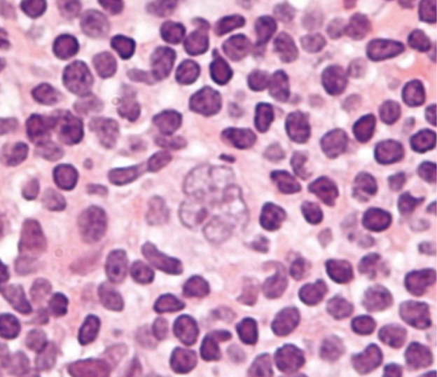
Image Source: Wikimedia Commons (2010). Popcorn cell in nodular lymphocyte predominant Hodgkin lymphoma – very high mag cropped, Licensed under a creative commons licence. Available for free at: https://en.wikipedia.org/wiki/Nodular_lymphocyte_predominant_Hodgkin_lymphoma#/media/File:Popcorn_cell_in_nodular_lymphocyte_predominant_Hodgkin_lymphoma_-_very_high_mag_cropped.jpg (Accessed: 16 January 2024)
Background of HL
HL incidence demonstrates a bimodal age distribution, with the first peak between 15 and 34 years and the second peak over 60 years. There is a higher incidence in men.
The precise aetiology of HL remains unclear, but there are numerous associated risk factors:
- Epstein-Barr virus (EBV): this is detected in Reed-Sternberg cells in many cases of HL. It has been linked to the pathogenesis of HL, but the absolute risk of developing HL after EBV infection remains very low. Its presence varies by subtype, location and immune status:
- EBV is hardly found in NLPHL but nearly always found in LDCHL
- EBV is found in 20-50% of classical HL in developed countries but found in nearly all cases in developing countries
- Almost all cases of HL in HIV positive patients are EBV positive
- Immunodeficiency (congenital and acquired)
- Autoimmune conditions
- Familial risk
- Environmental risk (obesity and smoking)
Presentation and diagnosis of HL
Common presenting symptoms include:
- Asymptomatic lymphadenopathy: most common presentation; painless, commonly cervical or supraclavicular
- Mediastinal mass on chest X-ray: may be incidental or done for symptoms such as cough, dyspnoea, chest pain or superior vena cava obstruction (classic signs of this are facial swelling that improves through the day and enlarged veins in the neck, upper chest and arms)
- Constitutional symptoms: ‘B’ symptoms of fever, night sweats, weight loss
- Fatigue
- Pruritus
- Pain on alcohol ingestion: at affected LNs or sites of bony involvement
Diagnosis relies on tissue biopsy, imaging and immunohistochemistry (see diagnosis and staging of lymphoma). Classical HL cells are always CD30 positive, mostly CD15 positive, occasionally CD20 positive and CD45 negative. NLPHL cells are CD30 and CD15 negative and CD20 and CD45 positive.
Management and prognosis of HL
Management of HL is complicated. It varies between paediatric and adult populations, disease stage and HL subtype. A summary of the treatment of adults is as follows:
| Early-stage classical HL | Combination chemotherapy¹ then involved-field radiotherapy (IFRT) |
| Late-stage classical HL | Combination chemotherapy¹ |
| Early-stage NLPHL | IFRT (or active surveillance) |
| Late-stage NLPHL | Chemoimmunotherapy² |
¹ A regime used commonly for classical HL in adults is ABVD: doxorubicin, bleomycin, vinblastine, dacarbazine
² Most often R-CHOP: rituximab, cyclophosphamide, doxorubicin, vincristine, prednisolone
Prognosis is generally very good. Patients treated in the early stage have an 85-90% chance of long-term complete remission. In children, even advanced HL has cure rates >85%.
Prognosis in advanced HL in adults is determined by the International Prognostic Score, which consists of seven adverse variables with independent prognostic value. It predicts freedom from disease progression at five years.
Due to the success of treatment, patients can develop complications of treatment many years after completing it. For example, radiation-induced hypothyroidism, chemotherapy side effects such as cardiomyopathy (doxorubicin) or pulmonary fibrosis (bleomycin), and secondary malignancies (e.g. AML).
Non-Hodgkin lymphoma
Background of NHL
NHL refers to any lymphoma that is not HL. As such, it encompasses a large and heterogenous group of conditions, only a few of which will be discussed here. NHL can be split into indolent and aggressive lymphomas.
NHL incidence increases with age and there is a male predominance.
There are numerous risk factors including congenital and acquired immunodeficiencies, autoimmune conditions, familial risk and environmental factors (e.g. pesticide exposure). There are also subtype-specific risk factors.
Prognosis is very dependent on subtype. In general, markers of poor prognosis include age >60, raised serum LDH, disease stage III or IV, ≤2 extranodal disease sites and poor performance status.
Presentation and diagnosis of NHL
Indolent (low-grade) lymphomas present insidiously, sometimes with symptoms over months to years. They are often disseminated at presentation and less likely to be curable. Aggressive (high-grade) lymphomas present sub-acutely or acutely with localised rapidly growing lymphadenopathy with or without ‘B’ symptoms but are more likely to be curable.
Painless lymphadenopathy is present in >2/3 of patients at presentation. ‘B’ symptoms are more likely to present in aggressive lymphomas. Hepatosplenomegaly is more common in indolent lymphomas.
As indolent lymphomas may be disseminated at presentation, they may present with extranodal symptoms. Gastrointestinal involvement may cause anorexia, early satiety and vomiting. Central nervous system involvement may cause headaches, focal neurological deficits or seizures.
Definitive diagnosis relies on tissue biopsy, imaging and immunohistochemistry (see above).
Follicular lymphoma
This is an indolent B-cell lymphoma. It is the most common indolent NHL and the second most common NHL. Median age at diagnosis is 65 years. The majority exhibit t(14;18), causing overexpression of the BCL2 oncogene. Lymphadenopathy is the most common presenting complaint; around 70% present with stage IV disease with BM involvement. Early-stage disease may be treated with IFRT. Advanced-stage disease may be treated with active surveillance if asymptomatic or if symptomatic, chemoimmunotherapy (e.g. R-CHOP). Median survival is 10 years.
Extranodal marginal zone lymphoma of mucosa-associated lymphoid tissue (MALT)
This is more commonly known as a MALT lymphoma. It is an indolent B-cell lymphoma. Median age of diagnosis is 66 years. MALT lymphomas can occur in any tissue with MALT e.g. stomach, small bowel, lung. They often arise on a background of chronic immune stimulation.
Gastric MALT lymphomas frequently develop following chronic gastritis caused by Helicobacter pylori. Over 90% of patients are H. pylori positive. They can present with dyspepsia, epigastric pain or gastrointestinal bleeding. Diagnosis is made with endoscopy and biopsy. Most patients present with localised disease. First line treatment for local disease in H. pylori positive patients is eradication therapy. In H. pylori negative patients or in H. pylori positive patients not responding to first line therapy, IFRT is used. 5-year survival is >80%.
H. pylori eradication therapy consists of a proton pump inhibitor (PPI) and antibiotics (first line: amoxicillin and clarithromycin/metronidazole; second line: amoxicillin and a tetracycline/quinolone, e.g. levofloxacin).
Diffuse large B-cell lymphoma (DLBCL)
This is an aggressive lymphoma and the most common NHL. Median age of diagnosis is 64 with a slight male predominance. DLBCL can occur de novo or from transformation of a low-grade lymphoma or of CLL (Richter’s transformation). It presents as rapidly progressing stage I or II disease in 50% of cases; extranodal disease is common. Early-stage disease is treated with R-CHOP chemoimmunotherapy +/- IFRT. Advanced-stage disease is treated with R-CHOP.
Mantle cell lymphoma
This is an uncommon, aggressive B-cell lymphoma, although there is a subset of patients who experience a more indolent disease course. Median age of diagnosis is 63 years, and it has a strong male predominance. It is highly associated with t(11;14). Around 70% of patients present with stage IV disease. There is no standardised first-line therapy; conventional chemoimmunotherapy with R-CHOP can be used. Median survival is 3-4 years.
Burkitt lymphoma
This is a highly aggressive B-cell lymphoma. There are three forms of the disease: endemic, sporadic and immunodeficiency associated. All three are clinically similar and dependent on dysregulation of the MYC gene on chromosome 8 through translocations.
- Endemic disease is seen primarily in Africa, presenting with extranodal disease of the jaw/facial bones (most common) or abdominal organs. Peak incidence is 4-7 years; 90% are associated with EBV infection.
- Sporadic disease is seen more in developed nations, usually presenting with abdominal symptoms, but a smaller percentage can present with jaw/facial bone disease. Median age of presentation is 31 years; 30% are associated with EBV infection.
- Immunodeficiency-associated disease is primarily seen in patients with HIV infection but also in other causes of acquired or congenital immunodeficiency.
Adult T-cell leukaemia-lymphoma (ATLL)
This is an aggressive lymphoma caused by chronic infection with human T-lymphotropic virus type 1 (HTLV-1). Incidence is highest where HTLV-1 infection is endemic: islands in southwest Japan, the Caribbean, parts of West Africa and Central and South America. There is often a long latency period of several decades between infection and development of ATLL; median age of diagnosis is 58 years. Median survival is very poor, ranging from months to a year, depending on subtype. Treatment is with combination chemotherapy.
Enteropathy-associated T-cell lymphoma (EATL)
EATL is an aggressive lymphoma highly associated with untreated coeliac disease. Management of coeliac disease with a gluten-free diet prevents development of EATL. Patients present with abdominal pain, often associated with gastrointestinal bleeding, bowel obstruction or perforation. Treatment is with combination chemotherapy with or without autologous SCT. Overall 5-year survival is 10-20%.
| Subtype | Cell type | Aggressiveness | Key features |
|---|---|---|---|
| Follicular | B-cell | Indolent | ~85% exhibit t(14;18)
Lymphadenopathy is the most common presenting complaint |
| MALT | B-cell | Indolent | >90% with gastric MALT are H. pylori positive |
| DLBCL | B-cell | Aggressive | Can occur de novo or from transformation of a low-grade lymphoma or CLL (Richter’s transformation) |
| Mantle cell | B-cell | Aggressive | Highly associated with t(11;14) |
| Burkitt | B-cell | Aggressive | Endemic: primarily in Africa, jaw/facial bone disease, 90% associated with EBV; sporadic: more in developed nations, primarily abdominal symptoms; immunodeficiency associated: mainly seen in HIV |
| ATLL | T-cell | Aggressive | Caused by chronic infection with HTLV-1 |
| EATL | T-cell | Aggressive | Highly associated with untreated coeliac disease |
References
- Provan D, Baglin T, Dokal I, de Vos J. Oxford Handbook of Clinical Haematology. 4th ed. Oxford: Oxford University Press; 2015.
- LaCasce AS, Ng AK. Hodgkin lymphoma: Epidemiology and risk factors [Internet]. Freedman AS, Rosmarin AG, editors. UpToDate. 2020 [cited 2021 Dec 20]. Available from: https://www.uptodate.com/contents/hodgkin-lymphoma-epidemiology-and-risk-factors
- LaCasce AS, Ng AK. Pretreatment evaluation, staging, and treatment stratification of classic Hodgkin lymphoma [Internet]. Freedman AS, Rosmarin AG, editors. UpToDate. 2020 [cited 2021 Dec 20]. Available from: https://www.uptodate.com/contents/pretreatment-evaluation-staging-and-treatment-stratification-of-classic-hodgkin-lymphoma
- Freedman AS, Friedberg JW. Pretreatment evaluation, staging, and response assessment of non-Hodgkin lymphoma [Internet]. Lister A, Rosmarin AG, editors. UpToDate. 2020 [cited 2021 Dec 20]. Available from: https://www.uptodate.com/contents/pretreatment-evaluation-staging-and-response-assessment-of-non-hodgkin-lymphoma
- Aggarwal P, Limaiem F. Reed Sternberg Cells [Internet]. NCBI. 2021 [cited 2021 Dec 20]. Available from: https://www.ncbi.nlm.nih.gov/books/NBK542333/
- Moskowitz A. Hodgkin’s lymphoma [Internet]. BMJ Best Practice. 2019 [cited 2021 Dec 21]. Available from: https://bestpractice.bmj.com/topics/en-gb/311
- McClain KL, Kamdar K. Overview of Hodgkin lymphoma in children and adolescents [Internet]. Park JR, Rosmarin AG, editors. UpToDate. 2021 [cited 2021 Dec 21]. Available from: https://www.uptodate.com/contents/overview-of-hodgkin-lymphoma-in-children-and-adolescents
- LaCasce AS, Ng AK, Aster JC. Clinical presentation and diagnosis of classic Hodgkin lymphoma in adults [Internet]. Freedman AS, Rosmarin AG, editors. UpToDate. 2020 [cited 2021 Dec 21]. Available from: https://www.uptodate.com/contents/clinical-presentation-and-diagnosis-of-classic-hodgkin-lymphoma-in-adults
- Canellos GP, Ng AK. Overview of the treatment of classic Hodgkin lymphoma in adults [Internet]. Freedman AS, Rosmarin AG, editors. UpToDate. 2020 [cited 2021 Dec 21]. Available from: https://www.uptodate.com/contents/overview-of-the-treatment-of-classic-hodgkin-lymphoma-in-adults
- LaCasce AS, Ng AK. Treatment of nodular lymphocyte-predominant Hodgkin lymphoma [Internet]. Freedman AS, Rosmarin AG, editors. UpToDate. 2021 [cited 2021 Dec 21]. Available from: https://www.uptodate.com/contents/treatment-of-nodular-lymphocyte-predominant-hodgkin-lymphoma
- Chan E, Ng CH, Ooi M, Poon M. Non-Hodgkin’s lymphoma [Internet]. BMJ Best Practice. 2021 [cited 2021 Dec 21]. Available from: https://bestpractice.bmj.com/topics/en-gb/312
- Freedman AS, Friedberg JW, Aster JC. Clinical presentation and initial evaluation of non-Hodgkin lymphoma [Internet]. Lister A, Rosmarin AG, editors. UpToDate. 2020 [cited 2021 Dec 21]. Available from: https://www.uptodate.com/contents/clinical-presentation-and-initial-evaluation-of-non-hodgkin-lymphoma
- Freedman AS, Aster JC. Clinical manifestations, pathologic features, diagnosis, and prognosis of follicular lymphoma [Internet]. Lister A, Connor RF, editors. UpToDate. 2020 [cited 2021 Dec 21]. Available from: https://www.uptodate.com/contents/clinical-manifestations-pathologic-features-diagnosis-and-prognosis-of-follicular-lymphoma
- Freedman AS, Aster JC. Clinical manifestations, pathologic features, and diagnosis of extranodal marginal zone lymphoma of mucosa associated lymphoid tissue (MALT) [Internet]. Lister A, Connor RF, editors. UpToDate. 2021 [cited 2021 Dec 21]. Available from: https://www.uptodate.com/contents/clinical-manifestations-pathologic-features-and-diagnosis-of-extranodal-marginal-zone-lymphoma-of-mucosa-associated-lymphoid-tissue-malt
- Freedman AS, Friedberg JW, Ng AK. Treatment of extranodal marginal zone lymphoma of mucosa associated lymphoid tissue (MALT lymphoma) [Internet]. Lister A, Connor RF, editors. UpToDate. 2021 [cited 2021 Dec 21]. Available from: https://www.uptodate.com/contents/treatment-of-extranodal-marginal-zone-lymphoma-of-mucosa-associated-lymphoid-tissue-malt-lymphoma
- Freedman AS, Aster JC. Epidemiology, clinical manifestations, pathologic features, and diagnosis of diffuse large B cell lymphoma [Internet]. Lister A, Rosmarin AG, editors. UpToDate. 2021 [cited 2021 Dec 21]. Available from: https://www.uptodate.com/contents/epidemiology-clinical-manifestations-pathologic-features-and-diagnosis-of-diffuse-large-b-cell-lymphoma
- Freedman AS, Aster JC. Clinical manifestations, pathologic features, and diagnosis of mantle cell lymphoma [Internet]. Lister A, Rosmarin AG, editors. UpToDate. 2021 [cited 2021 Dec 21]. Available from: https://www.uptodate.com/contents/clinical-manifestations-pathologic-features-and-diagnosis-of-mantle-cell-lymphoma
- Freedman AS, Aster JC. Epidemiology, clinical manifestations, pathologic features, and diagnosis of Burkitt lymphoma [Internet]. Lister A, Rosmarin AG, editors. UpToDate. 2021 [cited 2021 Dec 21]. Available from: https://www.uptodate.com/contents/epidemiology-clinical-manifestations-pathologic-features-and-diagnosis-of-burkitt-lymphoma
- Freedman AS, Aster JC, Suzuki R. Clinical manifestations, pathologic features, and diagnosis of adult T cell leukemia-lymphoma [Internet]. Lister A, Rosmarin AG, editors. UpToDate. 2020 [cited 2021 Dec 22]. Available from: https://www.uptodate.com/contents/clinical-manifestations-pathologic-features-and-diagnosis-of-adult-t-cell-leukemia-lymphoma
- Matsuoka M, Suzuki R. Treatment and prognosis of adult T cell leukemia-lymphoma [Internet]. Freedman AS, Rosmarin AG, editors. UpToDate. 2020 [cited 2021 Dec 22]. Available from: https://www.uptodate.com/contents/treatment-and-prognosis-of-adult-t-cell-leukemia-lymphoma
- Freedman AS, Aster JC. Clinical manifestations, pathologic features, and diagnosis of enteropathy-associated T cell lymphoma [Internet]. Lister A, Rosmarin AG, editors. UpToDate. 2021 [cited 2021 Dec 22]. Available from: https://www.uptodate.com/contents/clinical-manifestations-pathologic-features-and-diagnosis-of-enteropathy-associated-t-cell-lymphoma
Complete Guide to Clinical Genetics ST3 Application and Recruitment (2027)
Written by Medibuddy. Last updated: October 2026. Clinical Genetics is

Complete Guide to Clinical Radiology ST3 Application and Recruitment (2027)
Recruitment paused: NHS England is not running Clinical Radiology ST3+ recru

Complete Guide to Paediatric Surgery ST3 Application and Recruitment in United Kingdom (2026)
Written by Medibuddy. Last updated: October 2026. If you’re planning


Take your subscriptions with you
Our mobile app allows you to access your interview and exam question banks wherever you are.

