


 Close
Close
 23rd July 2024
Hodgkin lymphoma
Categories of HL
HL can be further sub-categorised:
- Classical HL (~90% of HL)
- Nodular sclerosis classical HL (NSCHL)
- Mixed cellularity classical HL (MCCHL)
- Lymphocyte rich classical HL (LRCHL)
- Lymphocyte depleted classical HL (LDCHL)
- Nodular lymphocyte predominant HL (NLPHL) (~10% of HL)
Hodgkin lymphoma is characterised by the presence of Reed-Sternberg cells. They often have bilobed nuclei with prominent nucleoli (described as having an ‘owl’s eyes’ appearance) but can also be multinucleated.
Lymphocytic and histiocytic (‘popcorn’) cells are a variant of Reed-Sternberg cells characterised by a single multilobulated nucleus and are required to make a diagnosis of NLPHL.

Image Source: Ncbi (2022). Reed Sternberg Cells, Licensed under a creative commons licence. Available for free at: https://www.ncbi.nlm.nih.gov/books/NBK542333/ (Accessed: 16 January 2024)
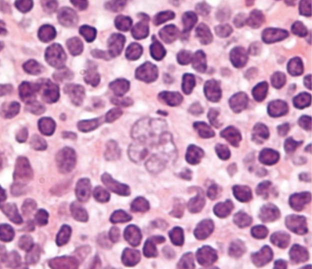
Image Source: Wikimedia Commons (2010). Popcorn cell in nodular lymphocyte predominant Hodgkin lymphoma – very high mag cropped, Licensed under a creative commons licence. Available for free at: https://en.wikipedia.org/wiki/Nodular_lymphocyte_predominant_Hodgkin_lymphoma#/media/File:Popcorn_cell_in_nodular_lymphocyte_predominant_Hodgkin_lymphoma_-_very_high_mag_cropped.jpg (Accessed: 16 January 2024)
Background of HL
HL incidence demonstrates a bimodal age distribution, with the first peak between 15 and 34 years and the second peak over 60 years. There is a higher incidence in men.
The precise aetiology of HL remains unclear, but there are numerous associated risk factors:
- Epstein-Barr virus (EBV): this is detected in Reed-Sternberg cells in many cases of HL. It has been linked to the pathogenesis of HL, but the absolute risk of developing HL after EBV infection remains very low. Its presence varies by subtype, location and immune status:
- EBV is hardly found in NLPHL but nearly always found in LDCHL
- EBV is found in 20-50% of classical HL in developed countries but found in nearly all cases in developing countries
- Almost all cases of HL in HIV positive patients are EBV positive
- Immunodeficiency (congenital and acquired)
- Autoimmune conditions
- Familial risk
- Environmental risk (obesity and smoking)
Presentation and diagnosis of HL
Common presenting symptoms include:
- Asymptomatic lymphadenopathy: most common presentation; painless, commonly cervical or supraclavicular
- Mediastinal mass on chest X-ray: may be incidental or done for symptoms such as cough, dyspnoea, chest pain or superior vena cava obstruction (classic signs of this are facial swelling that improves through the day and enlarged veins in the neck, upper chest and arms)
- Constitutional symptoms: ‘B’ symptoms of fever, night sweats, weight loss
- Fatigue
- Pruritus
- Pain on alcohol ingestion: at affected LNs or sites of bony involvement
Diagnosis relies on tissue biopsy, imaging and immunohistochemistry (see diagnosis and staging of lymphoma). Classical HL cells are always CD30 positive, mostly CD15 positive, occasionally CD20 positive and CD45 negative. NLPHL cells are CD30 and CD15 negative and CD20 and CD45 positive.
Management and prognosis of HL
Management of HL is complicated. It varies between paediatric and adult populations, disease stage and HL subtype. A summary of the treatment of adults is as follows:
| Early-stage classical HL | Combination chemotherapy¹ then involved-field radiotherapy (IFRT) |
| Late-stage classical HL | Combination chemotherapy¹ |
| Early-stage NLPHL | IFRT (or active surveillance) |
| Late-stage NLPHL | Chemoimmunotherapy² |
¹ A regime used commonly for classical HL in adults is ABVD: doxorubicin, bleomycin, vinblastine, dacarbazine
² Most often R-CHOP: rituximab, cyclophosphamide, doxorubicin, vincristine, prednisolone
Prognosis is generally very good. Patients treated in the early stage have an 85-90% chance of long-term complete remission. In children, even advanced HL has cure rates >85%.
Prognosis in advanced HL in adults is determined by the International Prognostic Score, which consists of seven adverse variables with independent prognostic value. It predicts freedom from disease progression at five years.
Due to the success of treatment, patients can develop complications of treatment many years after completing it. For example, radiation-induced hypothyroidism, chemotherapy side effects such as cardiomyopathy (doxorubicin) or pulmonary fibrosis (bleomycin), and secondary malignancies (e.g. AML).
Complete Guide to Clinical Genetics ST3 Application and Recruitment (2027)
Written by Medibuddy. Last updated: October 2026. Clinical Genetics is

Complete Guide to Clinical Radiology ST3 Application and Recruitment (2027)
Recruitment paused: NHS England is not running Clinical Radiology ST3+ recru

Complete Guide to Paediatric Surgery ST3 Application and Recruitment in United Kingdom (2026)
Written by Medibuddy. Last updated: October 2026. If you’re planning


Take your subscriptions with you
Our mobile app allows you to access your interview and exam question banks wherever you are.

