


 Close
Close
 23rd July 2024
Myelodysplastic syndromes (MDS)
Background of MDS
MDSs are a heterogenous group of conditions characterised by ineffective haematopoiesis, causing impaired cell maturation and proliferation.
Median age of diagnosis is 70 years with a male predominance.
Around 80% of cases are primary. Common chromosomal abnormalities include deletions of chromosomes 5, 7, 11, 12, 13 and 20.
Secondary MDSs can arise from risk factors including previous exposure to chemotherapy (alkylating agents and topoisomerase II inhibitors), previous SCT and environmental factors (benzene and tobacco).
Presentation and diagnosis of MDS
Patients may be asymptomatic at diagnosis, being identified with abnormal blood tests.
They may present with signs and symptoms of anaemia, neutropenia and thrombocytopenia. Autoimmune conditions are more common in patients with MDS, most commonly rheumatic heart disease, rheumatoid arthritis and pernicious anaemia.
Diagnosis relies upon:
- Bloods: anaemia with variable thrombocytopenia and neutropenia
- Blood film: red cell dysplasia (e.g. oval macrocytic cells, basophilic stippling) and white cell dysplasia (e.g. pseudo-Pelger-Huet abnormality) (Figures 11 and 12)
- Bone marrow: hypercellular with single or multilineage dysplasia; further investigation of the sample with cytochemistry, immunocytochemistry, flow cytometry and cytogenetic analysis for diagnosis and prognosis
MDSs are classified based on the number of cell lineages demonstrating dysplasia, percentage of blast cells, presence of Auer rods, percentage of ringed sideroblasts and cytogenetics. Classification is important for treatment and prognosis.
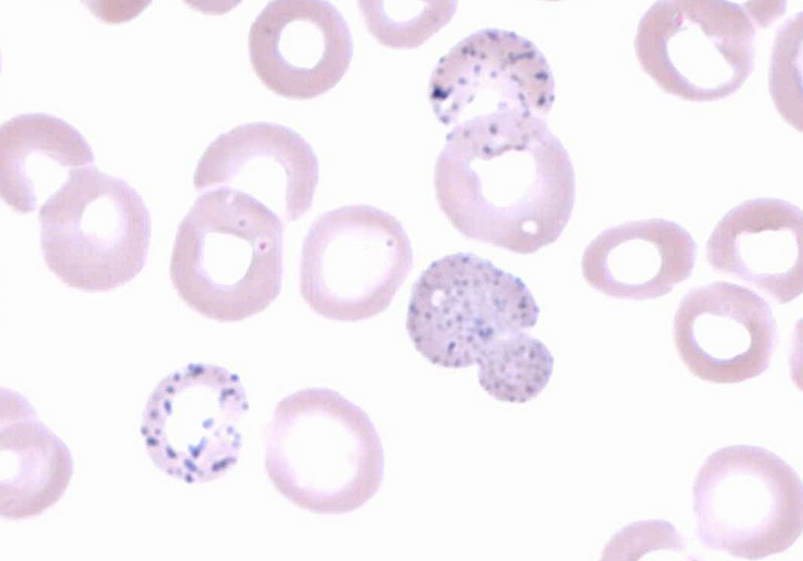
Image Source: Wikimedia Commons (2014). Basophilic stippling 2, Licensed under a creative commons licence. Available for free at: https://en.wikipedia.org/wiki/Basophilic_stippling#/media/File:Basophilic_stippling_2.jpg (Accessed: 16 January 2024)

Image Source: Wikimedia Commons (2008). Hypogranular neutrophil with a pseudo-Pelger-Huet nucleus in MDS, Licensed under a creative commons licence. Available for free at: https://commons.wikimedia.org/wiki/File:Hypogranular_neutrophil_with_a_pseudo-Pelger-Huet_nucleus_in_MDS.jpg (Accessed: 16 January 2024)
Management and prognosis of MDS
Risk is assessed using percentage of bone marrow blasts, cytogenetics and number and degree of cytopenias.
For lower-risk asymptomatic patients, active surveillance is used. Symptomatic lower-risk or higher-risk patients who are medically fit and willing may undergo intensive therapy (e.g. induction chemotherapy with allogeneic SCT).
If unable or unwilling to undergo an intensive regime, patients can have a lower intensity regime alongside supportive therapy (e.g. transfusions and growth factors).
Median overall survival ranges from 8.8 years in the lowest risk group to 9 months in the highest risk group.
Complete Guide to Clinical Genetics ST3 Application and Recruitment (2027)
Written by Medibuddy. Last updated: October 2026. Clinical Genetics is

Complete Guide to Clinical Radiology ST3 Application and Recruitment (2027)
Recruitment paused: NHS England is not running Clinical Radiology ST3+ recru

Complete Guide to Paediatric Surgery ST3 Application and Recruitment in United Kingdom (2026)
Written by Medibuddy. Last updated: October 2026. If you’re planning


Take your subscriptions with you
Our mobile app allows you to access your interview and exam question banks wherever you are.

