


 Close
Close
 11th August 2024
Nerve and Muscle Disorders
Myopathies
Overview of myopathies
Myopathies may be inherited or acquired.
Inherited forms include muscular dystrophies, mitochondrial diseases, metabolic and other congenital myopathies (e.g. McArdles or Von Gierke’s), and channelopathies.
Acquired forms have a variety of aetiologies including drug-induced, endocrine (Addison’s, Cushing’s, hypo- or hyperthyroidism, DM, hyperaldosteronism), metabolic (glycogen storage disease, lipid metabolism), toxic or inflammatory.
Muscular dystrophies
Myotonic dystrophy
This is the most common muscular dystrophy. It is an autosomal dominant disorder that generally develops in the 2nd-4th decades and is characterised by respiratory and skeletal muscle weakness (distal limb and facial/neck most commonly), cataracts, frontal balding, cardiac conduction problems and myotonia (delayed relaxation of muscles following contraction).
Duchenne (DMD) and Becker muscular dystrophy (BMD)
DMD and BMD are both caused by mutations of the dystrophin gene on the X chromosome. Males, therefore, are most affected, though female carriers can display mild symptoms.
DMD is the more severe and more common form (prevalence 3/100,000) and typically presents in the first years of life. Features include proximal muscle weakness (with walking problems including toe walking or waddling gait), cardiomyopathy or conduction issues, and scoliosis/bony fractures. The prognosis is poor, with most wheelchair bound by the early teens and death usually from respiratory compromise or cardiomyopathy, in the teens or twenties.
BMD, the rarer form (0.3/100,000), presents with milder symptoms in the teens or twenties. Many survive into their thirties.
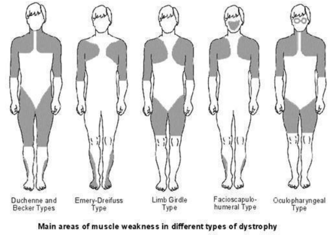
Image Source: Egton Medical Information Systems Limited. All rights reserved (2023). Bones joints muscles/muscular-dystrophies, Licensed under a creative commons licence. Available for free at: https://patient.info/bones-joints-muscles/muscular-dystrophies (Accessed: 17 January 2024)
Facioscapulohumeral muscular dystrophy (FSHD)
FSHD is an autosomal dominant condition with prevalence of 1-2/100,000. Patients generally present in their teens with weakness of facial muscles, scapula (upwards displacement), upper arm (with deltoid sparing), and lower leg. Weakness is generally asymmetric. Genetic testing confirms the diagnosis identifying mutations leading to inappropriate DUX4 gene expression.
Emery-Dreifuss muscular dystrophy
This has autosomal dominant or X-linked inheritance. Clinical features include scapula, humerus and peroneal weakness, and cardiac conduction defects often requiring pacing. Patients may also develop contractures of the cervical extensors, biceps and long finger extensors.
Mitochondrial disorders
MERRF: Myoclonic epilepsy with ragged red fibres
The principal clinical features of MERRF are myoclonus, epilepsy, myopathy and ataxia, with neuropathy, visual problems, lipomas, spasticity, deafness and Wolff-Parkinson-White (WPW) syndrome. The characteristic ragged red fibres are found on muscle biopsy.
MELAS: Mitochondrial encephalomyopathy, lactic acidosis and stroke-like episodes
Clinical features include stroke-like episodes, seizures, migraine-like headaches, deafness, short stature and myopathy.
MIDD: Maternally inherited diabetes and deafness
MIDD is characterised by insulin-dependent diabetes and sensorineural deafness, often alongside myopathy, visual problems, short stature, and cardiac and renal issues (focal segmental glomerulosclerosis).
MNGIE: Mitochondrial neurogastrointestinal encephalomyopathy
Clinical features include gastrointestinal dysmotility (secondary to visceral myopathy), ptosis, ophthalmoplegia, polyneuropathy and distal myopathy. Onset is from childhood to the 4th decade.
CPEO: Chronic progressive external ophthalmoplegia and KSS: Kearns-Sayre syndrome
Patients with CPEO develop ophthalmoplegia and ptosis. KSS has similar features with the addition of retinitis pigmentosa. Onset of KSS, generally in childhood, is earlier than that of CPEO (usually 4th decade).
Glycogen storage diseases (GSD)
The glycogen storage diseases are inborn errors of metabolism that lead to issues with glucose processing and energy release.
Muscle symptoms include pain and stiffness following exercise, elevated CK (creatine kinase) and muscle transaminases and myoglobinuria. In McArdle’s, the ‘second wind’ phenomenon may be noted in which after an initial barrier (with fatigue, tachycardia and breathlessness) is surmounted, exercise tolerance improves. This is due to activation of other energy sources (e.g. fatty acids, hepatic glucose production) to supplement ineffective muscle energy release due to myophosphorylase deficiency.
Dermatomyositis and polymyositis (DM, PM)
Dermatomyositis and polymyositis both present with symmetrical proximal muscle weakness and evidence of inflammation on muscle biopsy (fibre necrosis, degeneration and regeneration, and inflammatory cell infiltrates) which is the gold standard for diagnosis.
Dermatomyositis has additional dermatologic features which may include: Gottron’s papules (scaly purple/red knuckles), periorbital heliotrope rash, shawl-sign (an erythematous rash over the neck and chest), nailfold erythema and periungual telangiectasia, subcutaneous calcification (calcinosis cutis), and ‘mechanic’s hands’ (rough, cracked, darkened areas). Interstitial lung disease occurs in 30-40% of patients.
Both are treated with immunosuppression, usually initially with prednisolone.
Both PM and dermatomyositis may be associated with malignancy via a paraneoplastic mechanism. Cervical, lung, ovarian, pancreatic, bladder and gastric adenocarcinomas are most strongly linked.
Inclusion-body myositis (IBM)
IBM causes a specific pattern of muscle wasting, with weakness of the quadriceps and forearm muscles. Swallow function may also be affected. Onset is generally in the 4th to 6th decade. There is no current treatment.
Muscle enzymes and CK are often mildly elevated. Diagnosis is on the basis of typical examination findings, lab tests and muscle biopsy which may show the characteristic rimmed-vacuoles, along with mononuclear cell inflammatory infiltrates and non-necrotic fibres.
Drug-induced myopathies
Common drug causes of myopathies include:
| Steroids | Alcohol |
| Statins | Cocaine |
| Penicillamine | Antipsychotics |
| Colchicine | Antiretrovirals – AZT (azidothymidine) and other NRTIs (nucleoside reverse transcriptase inhibitor) |
References
- Manji H. Oxford Handbook of Neurology. Oxford Medical Publications; 2020
- Stone WL, Basit H, Adil A. Glycogen Storage Disease [Internet]. PubMed. Treasure Island (FL): StatPearls Publishing; 2020. [Accessed 2022 Jan 22] Available from: https://www.ncbi.nlm.nih.gov/books/NBK459277/
- Darras BT. Duchenne and Becker Muscular Dystrophy: Clinical Features and Diagnosis [Internet]. Patterson MC (Ed), Firth HV (Ed). UpToDate. 2020 [cited 2022 Feb 14]. Available from: https://www.uptodate.com/contents/duchenne-and-becker-muscular-dystrophy-clinical-features-and-diagnosis
- Darras BT. Limb-girdle muscular dystrophy [Internet]. Nordli DR (Ed), Shefner JM (Ed). UpToDate. 2022 [cited 2022 Feb 14]. Available from: https://www.uptodate.com/contents/limb-girdle-muscular-dystrophy
- Darras BT. Emery-Dreifuss muscular dystrophy [Internet]. Patterson MC (Ed). UpToDate. 2020 [cited 2022 Feb 14]. Available from: https://www.uptodate.com/contents/emery-dreifuss-muscular-dystrophy
- Darras BT. Facioscapulohumeral muscular dystrophy [Internet]. Nordli DR (Ed), Shefner JM (Ed). UpToDate. 2022 [cited 2022 Feb 14]. Available from: https://www.uptodate.com/contents/facioscapulohumeral-muscular-dystrophy
- Darras BT. Myotonic dystrophy: Aetiology, clinical features, and diagnosis [Internet]. Shefner JM (Ed), Nordli DR (Ed). UpToDate. 2021 [cited 2022 Feb 14]. Available from: https://www.uptodate.com/contents/myotonic-dystrophy-etiology-clinical-features-and-diagnosis
- Vydra DG, Rayi A. Myotonic Dystrophy [Internet]. PubMed. Treasure Island (FL): StatPearls Publishing; 2020 [accessed 2022 Jan 22]. Available from: https://www.ncbi.nlm.nih.gov/books/NBK557446/
- Craigen WJ. Overview of inherited disorders of glucose and glycogen metabolisms [Internet]. Hahn S (Ed). UpToDate. 2021 [cited 2022 Feb 14]. Available from: https://www.uptodate.com/contents/overview-of-inherited-disorders-of-glucose-and-glycogen-metabolism
Neuromuscular junction disorders
Myasthenia gravis (MG)
Overview of MG
The most common disorder of the neuromuscular junction (NMJ) (incidence 7-23 per million), myasthenia gravis has a bimodal age distribution with peaks at 20-30 years, predominantly in females, and 60-80 years, predominantly in males.
It is caused by antibody-mediated disruption of signalling at the acetylcholine receptor (AChR), with 80-90% of patients having direct IgG antibodies to the AChR. Other antibodies that may be identified are anti-muscle-specific receptor tyrosine kinase (MuSK) and, more rarely, LRP4 antibodies. In some cases however, no antibodies are identified. Those with generalised disease are more likely to have identifiable antibodies than those with purely ocular disease, only half of whom are antibody positive.
Clinical features of MG
Patients most commonly present with ocular symptoms including ptosis or diplopia, along with muscle weakness. Symptoms typically worsen throughout the day and show fatigue. Fifteen percent present with bulbar symptoms such as difficulty chewing and weakness or fatigability of the voice. Fewer than 5% present with limb weakness alone.
Common drugs that may exacerbate MG include the following:
| Avoid | Use with caution |
|---|---|
| Antibiotics: aminoglycosides, fluoroquinolones, macrolides | Anaesthetic agents: isoflurane, halothane, procaine, lidocaine |
| Hydroxychloroquine, chloroquine, quinine | Corticosteroids |
| NMJ-blocking anaesthetics, e.g. rocuronium | Beta-blockers |
| Magnesium | Iodinated radiologic contrast agents |
| Botulinum toxin | Phenytoin |
| Penicillamine | Lithium |
| Antiarrhythmics – procainamide, quinidine | PD-1 inhibitors, e.g. nivolumab, pembrolizumab |
Abbreviations: PD-1 (Programmed cell death protein 1)
Management of MG
The mainstay of MG treatment are the acetylcholinesterase inhibitors, such as pyridostigmine. Many patients will also require additional treatments such as glucocorticoids or steroid-sparing agents (azathioprine or mycophenolate). Thymectomy is another important modality in management and may be considered in those with or without thymoma in certain cases.
Side effects of acetylcholinesterase inhibitors are largely cholinergic and can be managed by dose reduction, taking doses with food, or with the addition of propantheline or another anticholinergic.
Myasthenic crisis is a medical emergency characterised by life-threatening compromise of respiratory muscle function. All patients should be admitted and closely monitored, with vital capacity (VC) the most important modality. For those with falling VC or respiratory distress, intubation should be urgently considered. Plasma exchange and IVIG have similar efficacy in the acute treatment of MG so are both used, often in combination with steroids.
| Curare
Curare is the name given to a diverse group of poisons once used in South and Central America, often on the tips of poison darts or arrows. The poisons inhibit the acetylcholine receptor at the NMJ causing muscular paralysis. These properties made curares useful both for warfare and for medicine, where interest in their use for muscle relaxation for surgery dates back to the early twentieth century. The modern descendants of these agents, including rocuronium and pancuronium, still bear within them a trace of their origin: the ‘cur’ in their names. |
Lambert-Eaton myasthenic syndrome (LEMS)
LEMS is caused by antibodies to voltage-gated calcium channels (VGCC) on the presynaptic membrane, leading to decreased release of acetylcholine into the synapse. It can occur as a primary autoimmune disorder, but the majority of cases are paraneoplastic, with more than half related to small cell lung cancer (SCLC). Most (60-75%) patients are male, with mean age of onset in the 6ᵗʰ decade.
It is characterised by predominantly proximal muscle weakness which improves with exercise (post-exercise facilitation). Oculobulbar symptoms are common – diplopia, ptosis, dysarthria and dysphagia are all seen. Autonomic symptoms are also prominent, with constipation, dry mouth, altered perspiration and erectile dysfunction in men reported. Reflexes are typically absent.
In addition to antibody testing, EMG may be carried out demonstrating increasing compound muscle action potentials (CMAPs).
The mainstay of treatment should be identification and treatment of the underlying malignancy. Symptomatic relief may be afforded by 3,4-diaminopyridine (amifampridine) and immunosuppression.
Botulism and tetanus
The toxin released by Clostridium botulinum also works at the NMJ by blocking acetylcholine release. Exposure leads to an acute-onset descending flaccid paralysis.
Tetanus toxin also binds to the NMJ but is transported up the neuron to the CNS where it blocks neurotransmitter release from spinal inhibitory interneurons, thereby causing a spastic rather than flaccid paralysis.
References
- Bird SJ. Diagnosis of myasthenia gravis [Internet]. Shefner JM (Ed). UpToDate. 2021 [cited 2022 Feb 14]. Available from: https://www.uptodate.com/contents/diagnosis-of-myasthenia-gravis
- Jayarangaiah A, Theetha Kariyanna P. Lambert Eaton Myasthenic Syndrome [Internet]. PubMed. Treasure Island (FL): StatPearls Publishing; 2020. [Accessed 2022 Feb 09]. Available from: https://www.ncbi.nlm.nih.gov/books/NBK507891/
- Bird SJ. Overview of the treatment of myasthenia gravis. UpToDate [Internet]. Shefner JM (Ed). UpToDate. 2021 [cited 2022 Feb 14]. Available from: https://www.uptodate.com/contents/overview-of-the-treatment-of-myasthenia-gravis
- Manji H. Oxford Handbook of Neurology. Oxford Medical Publications; 2020
- Pellizzari R, Rossetto O, Schiavo G, Montecucco C. Tetanus and botulinum neurotoxins: mechanism of action and therapeutic uses. Clementi F, Fesce R, Meldolesi J, Valtorta F, editors. Philosophical Transactions of the Royal Society of London Series B: Biological Sciences [Internet]. 1999 Feb 28;354(1381):259–68. [Accessed 2022 Feb 09]. Available from: https://www.ncbi.nlm.nih.gov/pmc/articles/PMC1692495/
- Wendell LC, Levine JM. Myasthenic Crisis. The Neurohospitalist [Internet]. 2011 Jan;1(1):16–22. [Accessed 2022 Feb 14]. Available from: https://www.ncbi.nlm.nih.gov/pmc/articles/PMC3726100/
- Sussman J, Farrugia ME, Maddison P, Hill M, Leite MI, Hilton-Jones D. The Association of British Neurologists’ myasthenia gravis guidelines. Annals of the New York Academy of Sciences. 2017 Nov 9;1412(1):166–9
Neuropathies
Terminology
Neuropathies affecting a single nerve are termed mononeuropathies and are generally secondary to trauma or compression. Specific deficits and causes are discussed below.
Mononeuritis multiplex involves multiple, asymmetric, non-contiguous nerves. It is most commonly caused by a vasculitic process (polyarteritis nodosa, rheumatoid arthritis, systemic lupus erythematosus, or the ANCA-associated vasculitides) but can also have infectious (HIV, hepatitis), drug-related or infiltrative (sarcoidosis) aetiologies.
Polyneuropathy refers to a generally systemic, relatively homogenous process affecting multiple peripheral nerves. They can be divided by aetiology or by pathological process (axonal or demyelinating), as in the table below. Nerve conduction studies (NCS) can be used to differentiate these latter distinctions, with demyelination giving a slowed conduction velocity with or without conduction blocks. An axonal neuropathy, by contrast, decreases the amplitude of potentials (compound muscle action potentials, CMAPs, and sensory nerve action potentials, SNAPs) due to damage to axons, while conduction velocity is generally relatively preserved until the disease is severe.
| Predominantly axonal | Predominantly demyelinating |
|---|---|
| Diabetes
Alcohol CKD (chronic kidney disease) B12 deficiency Infections: HIV, leprosy, Lyme disease Hypothyroidism Vasculitides, e.g. PAN (polyarteritis nodosa), GPA(granulomatosis with polyangiitis) Drugs (see below) Critical illness Amyloidosis Acute motor axonal neuropathy (AMAN) |
Guillain-Barré syndrome (GBS), chronic inflammatory demyelinating polyneuropathy (CIDP)
Infections: Lyme disease, diphtheria, leprosy Charcot-Marie-Tooth disease (CMT) Paraproteins Lead poisoning, porphyria Drugs, e.g. amiodarone, N-hexane Multifocal motor neuropathy (MMN) Hereditary neuropathy with pressure palsies (HNPP) Hereditary sensory and autonomic neuropathy Mitochondrial disorders (In rare cases, B12 deficiency and diabetes may cause a demyelinating neuropathy) |
Inherited neuropathies
Charcot-Marie-Tooth
CMT, also known as the hereditary motor sensory neuropathies (HMSN), refers to a group of disorders with inherited mutations to myelin (or rarely axonal) proteins.
- CMT1, the most common form, is characterised by distal lower limb sensory and motor loss, bony foot deformities (pes cavus, often with fractures followed by remodelling). Reflexes are typically diminished or absent. Biopsy may show characteristic ‘onion-bulb’ demyelination.
- CMT2 is a rare axonal form, characterised by sensory loss, distal weakness, atrophy, hyporeflexia and variable foot deformity. It presents in the teens to early twenties.
The mainstay of treatment is supportive, with orthotics and physiotherapy playing a large role.
Hereditary neuropathy with pressure palsies
HNPP describes a recurrent, transient, acute demyelinating mononeuropathy at sites of compression or trauma. Nerves commonly affected include radial, peroneal, median, axillary, ulnar, or brachial plexus.
Metabolic and toxic neuropathies
Diabetic neuropathy
This can manifest in multiple different ways, the most common of which is a length-dependent (i.e. distal onset ‘glove-and-stocking’), symmetric, sensory-predominant neuropathy. Other forms include autonomic neuropathy, polyradiculopathies, cranial mononeuropathies (typically oculomotor nerve), and mononeuritis multiplex.
Alcoholic neuropathy
This symmetric distal-onset neuropathy is due to direct axonal toxicity, often complicated by nutritional deficiency causing demyelination. Sensory symptoms generally precede motor. Dry beriberi is a term used to describe a demyelinating polyneuropathy caused by thiamine deficiency, which may occur in the context of chronic alcohol misuse.
Porphyria
Acute intermittent porphyria (AIP) is a rare autosomal dominant metabolic disorder resulting from a deficiency of porphobilinogen deaminase. Most patients present in their 20-30s, and females are much more likely to manifest symptoms, though gene frequency is equal between males and females. Exacerbating factors include smoking, medications, stress and sex hormones.
The constellation of symptoms includes abdominal pain, vomiting, psychiatric symptoms, peripheral neuropathies (motor predominance), hypertension, tachycardia and hyponatraemia. Severe manifestations include seizures, quadriplegia and respiratory failure. In an acute attack, urine may turn deep red on exposure to light and is tested for elevated urinary porphobilinogen. Treatment is with haem arginate (hemin).
Porphyria cutanea tarda (PCT) is caused by reduced uroporphyrinogen decarboxylase activity. Presentation is generally with a photosensitive rash. In later stages of the disease, a liver impairment may be seen. Testing shows increased plasma and urine porphyrins.
Lead poisoning
This may cause a similar pattern of symptoms to porphyria. The blood lead level is used to guide whether chelation therapy with DMSA (dimercaptosuccinic acid) or EDTA (ethylenediaminetetraacetic acid) is indicated.
Drug-induced polyneuropathy
Many drugs may cause peripheral neuropathy. Some of the most strongly associated are listed here:
- Antimicrobials: fluoroquinolones, nitrofurantoin, metronidazole, isoniazid and dapsone
- Chemotherapy agents: vinca alkaloids, taxanes and platinum-based regimens
- TNF (tumor necrosis factor) or immune checkpoint inhibitors
- Phenytoin
- Amiodarone
- Colchicine
Inflammatory neuropathies
Guillain-Barré Syndrome (GBS)
GBS is an acute inflammatory polyneuropathy which causes an ascending, progressive, symmetric weakness with loss of deep tendon reflexes. Sensory involvement (commonly hand and feet paraesthesias) is generally mild. It develops acutely or subacutely with a nadir at a maximum of 4 weeks and often follows infection – C. jejuni, EBV, CMV, influenza, HIV, or COVID-19 have all been implicated. CSF is generally normal apart from elevated protein, and anti-GM1 antibodies may be found, particularly in cases associated with C. jejuni.
Treatment, as with the variants below, is with IVIG or plasma exchange. It has a 5% mortality rate and 15% are unable to walk unaided at 1 year.
Miller Fisher syndrome (MFS)
MFS is a variant of GBS with eye involvement (ophthalmoplegia), ataxia and areflexia. It is associated with GQ1b antibodies.
Acute motor axonal neuropathy (AMAN)
AMAN is an axonal variant of GBS with no sensory involvement and often more rapid progression. It is strongly associated with C.jejuni and with anti-ganglioside antibodies (GD1a, GM1).
Chronic inflammatory demyelinating polyneuropathy (CIDP)
CIDP has a similar presentation to GBS but is chronically progressive (over several months).
Multifocal motor neuropathy (MMN)
MMN, or multifocal motor neuropathy with conduction block, is characterised by asymmetric motor deficits in peripheral nerves occurring over months to years, typically affecting the hands or legs. Nerve conduction studies show conduction blocks, anti-GM1 antibodies are generally positive, and treatment is with IVIG.
Mononeuropathies
Carpal tunnel syndrome
Compression of the median nerve at the wrist causes thenar wasting, weakness of ‘LOAF’ muscles (lateral lumbricals, opponens pollicis, abductor pollicis brevis and flexor pollicis brevis), and sensory loss over the palm and lateral three and a half fingers.
| Risk factors for carpal tunnel syndrome | |
|---|---|
| Diabetes mellitus
Rheumatoid arthritis Acromegaly Trauma Female sex Neoplasms |
Hypothyroidism
Pregnancy Peripheral oedema Tumours Sarcoidosis Enforced flexion (e.g. Colles splint) |
Other median nerve injuries
Damage at the elbow gives similar symptoms to those of carpal tunnel syndrome, with the addition of weakness of forearm pronation and wrist flexion.
Damage to the anterior interosseous branch nerve causes weakness of forearm pronation and flexion of the thumb and index finger. It most commonly occurs in the context of forearm trauma or neuralgic amyotrophy (a brachial plexopathy characterised by severe pain and weakness in the distribution of the upper brachial plexus).
Ulnar nerve injury
This generally occurs due to elbow trauma at the medial condyle or in Guyon’s canal in the wrist. It causes weakness of wrist flexors, interossei, medial two lumbricals, and hypothenar wasting. There is sensory loss over the ulnar border of the palm and medial 1.5 fingers.
Radial nerve injury
This is most commonly caused by humeral fracture or compression of the radial nerve in the spiral groove. Signs include weakness of wrist and finger extension and sensory loss over the dorsum of the hand.
Common peroneal nerve injury
Causes of common peroneal nerve injury include fibula fractures, leg crossing or stretching and leg casts. Signs on examination include weak dorsiflexion and eversion of the ankle and sensory loss to the dorsum of the foot. Inversion is innervated by the tibial nerve so is not affected, which helps to differentiate this from an L5 radiculopathy.
Posterior tibial nerve injury
Generally, this is injured in the ankle (tarsal tunnel) due to fractures of the talus, calcaneus or medial malleolus or rheumatoid disease. Signs include weak intrinsic foot muscles (e.g. inability to stand on tiptoes) and sensory loss over the sole of the foot.
Sciatic nerve injury
Injury in the gluteal region can damage the sciatic nerve, and this generally occurs secondary to hip dislocation or fracture, or hip surgery. Other causes include pelvic masses and piriformis syndrome. It presents with weakness of the hamstrings and muscles below the knee, sensory loss to the lower leg sparing the medial calf and foot arch and loss of the ankle reflex (with preserved patellar reflex).
Mid-femoral region injury can occur with femoral fracture or ischaemia. Signs are similar to the above but generally without hamstring involvement.
Femoral nerve injury
This has similar causes to gluteal-region sciatic injury. Typical signs are quadriceps weakness, sensory loss over the anterior and medial thigh, medial shin and arch, and a diminished patellar reflex with normal ankle jerk.
Lateral femoral cutaneous nerve
Meralgia paresthetica is a pure sensory syndrome caused by compression of the lateral femoral cutaneous nerve, characterised by anterior and lateral thigh pain. Risk factors include obesity, tight fitting clothes or belts, hip injuries or repetitive motion of the hip.
Morton’s neuroma
This describes nerve thickening between the 3ʳᵈ and 4ᵗʰ metatarsophalangeal joints, which can cause a burning pain.
Neuralgic amyotrophy
Also known as Parsonage-Turner syndrome, neuralgic amyotrphy is an inflammatory brachial plexopathy characterised by severe pain with later development of patchy weakness in the distribution of the upper brachial plexus.
References
- Manji H. Oxford Handbook of Neurology. Oxford Medical Publications; 2020
- Lewis RA. Chronic inflammatory demyelinating polyneuropathy: Aetiology, clinical features, and diagnosis [Internet]. Shefner JM (Ed). UpToDate. 2020 [cited 2022 Feb 14]. Available from: https://www.uptodate.com/contents/chronic-inflammatory-demyelinating-polyneuropathy-etiology-clinical-features-and-diagnosis
- Chandrashekhar S. Guillain-Barré syndrome in adults: Pathogenesis, clinical features, and diagnosis [Internet]. Shefner JM (Ed), Rabinstein AA (Ed). UpToDate. 2022 [cited 2022 Feb 14]. Available from: https://www.uptodate.com/contents/guillain-barre-syndrome-in-adults-pathogenesis-clinical-features-and-diagnosis
- Kang PB. Charcot-Marie-Tooth disease: Genetics, clinical features, and diagnosis [Internet]. Nordli DR (Ed), Firth HV (Ed), Shefner JM (Ed). UpToDate. 2022 [cited 2022 Feb 14]. Available from: https://www.uptodate.com/contents/charcot-marie-tooth-disease-genetics-clinical-features-and-diagnosis
- Kang PB. Charcot-Marie-Tooth disease: Management and prognosis [Internet]. Nordli DR (Ed), Firth HV (Ed), Shefner JM (Ed). UpToDate. 2022 [cited 2022 Feb 14]. Available from: https://www.uptodate.com/contents/charcot-marie-tooth-disease-management-and-prognosis
Spinal cord disorders
Overview of spinal cord disorders
The diseases of the spinal cord are a diverse group that can be divided by aetiology:
| Degenerative | Degeneration of the discs or vertebra (spondylosis) leads to pain and stiffness and can cause neurological symptoms by causing compression of the cord (myelopathies), the cauda equina, or exiting nerve roots (radiculopathies). |
| Inflammatory | Spinal inflammatory diseases include transverse myelitis, neurosarcoid and paraneoplastic syndromes. |
| Infectious | Infections of the spine include epidural abscesses, viral myelitis, tabes dorsalis (tertiary syphilis), Potts’s disease (tuberculous vertebral body destruction) and schistosomiasis. |
| Vascular | Thrombotic or haemorrhagic disease of the vasculature supplying the spine is less common than intracerebrally but can be devastating when it occurs. Arterio-venous malformations may also develop in the spine causing myelo- or radiculopathic symptoms. |
| Malignant | Extramedullary, or more rarely intramedullary, tumour growth (most often metastatic) may lead to compressive symptoms, pain or structural compromise. |
| Toxic or metabolic | Includes subacute combined degeneration of the cord, radiation exposure, intrathecal chemotherapy, electrical injury, hepatic dysfunction, and decompression sickness after diving. |
| Other | Syringomyelia. Ossification of the posterior longitudinal ligament. Pannus formation in rheumatological disease. |
Terminology for spinal cord disorders
Neurological presentations of spinal disease include myelopathies (disruption of the spinal cord) and radiculopathies (disruption of an exiting nerve root). The former leads to loss of function below the level of the lesion with upper motor neuron symptoms. LMN symptoms may be seen around the level of the lesion. Radiculopathies cause symptoms confined to the dermatome/myotome supplied by that nerve. Many of the disorders in the table above can lead to either pathology, for example spinal tumours may cause radiculopathic or myelopathic symptoms depending on location. Finally, inflammation of the spinal cord is termed myelitis.
Anatomy

Image Source: Wikimedia Commons (1962). index.php?curid=30017222, Licensed under a creative commons licence. Available for free at: https://commons.wikimedia.org/w/index.php?curid=30017222 (Accessed: 17 January 2024).
Upper limb: movements, nerves and roots
| Movement | Nerve | Root(s) |
|---|---|---|
| Shoulder Abduction | Axillary | C5 |
| Elbow Flexion | Musculocutaneous | C5,6 |
| Elbow Extension | Radial | C7,8 |
| Wrist Flexion | Median, some ulnar | C6,7 |
| Wrist Extension | Radial | C7,8 |
| Finger Flexion | Median, Ulnar | C8, T1 |
| Finger Extensors | Posterior Interosseous (Radial branch) | C7,8 |
| Finger AB/ADduction | Ulnar | T1 |
Lower limb: movements, nerves and roots
| Movement | Nerve | Root(s) |
|---|---|---|
| Hip Flexion | Femoral | L1,2 |
| Hip Extension | Inferior gluteal | L5, S1 |
| Knee Flexion | Sciatic | L5, S1 |
| Knee Extension | Femoral | L3,4 |
| Ankle Dorsiflexion | Peroneal | L5, S1 |
| Ankle Plantarflexion | Tibial | S1,2 |
| Ankle Eversion | Peroneal | L5, S1 |
| Ankle Inversion | Tibial | L4,5 |
| Hallux Extension | Peroneal | L5, S1 |
Degenerative spinal disease
Degenerative spinal disease, or spondylosis, may affect the discs, facet or uncovertebral joints, ligaments or vertebral bodies. This may lead to narrowing of the foramina causing radiculopathies, or narrowing of the canal causing myelopathic symptoms from cord compression. Patients with cervical myelopathy most commonly present with clumsiness or weakness in the hands and spasticity and gait disturbance in the legs. Symptoms may worsen with neck flexion as this further narrows the canal. In the cervical spine, the most commonly affected region is C6-7 (C7 root), while in the lumbar spine, L4-5 (L5 root) and L5-S1 (S1 root) are most implicated. Definitive management is with spinal decompressive surgery.
Spinal inflammatory diseases
Transverse myelitis
This is a focal inflammation of the spinal cord, generally affecting one or two segments of the thoracic cord. The aetiology may be inflammatory (MS, NMO [neuromyelitis optica], ADEM), infectious (enteroviruses, West Nile virus, Herpes viruses, HIV, HTLV-1, Zika, Lyme, mycoplasma and syphilis) or due to connective tissue disorders (RA, SLE, Behçet’s, ankylosing spondylitis).
Neurosarcoid
Extramedullary or intramedullary granulomatous lesions can cause an acute or subacute myelopathy.
Spinal infections
Spinal epidural abscess (SEA)
Infection generally results from iatrogenic introduction (via indwelling lines or catheters or surgical procedures) or contiguously from skin or soft tissue infection. In up to two thirds of cases, Staph. aureus is the culprit. Presentation is generally with a combination of severe back pain, fevers, and neurological symptoms below and at the level of the lesion.
Viral myelitis
Multiple viruses may cause disruption of spinal cord function. A transverse myelitis-like picture may be caused by herpes viruses (EBV, CMV [cytomegalovirus], HSV, VZV) or hepatitis C; a GBS-like pathology is seen with some enteroviruses (e.g. Coxsackie, polio) or flaviviruses (e.g. West Nile or Japanese encephalitis). Poliomyelitis is a rare complication of polio infection characterised by variable degrees of acute, asymmetric, progressive, flaccid muscle weakness with decreased or absent reflexes, often with a viral meningitis-like prodrome.
Tabes dorsalis
Tabes dorsalis is one of many manifestations of tertiary syphilis, with primarily dorsal column involvement. It typically presents with lancinating nerve root pain and sensory ataxia. Though onset may be decades after the original infection, treatment is still with penicillins to clear the Treponema.
Spinal metabolic disease
Subacute combined degeneration of the cord and copper deficiency myelopathy
Vitamin B12 deficiency leads to degeneration of the dorsal and lateral tracts of the cord in SCD. Symptoms include numbness and paraesthesias, weakness, ataxia and gait disturbance. Untreated, it may progress to spasticity and paraplegia. Risk factors include dietary deficiency, pernicious anaemia, drugs (alcohol, nitrous oxide, metformin, PPIs, H2 receptor antagonists), IBD (inflammatory bowel disease), coeliac disease, and gastric/small bowel resection.
Copper deficiency myelopathy presents similarly to SCD.
Radiation myelopathy and chemotherapy
Radiation myelopathy primarily affects the lateral tracts.
Some chemotherapy agents can also induce a myelopathy, especially when given intrathecally, e.g. methotrexate.
Syringomyelia
This describes a fluid-filled cavity within the spinal cord, generally found between C2 and T9 but sometimes extending further (syringobulbia denotes extension into the brainstem). When occurring as a congenital abnormality, it is strongly associated with Chiari I malformations. Other causes include infectious, inflammatory, traumatic, malignant, and other congenital abnormalities.
Malignant spinal cord compression (MSCC)
MSCC affects up to 5% of cancer patients and is a medical emergency. Patients may present with pain alone, myelopathic or radiculopathic symptoms. Urgent imaging with MRI is essential. Patients should be initiated on high-dose dexamethasone and analgesia. Patients often receive radiotherapy as treatment and spinal surgery may be considered in some cases.
Incomplete syndromes of the spinal cord
Central cord syndrome
Commonly caused by trauma, syringomyelia or central tumours, this presents with spinothalamic tract disruption (pain/temperature/tickle/itch) with dorsal column sparing. Motor disruption may occur and generally affects the upper limbs before lower.
Anterior cord syndrome
Most often caused by anterior spinal artery thrombosis, this presents with motor and sensory loss, with dorsal column sparing (fine touch/2-point discrimination/proprioception/vibration).
Brown-Séquard syndrome
Also known as hemicord syndrome, this presents with ipsilateral dorsal column and pyramidal tract dysfunction, with contralateral spinothalamic tract loss. Causes include trauma, especially penetrating wounds, compression (tumours, spondylosis, collections) and MS.
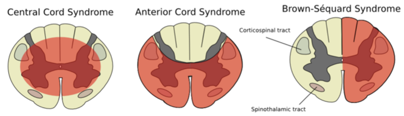
Image Source: Wikimedia Commons (2006). Cord s., Licensed under a creative commons licence. Available for free at: https://commons.wikimedia.org/wiki/File:Cord_s.svg (Accessed: 17 January 2024)
Cauda equina syndrome (CES)
CES is a neurosurgical emergency caused by canal stenosis compressing 2 or more of the 18 nerve roots making up the cauda equina. Though involving the canal, it is not a myelopathy, as it occurs below the conus medullaris. The most common causes are degenerative spinal disease, abscesses, tumours, or inflammatory conditions. It presents with symptoms which can include back or leg pain, sensory impairment, weakness, sphincter and sexual dysfunction, and saddle anaesthesia. Diagnosis is with MRI, and treatment is with urgent surgical decompression.
Motor neurone disease (MND)
MND refers to a group of diseases which can affect both upper and lower motor neurons. Extrinsic eye muscles are not affected. There are few sensory symptoms and no sensory signs. Median age of onset is 60 years.
The mainstay of therapy is supportive with early involvement of the multidisciplinary team and medications for symptom control. NIV has been shown to improve survival, while riluzole is the only medication currently licensed with prognostic benefit.
| Symptom | Management |
|---|---|
| Dyspnoea | NIV is first line. Further symptomatic relief may be offered by opioids or benzodiazepines. |
| Muscle spasms | Mexiletine or quinine sulphate are typically offered. |
| Sialorrhoea | Atropine, hyoscyamine, amitriptyline or glycopyrrolate. |
| Spasticity | Baclofen or tizanidine. |
| Emotional lability | Dextromethorphan-quinidine, amitriptyline or SSRIs. |
Amyotrophic lateral sclerosis (ALS) or Lou Gehrig’s disease
ALS makes up 50-70% of cases of MND. Onset is spinal in 75% of cases, bulbar in 20% and respiratory in 5%. The most common initial features include asymmetric limb weakness (thenar wasting in the hand with sparing of hypothenar muscles, foot drop), fasciculations, and stiffness leading to gait abnormalities.
Progressive bulbar palsy
This accounts for ~20% of cases and carries the poorest prognosis. Bulbar symptoms (presenting with dysphagia and/or dysarthria) are present, often with higher cognitive involvement.
Progressive muscular atrophy
Around 10% of cases of MND. LMN signs only, distal before proximal.
Primary lateral sclerosis
This is the rarest subtype and exhibits very slow progression. Patients have UMN signs only.
References
- Manji H. Oxford Handbook of Neurology. Oxford Medical Publications; 2020
- Eisen A. Disorders affecting the spinal cord [Internet]. Aminoff MJ (Ed). UpToDate. 2021. [Accessed 2022 Jan 12]. Available from: https://www.uptodate.com/contents/disorders-affecting-the-spinal-cord
- Means RT. Clinical manifestations and diagnosis of vitamin B12 and folate deficiency [Internet]. Fairfield KM, Takemoto CM (Ed). UpToDate. 2022 [cited 2022 Feb 14]. Available from: https://www.uptodate.com/contents/clinical-manifestations-and-diagnosis-of-vitamin-b12-and-folate-deficiency
- Magnus W, Viswanath O, Viswanathan VK, Mesfin FB. Cervical Radiculopathy [Internet]. PubMed. Treasure Island (FL): StatPearls Publishing; 2020 [cited 2022 Jan 29]. Available from: https://www.ncbi.nlm.nih.gov/books/NBK441828/
- Ameer MA, Stobart Gallagher MA, Gillis CC. Central Cord Syndrome [Internet]. PubMed. Treasure Island (FL): StatPearls Publishing; 2020. [Accessed 2022 Jan 25]. Available from: https://www.ncbi.nlm.nih.gov/books/NBK441932/
- Murahashi Y, Takebayashi T, Terashima Y, Tsuda H, Yoshimoto M, Yamashita T. Clinical Presentation of Cervical Myelopathy at C1–2 Level. Asian Spine Journal. 2016;10(4):755
- Sandoval JI, De Jesus O. Anterior Spinal Artery Syndrome [Internet]. PubMed. Treasure Island (FL): StatPearls Publishing; 2020 [cited 2022 Jan 22]. Available from: https://www.ncbi.nlm.nih.gov/books/NBK560731/
Complete Guide to Clinical Genetics ST3 Application and Recruitment (2027)
Written by Medibuddy. Last updated: October 2026. Clinical Genetics is

Complete Guide to Clinical Radiology ST3 Application and Recruitment (2027)
Recruitment paused: NHS England is not running Clinical Radiology ST3+ recru

Complete Guide to Paediatric Surgery ST3 Application and Recruitment in United Kingdom (2026)
Written by Medibuddy. Last updated: October 2026. If you’re planning


Take your subscriptions with you
Our mobile app allows you to access your interview and exam question banks wherever you are.

