


 Close
Close
 12th August 2024
Spinal cord disorders
Overview of spinal cord disorders
The diseases of the spinal cord are a diverse group that can be divided by aetiology:
| Degenerative | Degeneration of the discs or vertebra (spondylosis) leads to pain and stiffness and can cause neurological symptoms by causing compression of the cord (myelopathies), the cauda equina, or exiting nerve roots (radiculopathies). |
| Inflammatory | Spinal inflammatory diseases include transverse myelitis, neurosarcoid and paraneoplastic syndromes. |
| Infectious | Infections of the spine include epidural abscesses, viral myelitis, tabes dorsalis (tertiary syphilis), Potts’s disease (tuberculous vertebral body destruction) and schistosomiasis. |
| Vascular | Thrombotic or haemorrhagic disease of the vasculature supplying the spine is less common than intracerebrally but can be devastating when it occurs. Arterio-venous malformations may also develop in the spine causing myelo- or radiculopathic symptoms. |
| Malignant | Extramedullary, or more rarely intramedullary, tumour growth (most often metastatic) may lead to compressive symptoms, pain or structural compromise. |
| Toxic or metabolic | Includes subacute combined degeneration of the cord, radiation exposure, intrathecal chemotherapy, electrical injury, hepatic dysfunction, and decompression sickness after diving. |
| Other | Syringomyelia. Ossification of the posterior longitudinal ligament. Pannus formation in rheumatological disease. |
Terminology for spinal cord disorders
Neurological presentations of spinal disease include myelopathies (disruption of the spinal cord) and radiculopathies (disruption of an exiting nerve root). The former leads to loss of function below the level of the lesion with upper motor neuron symptoms. LMN symptoms may be seen around the level of the lesion. Radiculopathies cause symptoms confined to the dermatome/myotome supplied by that nerve. Many of the disorders in the table above can lead to either pathology, for example spinal tumours may cause radiculopathic or myelopathic symptoms depending on location. Finally, inflammation of the spinal cord is termed myelitis.
Anatomy

Image Source: Wikimedia Commons (1962). index.php?curid=30017222, Licensed under a creative commons licence. Available for free at: https://commons.wikimedia.org/w/index.php?curid=30017222 (Accessed: 17 January 2024).
Upper limb: movements, nerves and roots
| Movement | Nerve | Root(s) |
|---|---|---|
| Shoulder Abduction | Axillary | C5 |
| Elbow Flexion | Musculocutaneous | C5,6 |
| Elbow Extension | Radial | C7,8 |
| Wrist Flexion | Median, some ulnar | C6,7 |
| Wrist Extension | Radial | C7,8 |
| Finger Flexion | Median, Ulnar | C8, T1 |
| Finger Extensors | Posterior Interosseous (Radial branch) | C7,8 |
| Finger AB/ADduction | Ulnar | T1 |
Lower limb: movements, nerves and roots
| Movement | Nerve | Root(s) |
|---|---|---|
| Hip Flexion | Femoral | L1,2 |
| Hip Extension | Inferior gluteal | L5, S1 |
| Knee Flexion | Sciatic | L5, S1 |
| Knee Extension | Femoral | L3,4 |
| Ankle Dorsiflexion | Peroneal | L5, S1 |
| Ankle Plantarflexion | Tibial | S1,2 |
| Ankle Eversion | Peroneal | L5, S1 |
| Ankle Inversion | Tibial | L4,5 |
| Hallux Extension | Peroneal | L5, S1 |
Degenerative spinal disease
Degenerative spinal disease, or spondylosis, may affect the discs, facet or uncovertebral joints, ligaments or vertebral bodies. This may lead to narrowing of the foramina causing radiculopathies, or narrowing of the canal causing myelopathic symptoms from cord compression. Patients with cervical myelopathy most commonly present with clumsiness or weakness in the hands and spasticity and gait disturbance in the legs. Symptoms may worsen with neck flexion as this further narrows the canal. In the cervical spine, the most commonly affected region is C6-7 (C7 root), while in the lumbar spine, L4-5 (L5 root) and L5-S1 (S1 root) are most implicated. Definitive management is with spinal decompressive surgery.
Spinal inflammatory diseases
Transverse myelitis
This is a focal inflammation of the spinal cord, generally affecting one or two segments of the thoracic cord. The aetiology may be inflammatory (MS, NMO [neuromyelitis optica], ADEM), infectious (enteroviruses, West Nile virus, Herpes viruses, HIV, HTLV-1, Zika, Lyme, mycoplasma and syphilis) or due to connective tissue disorders (RA, SLE, Behçet’s, ankylosing spondylitis).
Neurosarcoid
Extramedullary or intramedullary granulomatous lesions can cause an acute or subacute myelopathy.
Spinal infections
Spinal epidural abscess (SEA)
Infection generally results from iatrogenic introduction (via indwelling lines or catheters or surgical procedures) or contiguously from skin or soft tissue infection. In up to two thirds of cases, Staph. aureus is the culprit. Presentation is generally with a combination of severe back pain, fevers, and neurological symptoms below and at the level of the lesion.
Viral myelitis
Multiple viruses may cause disruption of spinal cord function. A transverse myelitis-like picture may be caused by herpes viruses (EBV, CMV [cytomegalovirus], HSV, VZV) or hepatitis C; a GBS-like pathology is seen with some enteroviruses (e.g. Coxsackie, polio) or flaviviruses (e.g. West Nile or Japanese encephalitis). Poliomyelitis is a rare complication of polio infection characterised by variable degrees of acute, asymmetric, progressive, flaccid muscle weakness with decreased or absent reflexes, often with a viral meningitis-like prodrome.
Tabes dorsalis
Tabes dorsalis is one of many manifestations of tertiary syphilis, with primarily dorsal column involvement. It typically presents with lancinating nerve root pain and sensory ataxia. Though onset may be decades after the original infection, treatment is still with penicillins to clear the Treponema.
Spinal metabolic disease
Subacute combined degeneration of the cord and copper deficiency myelopathy
Vitamin B12 deficiency leads to degeneration of the dorsal and lateral tracts of the cord in SCD. Symptoms include numbness and paraesthesias, weakness, ataxia and gait disturbance. Untreated, it may progress to spasticity and paraplegia. Risk factors include dietary deficiency, pernicious anaemia, drugs (alcohol, nitrous oxide, metformin, PPIs, H2 receptor antagonists), IBD (inflammatory bowel disease), coeliac disease, and gastric/small bowel resection.
Copper deficiency myelopathy presents similarly to SCD.
Radiation myelopathy and chemotherapy
Radiation myelopathy primarily affects the lateral tracts.
Some chemotherapy agents can also induce a myelopathy, especially when given intrathecally, e.g. methotrexate.
Syringomyelia
This describes a fluid-filled cavity within the spinal cord, generally found between C2 and T9 but sometimes extending further (syringobulbia denotes extension into the brainstem). When occurring as a congenital abnormality, it is strongly associated with Chiari I malformations. Other causes include infectious, inflammatory, traumatic, malignant, and other congenital abnormalities.
Malignant spinal cord compression (MSCC)
MSCC affects up to 5% of cancer patients and is a medical emergency. Patients may present with pain alone, myelopathic or radiculopathic symptoms. Urgent imaging with MRI is essential. Patients should be initiated on high-dose dexamethasone and analgesia. Patients often receive radiotherapy as treatment and spinal surgery may be considered in some cases.
Incomplete syndromes of the spinal cord
Central cord syndrome
Commonly caused by trauma, syringomyelia or central tumours, this presents with spinothalamic tract disruption (pain/temperature/tickle/itch) with dorsal column sparing. Motor disruption may occur and generally affects the upper limbs before lower.
Anterior cord syndrome
Most often caused by anterior spinal artery thrombosis, this presents with motor and sensory loss, with dorsal column sparing (fine touch/2-point discrimination/proprioception/vibration).
Brown-Séquard syndrome
Also known as hemicord syndrome, this presents with ipsilateral dorsal column and pyramidal tract dysfunction, with contralateral spinothalamic tract loss. Causes include trauma, especially penetrating wounds, compression (tumours, spondylosis, collections) and MS.
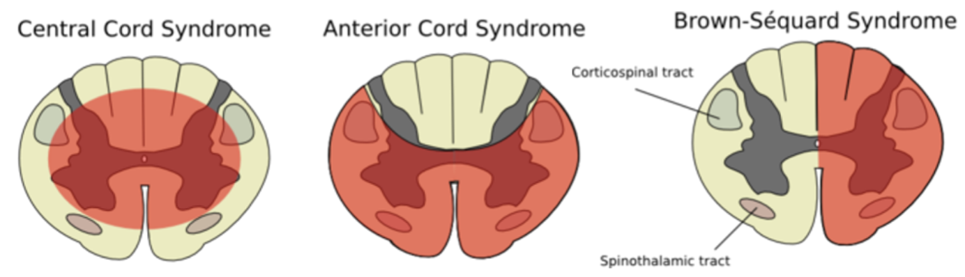
Image Source: Wikimedia Commons (2006). Cord s., Licensed under a creative commons licence. Available for free at: https://commons.wikimedia.org/wiki/File:Cord_s.svg (Accessed: 17 January 2024)
Cauda equina syndrome (CES)
CES is a neurosurgical emergency caused by canal stenosis compressing 2 or more of the 18 nerve roots making up the cauda equina. Though involving the canal, it is not a myelopathy, as it occurs below the conus medullaris. The most common causes are degenerative spinal disease, abscesses, tumours, or inflammatory conditions. It presents with symptoms which can include back or leg pain, sensory impairment, weakness, sphincter and sexual dysfunction, and saddle anaesthesia. Diagnosis is with MRI, and treatment is with urgent surgical decompression.
Motor neurone disease (MND)
MND refers to a group of diseases which can affect both upper and lower motor neurons. Extrinsic eye muscles are not affected. There are few sensory symptoms and no sensory signs. Median age of onset is 60 years.
The mainstay of therapy is supportive with early involvement of the multidisciplinary team and medications for symptom control. NIV has been shown to improve survival, while riluzole is the only medication currently licensed with prognostic benefit.
| Symptom | Management |
|---|---|
| Dyspnoea | NIV is first line. Further symptomatic relief may be offered by opioids or benzodiazepines. |
| Muscle spasms | Mexiletine or quinine sulphate are typically offered. |
| Sialorrhoea | Atropine, hyoscyamine, amitriptyline or glycopyrrolate. |
| Spasticity | Baclofen or tizanidine. |
| Emotional lability | Dextromethorphan-quinidine, amitriptyline or SSRIs. |
Amyotrophic lateral sclerosis (ALS) or Lou Gehrig’s disease
ALS makes up 50-70% of cases of MND. Onset is spinal in 75% of cases, bulbar in 20% and respiratory in 5%. The most common initial features include asymmetric limb weakness (thenar wasting in the hand with sparing of hypothenar muscles, foot drop), fasciculations, and stiffness leading to gait abnormalities.
Progressive bulbar palsy
This accounts for ~20% of cases and carries the poorest prognosis. Bulbar symptoms (presenting with dysphagia and/or dysarthria) are present, often with higher cognitive involvement.
Progressive muscular atrophy
Around 10% of cases of MND. LMN signs only, distal before proximal.
Primary lateral sclerosis
This is the rarest subtype and exhibits very slow progression. Patients have UMN signs only.
References
- Manji H. Oxford Handbook of Neurology. Oxford Medical Publications; 2020
- Eisen A. Disorders affecting the spinal cord [Internet]. Aminoff MJ (Ed). UpToDate. 2021. [Accessed 2022 Jan 12]. Available from: https://www.uptodate.com/contents/disorders-affecting-the-spinal-cord
- Means RT. Clinical manifestations and diagnosis of vitamin B12 and folate deficiency [Internet]. Fairfield KM, Takemoto CM (Ed). UpToDate. 2022 [cited 2022 Feb 14]. Available from: https://www.uptodate.com/contents/clinical-manifestations-and-diagnosis-of-vitamin-b12-and-folate-deficiency
- Magnus W, Viswanath O, Viswanathan VK, Mesfin FB. Cervical Radiculopathy [Internet]. PubMed. Treasure Island (FL): StatPearls Publishing; 2020 [cited 2022 Jan 29]. Available from: https://www.ncbi.nlm.nih.gov/books/NBK441828/
- Ameer MA, Stobart Gallagher MA, Gillis CC. Central Cord Syndrome [Internet]. PubMed. Treasure Island (FL): StatPearls Publishing; 2020. [Accessed 2022 Jan 25]. Available from: https://www.ncbi.nlm.nih.gov/books/NBK441932/
- Murahashi Y, Takebayashi T, Terashima Y, Tsuda H, Yoshimoto M, Yamashita T. Clinical Presentation of Cervical Myelopathy at C1–2 Level. Asian Spine Journal. 2016;10(4):755
- Sandoval JI, De Jesus O. Anterior Spinal Artery Syndrome [Internet]. PubMed. Treasure Island (FL): StatPearls Publishing; 2020 [cited 2022 Jan 22]. Available from: https://www.ncbi.nlm.nih.gov/books/NBK560731/
Complete Guide to Clinical Genetics ST3 Application and Recruitment (2027)
Written by Medibuddy. Last updated: October 2026. Clinical Genetics is

Complete Guide to Clinical Radiology ST3 Application and Recruitment (2027)
Recruitment paused: NHS England is not running Clinical Radiology ST3+ recru

Complete Guide to Paediatric Surgery ST3 Application and Recruitment in United Kingdom (2026)
Written by Medibuddy. Last updated: October 2026. If you’re planning


Take your subscriptions with you
Our mobile app allows you to access your interview and exam question banks wherever you are.

