


 Close
Close
 11th August 2024
Dementia and other cognitive disorders
Alzheimer’s disease
Overview of Alzheimer’s disease
Alzheimer’s disease is the most common dementia in adults of all ages. There is an initial deficit in episodic memory that progresses to include executive functioning, attention, language and visuospatial skills as the disease advances. Neuropsychiatric symptoms can be prominent, including mood disturbance, anxiety, hallucinations, agitation and restlessness. End-stage disease resulting additionally in reduced mobility, incontinence and inability to communicate can require high levels of care.
The histopathological hallmark of Alzheimer’s disease is cerebral cortical accumulation of beta amyloid (Aβ) plaques and neurofibrillary tangles.
Diagnosis of Alzheimer’s disease
Diagnosis is clinical and made from history, examination, investigations to exclude reversible causes and cognitive testing such as mini-mental state examination (MMSE) or Montreal Cognitive Assessment (MoCA) demonstrating deficits in short-term memory with reduction of category fluence (naming as many items of a category beginning a specific letter) and deficit in visuospatial tasks (copying 3D drawings/clocks) in more advanced disease.
Cognitive testing is complemented by structural correlates on MRI brain, with hallmark symmetrical medial temporal lobe and hippocampal atrophy in early disease and later inclusion of the parietal lobes.
CSF examination has a typical pattern of elevated tau protein and reduced Aβ1-42, but this is not routinely required.
Histological diagnosis is rarely acquired outside of post-mortem. Silver staining and specific tau protein immunohistochemistry demonstrate neurofibrillary tangles within neurons.
Management of Alzheimer’s disease
The mainstay of management is supportive and multidisciplinary. In early stages of disease, management should be proactive in providing education to patients and family, support for advanced care and financial planning whilst capacity is retained and facilitating everyday activities with assistance. Coordination across services such as a community dementia team, day centres, care agencies and GPs is key. With further disease progression and in advanced dementia, care may be better provided in a residential or nursing home setting with consideration of carer respite.
Pharmacological management aims to slow disease progression and prolong time before requiring 24-hour care. The main classes of drugs are anticholinesterases and NMDA receptor antagonists.
Donepezil, galantamine and rivastigmine are acetylcholinesterase inhibitors (AChEi) that are licensed for treatment of mild-to-moderate Alzheimer’s disease. Treatment should be initiated by a clinician with relevant specialist expertise in diagnosis and management of Alzheimer’s. A small but consistent benefit to cognitive function with AChEis compared to placebo has been demonstrated in meta-analyses with some evidence to suggest that withdrawal of treatment may be associated with worsening cognition. Common side effects of AChEis include gastrointestinal manifestations – diarrhoea, vomiting, nausea, reduced appetite, dyspepsia – as well as dizziness, muscle cramps and insomnia. Significant caution should be exercised in patients with bradycardia, heart block or recurrent syncope.
Memantine, an NMDA receptor antagonist, can be considered as an additional agent in individuals already on AChEis with disease progression or as a single agent in individuals with severe Alzheimer’s or those with moderate Alzheimer’s for whom AChEis is contraindicated.
The management of behavioural and psychological symptoms such as aggression or depression is less well evidenced. Conservative approaches addressing surroundings and care should be attempted before consideration of antidepressants and antipsychotics, under specialist guidance, particularly as atypical antipsychotics are associated with increased risk of stroke and mortality in this cohort.
Vascular dementia
Overview of vascular dementia
Vascular dementia is the second most common cause of dementia in older people. It is a dementia syndrome recognised as the decompensation of cognitive function resulting from the accumulation of vascular insults and can be considered on a spectrum of cognitive impairment of vascular aetiology.
The presentation is varied, and the affected domains and severity of cognitive deficits relate to the location and burden of cerebrovascular disease. Mechanisms of vascular insult include large vessel ischaemia resulting in discrete infarcts, small vessel disease of subcortical structures (basal ganglia, thalamus, brainstem), deep white matter disease (leukoaraiosis) and haemorrhage (cortical haemorrhage associated with cerebral amyloid angiopathy).
In contrast to Alzheimer’s disease, typically, executive and attention function is more affected than memory with earlier involvement of mobility and mood. Classically, a history of a stepwise progression is seen with repeated clinically significant vascular events, although sequential silent events will present with insidious cognitive decline. A mixed dementia syndrome with Alzheimer’s is common.
Diagnosis of vascular dementia
Diagnosis requires cognitive testing showing impairment in two or more domains and brain imaging demonstrating cerebrovascular disease with either history of cognitive symptoms related to vascular event(s) or cognitive impairment explained by the degree/pattern of disease seen on imaging.
Small vessel disease on MRI is seen as periventricular and subcortical white matter lesions, subcortical infarcts, and cerebral microbleeds (hypointense foci on T2-weighted sequences).
Management of vascular dementia
Antiplatelets and anticoagulation, respectively, are used as secondary prevention for arteriosclerotic and cardioembolic causes. Prevention is through modification of risk factors, which are hypertension, smoking, hypercholesterolaemia, diabetes and obesity; however, increasing age is the greatest risk factor.
Rare causes of vascular dementia
CADASIL
Cerebral autosomal dominant arteriopathy with subcortical infarcts and leukoencephalopathy (CADASIL) is caused by autosomal dominant inheritance of a mutation in the notch3 gene. There is typically a history of adult-onset migraine-like headaches and episodes of focal neurological symptoms, with an accumulation of subcortical infarcts resulting in dementia by the late forties.
Primary cerebral vasculitis
Primary cerebral vasculitis, isolated inflammation of cerebral vasculature, can cause a progressive dementia. It may present as cerebral amyloid-associated inflammation in some patients with cerebral amyloid angiopathy (CAA), presenting with headaches, seizures and progression of cognitive deficit. Neuroimaging can show microhaemorrhages, white matter changes and vasogenic oedema. Treatment is with immunosuppression.
Frontotemporal dementia
Overview of frontotemporal dementia
Frontotemporal dementia (FTD) is a syndrome that has a wide presentation that can be categorised into variants predominantly affecting behaviour or language. The peak incidence is between the ages of 45-65, and it affects both sexes equally. 40% of patients with FTD have a relevant family history.
In behavioural variants, there is a progressive history (usually verified by collateral) of deterioration in behaviour and cognition resulting in disinhibition, apathy, loss of empathy, hyperorality, perseveration and loss of executive functioning, with relative sparing of memory.
There are three language variants, termed collectively as primary progressive aphasias:
- Progressive non-fluent aphasia: impaired production of expressive speech with difficulties in repeating complex words or phrases, poor speech flow and fluency and incorrect grammatical formation of phrases with phonological substitutions for words. In early disease, recognition of objects and understanding of language is preserved.
- Semantic dementia: loss of semantic memory, i.e. naming of objects and understanding the meaning of words, but with production of language that is grammatically correct and fluent. Names of items are substituted with a generic reference such as ‘that thing’. Repetition of phrases is preserved, although comprehension can be impaired. In contrast with Alzheimer’s disease and in early stages, short-term memory for daily activities remains intact.
- Logopaenic aphasia: paucity of speech, particularly groping for words and inability to repeat phrases resulting from a deficit in working memory capacity.
Diagnosis of frontotemporal dementia
Imaging, either MRI or PET, shows frontal and/or anterior temporal atrophy or hypoperfusion, respectively. CSF examination excludes causes such as infection and inflammation, and Alzheimer’s disease.
Lewy body dementia
Overview of Lewy body dementia
Lewy body dementia (LBD) is conventionally understood to be on the same spectrum as Parkinson’s disease dementia (PDD), but nomenclature is dependent on whether cognitive impairment precedes motor symptoms of Parkinsonism (defined as LBD) or vice versa, i.e. PDD. It often co-exists with Alzheimer’s and vascular dementia.
The microscopic histopathological hallmark of LBD is intracellular neural Lewy bodies (formed of α-synuclein) distributed in three typical sites (cortex, brainstem and limbic areas). These stain with haematoxylin-eosin and α-synuclein histochemistry. Macroscopically, there is pallor of the substantia nigra and locus coeruleus and diffuse cerebral atrophy.
The cognitive presentation is similar to Alzheimer’s, however there is predominance of problems affecting executive, visuospatial and attentional functions with relatively less involvement of memory.
The key distinguishing features are:
- Fluctuations in cognition (attention and alertness)
- Visual hallucinations in up to 60% of patients – typically formed people or animals and usually not distressing (compared to psychiatric hallucinations); alternatively a sense of a ‘presence’
- Parkinsonism – eventually seen in most cases, depending on LBD/PDD, may predate or follow cognitive symptoms. Bradykinesia, rigidity, gait impairment and autonomic dysfunction
Other common features include:
- Rapid eye movement (REM) sleep behavioural disturbance
- Daytime somnolence
- Autonomic dysfunction (falls, syncope, postural hypotension, constipation, urinary urge, frequency and incontinence)
- Mood disturbance, including anxiety and depression
Diagnosis of Lewy body dementia
Although brain imaging is recommended at least once in dementia work up, there are no specific structural imaging features of LBD. Single-photon emission CT (SPECT) or PET can help differentiate between LBD and Alzheimer’s and will show reduced uptake of dopamine transporter uptake in the basal ganglia.
CSF examination is usually carried out in atypical presentations to exclude infection.
Referral to a sleep specialist and for polysomnography is advised as REM sleep without atonia is a suggestive feature of LBD.
Management of Lewy body dementia
Management is targeted at symptoms and supportive measures. Given the underlying deficits are of choline and dopamine neurotransmission, priority review of medications is important to avoid anticholinergics and dopamine agonists to reduce exacerbation of autonomic, motor and psychiatric symptoms. First-generation antipsychotics (haloperidol) should be avoided and patients tend to be very sensitive to atypical antipsychotics.
First-line treatments are anticholinesterase treatments such as donepezil and rivastigmine, which act to improve cognition and behavioural symptoms without significant worsening of motor symptoms, however side effects may include nausea and vomiting, runny nose, muscle cramps, hypersalivation, vivid dreams and postural hypotension.
Clonazepam in low doses is effective for REM sleep behaviour disturbance.
SSRIs are choice antidepressants, avoiding tricyclics that have anticholinergic effects.
Levodopa can be considered in specialist use if motor symptoms are significantly problematic.
Human prion disease – Creutzfeldt-Jakob disease
Creutzfeldt-Jakob disease (CJD) is a rare and inevitably fatal cause of progressive dementia with neurological symptoms. Intracellular human prion proteins are misfolded from an alpha-helical structure into a beta-pleated form, which in turn causes adjacent proteins to undergo this transformation. These ‘infectious proteins’ drive the pathophysiology of neuronal injury and eventual cell death as a result of accumulation of abnormal prion proteins in the CNS.
There are three main types of CJD:
- Sporadic (sCJD) – accounts for the majority of cases, up to 90% of CJD
- Acquired – include iatrogenic CJD (iCJD) (transmission of prion protein from contaminated surgical equipment or cadaveric tissue grafts) and variant CJD (vCJD) (ingestion of CJD-contaminated beef or blood transfusion)
- Familial – mutations in the gene coding for PRNP (endogenous prion protein) on chromosome 20; usually autosomal dominant with 100% penetrance, however, clinical presentation can be variable and less aggressive than sCJD and vCJD
| sCJD | Familial | vCJD | iCJD | |
|---|---|---|---|---|
| Onset | 60s | Younger onset than sCJD | Young adults – teenagers, 20s | Variable incubation period – also related to timing of inoculation event |
| Features
(in order of presentation) |
Cognitive symptoms – memory loss, aphasia and impaired executive functioning with progressive dementia
Ataxia Rigidity Limb myoclonus Familial can have a variable rate of progression, although it can present almost identically to sCJD |
Psychiatric symptoms – often severe depression
Dementia (initially mild cognitive impairment) Ataxia Painful sensory symptoms Involuntary movements – dystonia, chorea, myoclonus |
For neurosurgical inoculation – similar presentation to sCJD | |
Diagnosis of CJD
CSF examination – can be bland with mildly raised protein. Elevated 14-3-3 protein has high sensitivity and specificity for sCJD but is not useful for vCJD; real-time quaking-induced conversion for abnormal prions has high sensitivity and specificity.
EEG – periodic triphasic spikes
Imaging – MRI brain with FLAIR (fluid attenuated inversion recovery) and diffusion-weighted imaging (DWI) best demonstrates affected areas:
- sCJD – hyperintensities in the basal ganglia (caudate and putamen) and ‘cortical ribboning’ on DWI
- vCJD – bilateral periventricular thalamic hyperintensities, ‘hockey stick sign’
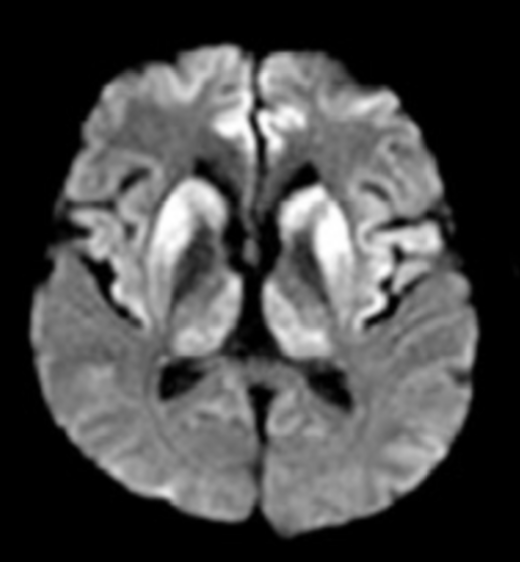
Image Source: ncbi.nlm.nih.gov 2025, PMC5619326, Licensed under a creative commons licence. Available for free at: https://www.ncbi.nlm.nih.gov/pmc/articles/PMC5619326/pdf/dn-09-04-0424.pdf (Accessed: 17 July 2024)
![MRCP Part 2 - [L] Axial T2 FLAIR MRI showing typical bilateral basal ganglia hyperintensity in sCJD [R] DWI MRI showing ‘cortical ribboning’ in sCJD](https://medibuddy.co.uk/wp-content/uploads/2024/08/Screenshot-at-Aug-08-17-55-24.png)
Image Source: ncbi.nlm.nih.gov 2025, PMC5619326, Licensed under a creative commons licence. Available for free at: https://www.ncbi.nlm.nih.gov/pmc/articles/PMC5619326/pdf/dn-09-04-0424.pdf (Accessed: 17 July 2024)
Normal pressure hydrocephalus
Normal pressure hydrocephalus (NPH) was historically described as the triad of gait disturbance, urinary incontinence and cognitive slowing as a result of hydrocephalus without raised cerebrospinal fluid pressure. The aide-memoire “wobbly, wet and wacky” may have come up in medical school! Clinical features typically develop gradually over years.
The reported incidence of NPH is variable as there are different diagnostic criteria. The pathophysiology is not well understood, but the underlying processes are thought to drive formation of a communicating hydrocephalus with resultant ischaemic insult to subcortical and periventricular structures including the basal ganglia.
Risk factors are predominantly age >65 and vascular, thought to be related to effects on compliance of cerebral blood vessels that, in turn, regulate the flow of CSF.
Diagnosis and management of NPH
Gait disturbance is the key and usually first symptom noticed and is typically small stepped, slow, hesitant, wide based and magnetic (‘stuck to the floor’), with difficulties in making 180 degree turns in 3 or fewer steps.
Lumbar puncture has both a diagnostic and therapeutic role – specifically, pressure measured by manometry should be normal. Large volume CSF removal (40-50 ml) can be done with cognitive testing and gait assessment pre- and post-therapeutic lumbar puncture; cases that respond to this may represent candidates for further lumbar drainage, although identifying this group of individuals can be challenging.
A trial of levodopa should not yield gait improvement (excluding Parkinson’s disease) and brain imaging can demonstrate ventriculomegaly which is disproportionate to sulcal atrophy, distinguishing it from global cerebral atrophy. There may also be evidence of periventricular white matter change, although imaging can also be normal. A space-occupying lesion with obstruction would exclude the diagnosis of NPH.
Movement disorders and dementia
Parkinson’s plus syndromes (including progressive supranuclear palsy and corticobasal degeneration) and Huntington’s disease that cause dementia as part of advanced disease are covered more thoroughly in the movements disorder section. These dementias typically present as frontotemporal or subcortical types.
Mimics of dementia
Depression (pseudodementia)
Cognitive deficits are common in psychiatric disorders, as is the coexistence of depression with dementia. It can be difficult to explicitly distinguish the degree of contribution of each factor, and occasionally, a trial of an antidepressant and/or referral for further psychiatric testing is considered. Typically, individuals present with distractibility, poor concentration and short term memory in the context of low mood (although not always experienced) and may also include biological symptoms of depression such as poor sleep and loss of libido and appetite. Cognitive screening tests often show reduced engagement with testing and inconsistent results with impaired attention and variably poor performance in memory and behavioural domains.
Delirium
Delirium is an acute state of rapidly fluctuating consciousness impacting orientation and attention, that is usually reversible (to a degree), precipitated by a homeostatic or metabolic disturbance. The diagnosis of dementia must exclude cognitive impairment that is exclusively in the context of a delirium. Management is driven by identification and treatment of the precipitant with supportive care to allow for delirium to resolve.
Hypothyroidism
Severe hypothyroidism can cause symptoms of apathy, slowing of cognition and speech, overlapping with the symptoms of dementia and depression.
Mild cognitive impairment
Mild cognitive impairment (MCI) is an acquired cognitive decline beyond that expected for age or level of education, however, which does not cause significant functional impairment. It is a differential to consider if dementia is suspected but the criteria are not met, or if it is of low severity. Mild cognitive impairment can be divided into amnestic (affecting memory) or non-amnestic (affecting cognitive domains such as language, executive function or visuospatial skills, but not memory). There is an increased incidence of progression of amnestic MCI to Alzheimer’s disease compared to the general population.
Encephalitis and infectious causes of dementia
HIV
Progressive cognitive impairment in HIV-infected individuals requires evaluation to exclude systemic illness or opportunistic infections of the central nervous system (and their long-term sequelae). Key factors to consider are an individual’s CD4 count and duration of/concordance with antiretrovirals. HIV-associated neurocognitive disorder (HAND) represents a broad spectrum of cognitive impairment from asymptomatic/mild cognitive impairment to HIV-associated dementia that meets criteria of affecting at least two cognitive domains with significant effect on activities of daily living.
Opportunistic cerebral infections in HIV are prevalent when the CD4 count is <100 cells/microlitre. Toxoplasma, herpes simplex and varicella zoster encephalitis, cryptococcal or tuberculous meningitis, progressive multifocal leukoencephalopathy secondary to JC (John Cunningham) virus infection and EBV infection with primary CNS lymphoma.
There can also be cognitive and neuropsychiatric side effects to certain antiretrovirals (commonly efavirenz in early treatment), however immune reconstitution inflammatory syndrome (IRIS) as a cause of progressive cognitive impairment in the context of recent initiation on antiretrovirals should be considered.
HIV-associated dementia (HAD) presents with logopaenic speech, slow and poor thought processes, with flat or labile affect in the absence of sadness. There can often be a degree of overlap with psychiatric morbidity. There are no specific neuroimaging changes seen in HAD and neurological examination can show gait disturbance, frontal release and pyramidal signs.
Neurosyphilis
Infection with syphilis that has spread to the meninges and cranial nerves causes the neurological signs of absent reflexes, Argyll-Robertson pupils, ataxia, peripheral motor/sensory deficits and associated cognitive impairment.
Nutritional causes of dementia
Wernicke’s encephalopathy
Typically presents as the triad of a complex ophthalmoplegia, delirium and ataxia due to severe B1 deficiency, most commonly seen in alcoholics. Needs urgent treatment with Vitamin B complex replacement to prevent permanent cognitive impairment.
B12 deficiency
Other features, in addition to cognitive impairment, include macrocytic anaemia, neurological signs (subacute combined degeneration of the cord) such as ataxia, and reduced proprioceptive and vibration sense in the lower limbs.
B3 deficiency – pellagra
Pellagra is a systemic disease best remembered using the 3 Ds: dermatitis, diarrhoea and dementia. It is caused by deficiency in niacin, most commonly due to diet in developing countries. It usually presents initially with characteristic skin changes and non-specific abdominal symptoms. Neuropsychiatric involvement with cognitive impairment is a late sign in untreated disease.
References
- National Institute for Health and Care Excellence (NICE). Donepezil, galantamine, rivastigmine and memantine for the treatment of Alzheimer’s disease. 2018 [cited 17 April 2023]. Available from: https://www.nice.org.uk/guidance/ta217/resources/donepezil-galantamine-rivastigmine-and-memantine-for-the-treatment-of-alzheimers-disease-pdf-82600254699973
- Firth, John, Christopher Conlon, and Timothy Cox (eds), Oxford Textbook of Medicine, 6 edn (Oxford, 2020; online edn, Oxford Academic, 1 Jan. 2020) Schott, J. Chapter 24.4.2, 24.11.5. [cited 16 March 2023]. Available from: https://doi.org/10.1093/med/9780198746690.001.0001
- Neugroschl J, Matthews B, Bhutto A, Gammack J. Alzheimer’s disease. BMJ Best Practice. Feb 2023. [cited 29 March 2023]. Available from: https://bestpractice.bmj.com/topics/en-gb/317
- Passmore P, Wilson D, Bateman G, Srikanth V. Vascular dementia. BMJ Best Practice. Nov 2022. [cited 29 March 2023]. Available from: https://bestpractice.bmj.com/topics/en-gb/319
- Lerner A, Elkasaby M, Appleby B, Gore E, Shrestha R, Wuerz T, Schneider J, Fahmy V, Banerjee S. Dementia with Lewy bodies. BMJ Best Practice. Jun 2023. [cited 29 March 2023] Available from: https://bestpractice.bmj.com/topics/en-gb/320
- Radhakrishnan R, Jauhari S, Whalley L, Onyike C. Frontotemporal dementia. BMJ Best Practice. Nov 2022. [cited 29 March 2023]. Available from: https://bestpractice.bmj.com/topics/en-gb/968
- Sim V, Geschwind M, Kuo A, Finley R. Prion disease. BMJ Best Practice. Feb 2020. [cited 29 March 2023]. Available from: https://bestpractice.bmj.com/topics/en-gb/484
- Grünewald R, Rowe J. Normal pressure hydrocephalus. BMJ Best Practice. Oct 2022. [cited 29 March 2023]. Available from: https://bestpractice.bmj.com/topics/en-gb/712
- Brouillette M-J, Hull M, Harris M, Repetto M. Assessment of HIV-related mental status changes. BMJ Best Practice. 11 Jul 2023. [cited 29 March 2023]. Available from: https://bestpractice.bmj.com/topics/en-gb/900
- Mead S, Rudge P. CJD mimics and chameleons. Pract Neurol 2017;17:113–121. [cited 13 April 2023]. Available from: https://www.ncbi.nlm.nih.gov/pmc/articles/PMC5520355
- V Ngan, Pellagra. DermnetNZ website. [cited 14 Jan 2024] Available from: https://dermnetnz.org/topics/pellagra
Complete Guide to Clinical Genetics ST3 Application and Recruitment (2027)
Written by Medibuddy. Last updated: October 2026. Clinical Genetics is

Complete Guide to Clinical Radiology ST3 Application and Recruitment (2027)
Recruitment paused: NHS England is not running Clinical Radiology ST3+ recru

Complete Guide to Paediatric Surgery ST3 Application and Recruitment in United Kingdom (2026)
Written by Medibuddy. Last updated: October 2026. If you’re planning


Take your subscriptions with you
Our mobile app allows you to access your interview and exam question banks wherever you are.

