


 Close
Close
 29th December 2024
Oncology Emergencies
Malignant hypercalcaemia
Overview of malignant hypercalcaemia
Malignant hypercalcaemia occurs in approximately 1 in 5 cancer patients and of all inpatients admitted with hypercalcaemia, cancer represents the most common cause.
Hypercalcaemia can be life threatening, resulting in progressive hypovolaemia, renal impairment, cognitive impairment and arrhythmia. It is, therefore, essential to detect and treat hypercalcaemia early, especially if free/adjusted Ca ≥ 3.0 mmol/L.
Note – a proportion of serum calcium is bound to albumin, and low serum albumin levels may result in a falsely low total calcium reading. Adjusted calcium (based on albumin) must be calculated to estimate free calcium levels.
Causes of malignant hypercalcaemia
Parathyroid hormone-related protein (PTHrP)
PTHrP production by cancer cells accounts for 80% of malignant hypercalcaemia. This is most commonly associated with squamous cell cancers (e.g. squamous cell lung cancer), renal, bladder, breast and ovarian cancers. The PTHrP stimulates osteoblasts to produce RANK-ligand, which then activates osteoclasts to resorb bone, resulting in hypercalcaemia. Of note, this ectopic production means that malignant hypercalcaemia may be present even in the absence of bony metastases.
Osteolytic bone metastases
Osteolytic bone metastases, commonly found in myeloma and breast cancer, account for 20% of malignant hypercalcaemia.
Other – calcitriol and PTH
Other causes of malignant hypercalcaemia make up less than 1% of cancer-related calcium release.
Ectopic 1,25-dihydroxyvitamin D (calcitriol) can be produced in ovarian and germ cell tumours that release 1-alpha hydroxylase or in lymphoma cells. Excess calcitriol release in either case will stimulate bone resorption.
PTH (parathyroid hormone) itself may be produced by tumours, in the case of parathyroid carcinoma, or through ectopic release by small cell lung cancers, lung adenocarcinomas and renal cell cancer.
Signs and symptoms of hypercalcaemia
Hypercalcaemia may present acutely, subacutely or may be asymptomatic and an incidental finding. High calcium levels can impact many organ systems and, if left untreated, can be fatal.
| Endocrine and renal | Polydipsia and polyuria – can lead to dehydration, hypovolaemia, and AKI. Untreated, AKI can lead to uraemia (itching, confusion).
High calcium can result in renal stones, causing renal colic and haematuria. Increased osteoclast activity can result in bone pain and aches. |
| Gastrointestinal | Constipation, abdominal pain, nausea, vomiting and loss of appetite.
Occasionally, it can contribute to pancreatitis and peptic ulcers. |
| Neurological and neuropsychiatric | Fatigue, malaise and generalised weakness.
Confusion, cognitive impairment and depression. Left untreated, it can lead to reduced GCS and coma. |
| Cardiac | Hypercalcaemia can lead to postural hypotension.
ECG changes can include shortened QT interval, J waves and, in severe cases, changes that mimic ST elevation. Left untreated, hypercalcaemia can cause fatal arrhythmias including VF (ventricular fibrillation) arrest. |
Investigations in malignant hypercalcaemia
A number of investigations should be carried out when investigating malignant hypercalcaemia:
- Routine blood tests for serum calcium, albumin, adjusted calcium, renal function
- Bone profile including PO4, PTH, 25-hydroxyvitamin D, calcitriol
- If PTH is normal or low in case of known malignancy, PTHrP. In PTHrP-producing tumours, PTHrP will be high, PTH suppressed, and PO4 low due to hyperphosphaturia
- In calcitriol-mediated hypercalcaemia, calcitriol will be elevated
- In cases of ectopic PTH production, PTH will be high
- ECG – may show shortened QT interval, J waves, or other abnormalities
- Bone scan or skeletal survey in patients with known or suspected osteolytic metastases, e.g. myeloma, known bone metastases, leukaemia
- Chest X-ray to look for lung cancer or other causes of hypercalcaemia including sarcoidosis or TB
Management of malignant hypercalcaemia
Emergency management is required if calcium ≥3.0 mmol/L (Table 2).
If appropriate, the underlying malignancy should be treated. In many cases, hypercalcaemia is related to already advanced cancer. In haematological malignancies including myeloma, steroids may be of benefit.
| First 72 hours |
|---|
| 1. Aggressive IV rehydration with normal saline, aiming 3-6 L in the first 24 h, guided by cardiac function and urine output. This improves hydration status, circulating volume and GFR (glomerular filtration rate) and increases urinary calcium excretion. |
| 2. Loop diuretics (e.g. furosemide) can be used alongside rehydration to promote calciuria and prevent overload. |
| 3. Calcium, albumin, renal function including potassium and magnesium should be checked daily. Potassium and magnesium may fall with improved hydration and may require IV replacement. |
| Ongoing management |
| 4. If calcium remains high at 24-72 h, IV bisphosphonates may be used (e.g. zoledronic acid 4 mg over 15 minutes or IV pamidronate over 60-90 minutes if renal function is poor) to inhibit osteoclast activity. First, check and optimise vitamin D levels to reduce the risk of rebound hypocalcaemia and osteonecrosis of the jaw. |
| 5. Rarely, IM calcitonin is used. Serum calcium is repeated at 4-6 hours – if there is a drop in calcium then the patient is calcitonin sensitive, and doses can be repeated 12 hourly for up to 48 hours. |
| 6. Avoid medications that may result in hypercalcaemia – calcium supplements, thiazide diuretics, lithium. |
Bisphosphonates are used if calcium remains high despite adequate IV rehydration. An important side effect of bisphosphonates is rebound hypocalcaemia, and it is therefore important to monitor calcium levels after administration and monitor for any clinical features of hypocalcaemia. Signs and symptoms include paraesthesia, muscle spasms or cramps, tetany and seizures. Eponymous signs include Trousseau’s sign – when inflating a blood pressure cuff to greater than systolic blood pressure, there is an involuntary carpopedal spasm of the ipsilateral wrist and hand and the Chvostek sign – when tapping in front of the ear overlying the course of the facial nerve, ipsilateral facial muscles will twitch.
References
- Cassidy J, Bissett D, Spence RAJ, Payne M, Morris-Stiff GJ. Oxford Handbook of Oncology. 4th ed. Oxford: Oxford University Press; 2015
- Shieh A, Martinez D. Hypercalcaemia of malignancy [Internet]. BMJ Best Practice. 2021 [cited 2022 Apr 17]. Available from: https://bestpractice.bmj.com/topics/en-gb/1204
- Mirrakhimov AE. Hypercalcemia of malignancy: An update on pathogenesis and management. North American Journal of Medical Sciences. 2015;7(11):483
- Shane E, Berenson JR. Treatment of hypercalcemia [Internet]. Rosen CJ, Mulder, MD JE, editors. UpToDate. 2022 [cited 2022 Apr 17]. Available from: https://www.uptodate.com/contents/treatment-of-hypercalcemia
Tumour lysis syndrome (TLS)
Overview of TLS
TLS is a syndrome characterised by biochemical and metabolic derangement driven by the rapid breakdown of tumour cells, causing the release of intracellular contents into the circulatory system. It is typically triggered by delivery of chemotherapy, however may also occur after any systemic anti-cancer treatment (SACT), after radiotherapy in radio-sensitive tumours, and even spontaneously in patients with a large tumour burden.
This is an emergency presentation, as the resultant renal impairment and electrolyte abnormalities can limit treatment options and can even result in death. It is crucial to identify TLS early, so that emergency treatment can be initiated, and also to identify patients at risk who may need prophylactic treatment and close monitoring.
Definition of TLS
TLS must be diagnosed through biochemical tests. It can be classified as laboratory or clinical TLS:
| Laboratory TLS | Clinical TLS |
|---|---|
Two or more of:
|
Laboratory TLS AND one or more of:
|
Risk factors for TLS
Risk stratifying allows the treating team to choose which patients to monitor closely or to offer prophylactic therapy.
Tumour type risk factors
TLS is generally associated with highly proliferative, chemosensitive cancers with a high burden of disease. It typically occurs in haematological malignancies, including ALL, AML, non-Hodgkin’s and Burkitt’s lymphoma. However, it can also occur in any solid tumour that has these risk factors, for example small cell lung cancer, breast cancer, renal cell cancer and metastatic testicular or germ cell cancers.
As options for SACT become more sophisticated and effective, the rates of TLS are likely to rise.
Patient risk factors
Patients with baseline biochemical abnormalities including high urate levels and chronic kidney disease are at higher risk of TLS. For the latter reason, the risk of TLS increases with age (thought to be due to worsening GFR). Exposure to nephrotoxic drugs may also increase the risk of TLS. Poor hydration status is also a risk factor, and some patients may require IV rehydration prior to commencing SACT.
Presentation of TLS
| Laboratory TLS | ||
|---|---|---|
| Hyperuricaemia | Nucleic acids within rapidly dividing cancer cells are metabolised to insoluble uric acid | Deposition within renal tubules results in uric acid nephropathy (oliguric AKI) |
| Hyperphosphataemia | Cancer cells have higher PO4 levels than normal tissues. Intracellular phosphate is released into bloodstream. Can precipitate with calcium and deposit as calcium phosphate in tissues | Deposits in renal tubules cause AKI
Deposits in skin can cause gangrene Deposits in the heart can cause arrhythmias |
| Hyperkalaemia | Intracellular potassium is released by cell breakdown, and worsened in concurrent TLS-driven AKI | Most dangerous electrolyte abnormality; can cause cardiac arrhythmias and sudden cardiac death |
| Hypocalcaemia (and hypomagnesaemia) | Calcium precipitates with phosphate as calcium phosphate and is depleted | Neuromuscular irritability; paraesthesias, muscle twitching, tetany, Trousseau’s sign, Chvostek’s sign, laryngospasm, bronchospasm
Hypotension Cardiac failure |
| AKI/renal failure | May be due to precipitation of uric acid crystals or calcium phosphate within renal tubules | Creatinine rise, oliguria, symptoms of uraemia (itch, nausea, vomiting, confusion, coma) |
| Metabolic acidosis | Related to declining renal function; acidosis then promotes uric acid crystallisation and can, in turn, worsen AKI | Nausea, vomiting, fatigue
Raised respiratory rate |
Management of TLS
TLS prophylaxis
When planning chemotherapy, it is important to consider strategies to prevent TLS in high risk patients.
All patients receiving chemotherapy should be monitored for biochemical and clinical features of TLS. Where possible, nephrotoxic drugs (such as NSAIDs and aminoglycosides) should be avoided, as well as drugs such as thiazide diuretics that increase serum uric acid levels.
In patients at high risk of TLS, there are a number of steps that may prevent TLS from developing.
- IV hydration prior to treatment.
- Phosphate binder to reduce serum phosphate levels, e.g. aluminium hydroxide, which binds and reduces bowel absorption of phosphate. Magnesium hydroxide is given to reduce the side effects of constipation.
- Reduce serum uric acid levels:
- Lower risk: allopurinol, a xanthine oxidase inhibitor, which reduces conversion of purine to uric acid. Will not reduce uric acid that has already been released by cells.
- Higher risk: rasburicase, a recombinant urate oxidase enzyme, which converts uric acid into water-soluble allantoin, which prevents crystallisation and is more easily removed by the kidney. Contraindicated in patients with G6PD deficiency, haemolytic anaemia, methaemoglobinaemia.
Management of acute TLS
Acute TLS is an emergency and requires urgent action and close monitoring. Emergency management is as follows:
1. Urgent correction of hyperkalaemia
|
| 2. Close fluid balance monitoring, with IV rehydration if hypovolaemic |
| 3. Close biochemical monitoring with twice daily bloods for renal function and urate |
| 4. If symptoms of hypocalcaemia, supplement calcium |
| 5. Some centres consider urinary alkalinisation (acetazolamide or sodium bicarbonate) |
| 6. Consider escalation to HDU (high dependency unit) or critical care for cardiac monitoring |
| 7. Consider need for haemodialysis to remove serum uric acid |
References
- Cassidy J, Bissett D, Spence RAJ, Payne M, Morris-Stiff GJ. Oxford Handbook of Oncology. 4th ed. Oxford: Oxford University Press; 2015
- Mughal T. Tumour lysis syndrome [Internet]. BMJ Best Practice. 2022 [cited 2022 Apr 17]. Available from: https://bestpractice.bmj.com/topics/en-gb/936
- Howard SC, Jones DP, Pui C-H. The Tumor Lysis Syndrome. The New England journal of medicine [Internet]. 2011 May 12;364(19):1844–54. Available from: https://www.ncbi.nlm.nih.gov/pmc/articles/PMC3437249/
Neutropenic sepsis
Overview of neutropenic sepsis
Chemotherapeutic drugs generally work by interrupting cell replication pathways. This can be highly effective as an anti-cancer therapy, however can lead to side effects in normal tissues with high replication rates. Bone marrow suppression is one such side effect.
Any cell line within the marrow may be suppressed; typically bone marrow failure secondary to cytotoxic drugs results in pancytopenia. Typically, neutropenic nadir occurs first, 7 to 14 days post chemotherapy (Table 6).
Neutropenia leads to significant risk of infection and sepsis and can be life threatening. It is essential to consider febrile neutropenia and initiate antibiotic therapy readily in patients receiving chemotherapy who present with fever.
| Days 7-10 | Days 10-14 | Days 14-21 |
|---|---|---|
| Leukopenia and neutropenia | Thrombocytopenia | Anaemia |
Definitions in neutropenic sepsis
| Febrile neutropenia | Neutropenic sepsis |
|---|---|
|
|
Risk factors for neutropenic sepsis
There is an increased risk of neutropenic sepsis in:
- Haematological malignancies
- High-dose/intensive chemotherapy regimes
- Receiving first cycle of chemotherapy or low first-cycle neutrophil nadir < 0.5 x 10⁹/L
- Previous neutropenic fever
- Long duration of, or severe neutropenia
- Performance status > 1
- Older age (> 65 years old )
- Comorbidities including diabetes mellitus, liver disease and renal disease
- Hypoalbuminaemia with (albumin < 35 g/dL)
- Pre-treatment anaemia (pre-treatment haemoglobin < 120 g/L)
Investigations and management of neutropenic sepsis
Investigation and management of patients with febrile neutropenia and neutropenic sepsis is broadly the same. Septic patients will require earlier close monitoring and early escalation to critical care or high dependency units.
Investigations in neutropenic sepsis
Admission investigations include:
- Blood gas for lactate
- Blood tests including FBC and differential, renal and liver function
- Septic screen, comprising blood cultures (peripheral and from lines), urine/sputum/stool/wound samples as appropriate, and chest X-ray
Emergency management of neutropenic sepsis
- Empirical broad-spectrum antibiotics (e.g. piperacillin-tazobactam) should be initiated in febrile patients receiving chemotherapy within 60 minutes of presentation to hospital, i.e. prior to results of FBC. Taking cultures should not delay delivery of antibiotics
- IV fluids are given if clinically hypovolaemic
- Granulocyte colony stimulating factor (G-CSF) can be started to support neutrophil count if neutropenia is confirmed. This should be discontinued after neutrophil count improves to > 1.0.
- Close monitoring and early escalation is essential, including sepsis management and early discussion with critical care teams
Ongoing management of neutropenic sepsis
- Monitor cell counts and support as necessary. Generally, platelet transfusions may be considered in discussion with the haematology team if platelet count is either < 10 x 10⁹/L, or < 20 x 10⁹/L and bleeding or septic. Alternative causes that may contribute to pancytopenia in septic patients should be considered, including disseminated intravascular coagulation.
- Consider escalation or rationalisation at 48 hours:
- With ongoing temperature spikes or deterioration, antibiotic therapy may be escalated.
- If cultures identify a culprit organism, antibiotics should be rationalised in discussion with microbiology. If line cultures are positive, line removal may be required.
- Of note, causative organisms are not identified in 2/3 cases, in which case a course of empirical antibiotics can be completed, depending on suspected source.
- Consider opportunistic infections at 4-6 days:
- Failure of fever to resolve after 5 days should prompt repeat cultures and consideration of an opportunistic infection caused by fungal or parasitic organisms, e.g. Pneumocystis carinii.
- These patients may require further imaging with CT scans and escalation of antimicrobial therapy.
- The patient’s oncology team should be informed, and further chemotherapy is generally delayed until the patient has recovered from the infection. Future chemotherapy can be managed with dose reductions, changing chemotherapy agent or regime, or prophylactic administration of G-CSF during treatment cycles.
| 1. Initiate broad-spectrum antibiotics – do not wait for blood results |
| 2. Full septic screen – ideally take cultures prior to antibiotic therapy, but should not delay antibiotics |
| 3. Start G-CSF if neutropenia is confirmed |
| 4. If septic: IV fluids, urinary catheter, monitor closely, discuss early with critical care team |
| 5. At 48 hours – rationalise antibiotics with results or re-culture and escalate antibiotics if ongoing fevers |
References
- Cassidy J, Bissett D, Spence RAJ, Payne M, Morris-Stiff GJ. Oxford Handbook of Oncology. 4th ed. Oxford: Oxford University Press; 2015
- Rolston KVI, Nesher L. Febrile neutropenia [Internet]. BMJ Best Practice. 2019 [cited 2022 Apr 17]. Available from: https://bestpractice.bmj.com/topics/en-gb/950
- National Institute for Health and Care Excellence. Neutropenic sepsis [Internet]. NICE. 2022 [cited 2022 Apr 17]. Available from: https://cks.nice.org.uk/topics/neutropenic-sepsis/
Malignant spinal cord compression (MSCC)
Overview of MSCC
Malignant spinal cord compression arises when a tumour infiltrates the spinal canal and compresses the spinal cord. It is a medical emergency that requires urgent initiation of treatment to prevent progressive neurological compromise. You should, therefore, have a high index of suspicion for MSCC in a patient with a history of cancer who presents with back pain and/or neurological signs or symptoms.
Pathophysiology of MSCC
MSCC tends to arise in tumour types that spread to or originate from bone (Table 9). It may be the first presenting feature of malignancy, particularly in tumour types that readily metastasise to bone, e.g. prostate, breast, myeloma.
Most commonly, MSCC is caused by tumour soft tissue extension into the canal space or by vertebral fractures and/or bone propulsion into the canal. Less commonly, the spinal cord may be compressed by other routes, such as progressive lymphadenopathy or extension from extradural (non bone-related) masses.
| Tumour types that metastasise to bone | Tumour types that arise from bone | |
|---|---|---|
| Commonly | Breast
Lung Prostate |
Myeloma
Lymphoma |
| Less commonly | Thyroid
Kidney Bladder Bowel Melanoma |
Primary bone tumours |
Presentation of MSCC
Pain
Back pain may precede neurological symptoms and may be constant and severe. This may be acute or subacute. In patients with known bony metastases with back pain, any sudden change in back pain should prompt investigation.
Neurological symptoms
The patient may notice any degree of weakness, sensory loss or paraesthesia in their arms (if cervical lesion) or legs (if lesion is thoracic or lower). They may describe new faecal incontinence with or without saddle anaesthesia. They may also have symptoms of urinary retention – difficulty passing urine, suprapubic pain, dribbling and overflow incontinence. These neurological symptoms are usually late signs of cord compromise.
Examination findings
In all patients presenting with suspected MSCC, it is essential to perform a full neurological examination, including a bladder scan to assess for urinary retention and a digital rectal examination to assess anal tone. Regular neurological examinations should be performed thereafter to monitor for any neurological deterioration.
On examination, there may be point tenderness on palpation of a vertebra overlying the compressive lesion.
There may be a pattern of complete or incomplete spinal cord neurological compromise. If complete, there will be a sensory level just below the level of compression, and below this there will be complete sensory loss and an upper motor neuron pattern of weakness. This would also be accompanied by bowel and bladder dysfunction.
The spinal cord ends at L1-2, and lesions below this may compress the cauda equina. This would characteristically result in sciatic pain, sexual dysfunction, urinary retention, saddle anaesthesia and loss of anal tone.
Investigation of MSCC
The gold standard investigation is an MRI of the whole spine, which should be performed within 24 hours of presentation. Even where there are localising signs, the whole spine should be imaged as there may be other sites of cord compression (or impending compression) above or below the symptomatic lesion.
In 15-20% of cases there may be no abnormalities on a plain X-ray, and this should not be used to rule out MSCC.
In addition, CT imaging of the spine may be considered, to guide decisions on bony stability and surgical options.
In cases where MRI is contraindicated or unavailable, CT may be considered with or without concurrent myelography. This should be performed only in specialist centres, as myelography may cause neurological deterioration, requiring urgent decompression.
Management of MSCC
Swift action is required when MSCC is suspected, to prevent further neurological decline. Treatment involves high-dose corticosteroids, ensuring spinal stability, and definitive treatment with surgery or radiotherapy (Table 10).
Corticosteroids
Immediate treatment with high-dose corticosteroids reduces perilesional oedema and should be initiated prior to confirmation of MSCC with imaging. The steroid of choice is dexamethasone, initially 16 mg stat. Ongoing dosing may be 16 mg OD (once daily) or 8 mg BD (twice daily) (8am and noon).
Thought should be given to co-prescription of gastroprotection (e.g. proton pump inhibitors such as omeprazole) and, in the longer term, bone protection with bisphosphonates and calcium supplementation. In breast cancer and myeloma with vertebral deposits, bisphosphonates should be started empirically to help with pain and stability.
Spinal surgery versus radiotherapy
Urgent discussion with the clinical oncology MSCC coordinator and neurosurgical team should follow. In confirmed MSCC, the optimal treatment is surgery (decompression of the cord and spinal fixation/stabilisation) followed by postoperative radiotherapy after surgical healing is complete.
Suitability for surgery depends on the following:
- The patient being fit enough for surgery
- An operation being technically possible
- Predicted prognosis from the underlying tumour likely to outlast operative recovery
- Prospect of neurological recovery; good chance if paralysis is acute onset (< 24-48 hours)
- In cases where MSCC is the presenting complaint, resection may provide a biopsy for tissue diagnosis
- Radioresistant tumour type or progression or relapse of MSCC after previous radiotherapy
- Patient choice or preference
Where spinal surgery is not possible, the patient should be considered for urgent (within 24 hours) radiotherapy.
After surgery and radiotherapy, high-dose dexamethasone can be weaned slowly.
Spinal stability
Spinal stability must also be considered; tumour burden, vertebral fractures and bone retropulsion may cause further cord compression with spinal movement. Until stability is confirmed, the patient should be nursed with full spinal precautions with flat bed rest and log-roll technique when repositioning.
If the spine is deemed unstable and spinal fixation is not appropriate or the patient is experiencing significant pain, the neurosurgical team may advise the use of external supports, collars or braces.
Supportive treatment
If there is pain, appropriate analgesia should be provided.
Patients are at high risk of venous thromboembolism due to malignancy and immobility, and appropriate prophylaxis should be prescribed – ideally prophylactic-dose low molecular weight heparin and compression stockings.
Bowel and bladder function should be monitored, with urgent catheterisation for urinary retention and bowel management systems considered for faecal incontinence. Prevent pressure sores with regular repositioning.
If there is significant neurological impairment, rehabilitation and appropriate discharge planning are paramount.
| 1. Treat with high-dose steroids – dexamethasone 16 mg daily |
| 2. Spinal precautions – bed rest, log roll |
| 3. MRI whole spine, urgently or within 24 hours |
| 4. In parallel, discuss with: |
| a) Neurosurgical team re. urgent surgical intervention and spinal stability |
| b) Clinical oncology team re. urgent radiotherapy |
| 5. If surgical fixation is not appropriate, consider external brace if spine is unstable or ongoing pain |
| 6. Throughout: supportive treatment – analgesia, VTE prophylaxis, catheterise if in urinary retention, bowel management system, pressure ulcer prevention, rehabilitation |
References
- Cassidy J, Bissett D, Spence RAJ, Payne M, Morris-Stiff GJ. Oxford Handbook of Oncology. 4th ed. Oxford: Oxford University Press; 2015
- National Institute for Health and Clinical Excellence. Metastatic spinal cord compression in adults [Internet]. NICE. 2008 [cited 2022 Apr 17]. Available from: https://www.nice.org.uk/guidance/cg75
- Patchell RA, Tibbs PA, Regine WF, Payne R, Saris S, Kryscio RJ, et al. Direct decompressive surgical resection in the treatment of spinal cord compression caused by metastatic cancer: a randomised trial. The Lancet. 2005 Aug;366(9486):643–8
Malignant superior vena cava obstruction (SVCO)
Overview of malignant SVCO
Superior vena cava obstruction is a medical emergency. Obstruction of venous delivery to the right atrium can cause increased venous pressures and reduced venous return from the head, neck and arms. This can result in life-threatening complications including laryngeal oedema or tracheal compression causing airway obstruction and cerebral oedema causing raised intracranial pressure.
It is, therefore, essential to recognise SVCO and any complications to allow immediate treatment (if life-threatening features) and further investigation.
Aetiology of SVCO
SVCO is predominantly caused by malignancy and its complications. Obstruction can be external, related to tumour mass or mediastinal lymphadenopathy, or secondary to thrombus. The causes of cancer related SVCO are summarised in Table 11.
External compression
The commonest cause of external compression is lung cancer. Non-small cell lung cancer (NSCLC) causes 50% of all SVCO, although less than 2% of patients with NSLC develop SVCO. Conversely, 15-20% of small cell lung cancer (SCLC) patients develop SVCO, however these only make up 25% of all SVCO cases, due to its lower prevalence.
Other malignancies that result in SVCO include non-Hodgkin’s lymphoma (usually driven by mediastinal lymphadenopathy) and, more rarely, thymomas or mediastinal germ cell tumours.
Superior vena cava (SVC) clots
Clots within the superior vena cava may also cause SVCO. Malignancy increases this risk due to its prothrombotic nature. Tumours that cause extrinsic compression of the SVC create turbulent flow, leading to thrombus formation. Patients receiving systemic anti-cancer therapy may have an indwelling intravascular device, such as Hickmann or PICC lines, which also increase risk of clotting.
| External compression (approximately 85% cases) |
|
| Thrombus |
|
| Benign causes |
|
Clinical features of SVCO
The signs and symptoms of SVCO (Table 12) are related to the back pressure in veins draining into the SVC.
Symptoms of SVCO
Patients may describe facial swelling and redness that may be worse in the mornings and when bending forward (due to poor venous return from the head and neck) and may improve on standing/sitting up due to gravity. These symptoms may be associated with a sense of pressure or discomfort.
They may have noticed progressive venous distension, or new veins, in their head, neck and upper chest.
The patient may have shortness of breath if there is associated laryngeal oedema or airway compression. There may be headache, nausea and, in some cases, confusion related to cerebral oedema.
Of note, there may be a subacute presentation, and patients may have noticed improvement in symptoms after a period of 2-3 weeks – once venous collaterals have been recruited.
Signs of SVCO
The patient may be plethoric in the head, neck and upper chest with or without associated oedema and distended veins. If there is laryngeal or bronchial oedema, stridor may be heard and the patient may be visibly dyspnoeic or cyanosed. If cerebral oedema has developed, papilloedema may be present.
Classically, there is a positive Pemberton’s sign: on holding both arms above their head for 2 minutes there is worsening plethora, venous distension, cyanosis and/or respiratory distress.
| Symptom(s) | Cause | Sign(s) |
|---|---|---|
| Facial swelling, redness – worse on waking and when leaning forward | Poor venous drainage from head, neck and upper limbs | Plethora of head, neck, arms and upper chest |
| New or enlarging veins in head/neck/chest | Recruitment of venous collaterals | Fixed and distended veins in head/neck/chest |
| Sensation of pressure build up in head/neck/arms, worse on leaning forwards or raising arms | Symptoms exacerbated in positions where gravity encourages greater venous return to SVC | Positive Pemberton’s sign |
| Shortness of breath, stridor | Laryngeal or bronchial oedema and/or tracheal compression | Dyspnoea, stridor, cyanosis |
| Headache, nausea | Cerebral oedema | Papilloedema |
Investigations in SVCO
Eighty percent of patients with SVCO have an abnormal CXR. Findings include mediastinal widening (right-sided paratracheal mass or mediastinal lymphadenopathy) or other features of lung cancer, e.g. unilateral effusion.
CT thorax with contrast is important to delineate the cause and may also help evaluate underlying malignancy. If no mass is identified, a venogram will identify any thrombus and allow planning for thrombolysis or stent insertion.
Up to 50-60% of patients presenting with malignant SVCO do not have a prior diagnosis of cancer. In these cases, urgent investigations will need to be completed to diagnose the primary cancer (further imaging and biopsy).
Management of SVCO
In most cases of SVCO there is time to organise a tissue biopsy prior to definitive treatment, however if there is evidence of respiratory compromise or raised intracranial pressure, definitive treatment must not be delayed.
Emergency management of SVCO
If there are features of respiratory compromise (stridor, desaturation or dyspnoea), then initially:
- Sit patient upright or elevate head and neck; deliver 100% FiO2 (fraction of inspired oxygen); establish IV access
- Start high-dose corticosteroids to reduce perilesional oedema (16 mg dexamethasone stat then continue OD). There is little evidence that steroids are beneficial in most non-haematological causes of SVCO, however they continue to be prescribed due to relatively low side effect profile in short duration
- Check arterial blood gas and monitor saturations; if in respiratory failure, involve ITU and ENT
- An emergency tracheostomy may be required if severe airway compromise
Ongoing and definitive management of SVCO
Ongoing management requires full investigation as discussed above, followed by treatment to relieve the obstruction (Table 13). Generally, stenting is preferred as it relieves symptoms quickly, is very successful and can be re-attempted if symptoms recur.
| Stenting |
|
| Radiotherapy |
|
| Chemotherapy |
|
| Removal of intravascular device ± thrombolysis |
|
References
- Cassidy J, Bissett D, Spence RAJ, Payne M, Morris-Stiff GJ. Oxford Handbook of Oncology. 4th ed. Oxford: Oxford University Press; 2015
- National Institute for Health and Clinical Excellence. Stent placement for vena caval obstruction [Internet]. NICE. 2004 [cited 2022 Apr 17]. Available from: https://www.nice.org.uk/guidance/ipg79
- Rowell NP, Gleeson FV. Steroids, Radiotherapy, Chemotherapy and Stents for Superior Vena Caval Obstruction in Carcinoma of the Bronchus: a Systematic Review. Clinical Oncology. 2002 Oct;14(5):338–51
- Drews RE, Rabkin DJ, Bruera E, Eidt JF, Mills, Sr JL, Muller NL, et al. UpToDate: Malignancy-related superior vena cava syndrome [Internet]. UpToDate. 2022 [cited 2022 Apr 17]. Available from: https://www.uptodate.com/contents/malignancy-related-superior-vena-cava-syndrome
Malignant raised intracranial pressure (ICP)
Overview of malignant raised ICP
Both primary and metastatic brain lesions can cause raised ICP due to mass effect or hydrocephalus.
Patients with cancer are also at increased risk of developing raised ICP due to non-neoplastic causes including intracranial haemorrhage (with thrombocytopenia or coagulopathy) and CNS infections (if immunocompromised).
Any of these causes of raised ICP can result in herniation and death and must be recognised and treated early.
Presentation of malignant raised ICP
Symptoms of malignant raised ICP
Patients usually complain of a headache that is characteristically worse in the mornings (or after lying flat) and relieved on standing up. It may be worsened by valsalva manoeuvres such as coughing or sneezing.
It is also common for patients to describe progressive nausea and vomiting.
There may be cognitive change or impairment, drowsiness, and in severe cases, seizures and reduced GCS.
Examination in malignant raised ICP
Fundoscopy may show papilloedema.
Neurological examination may reveal disturbed gait, weakness or sensory loss depending on tumour location.
There may be other ‘false localising’ signs due to raised ICP, including cranial nerve palsies, most commonly CN VI palsy (paralysis of the medial rectus and failure of the eye to abduct). There may also be palsies of CN V (facial numbness) and CN VII (facial weakness).
If there is transtentorial herniation, there may be a surgical CN III palsy (ipsilateral mydriasis), which may be followed by complete CN III palsy (ptosis and down-and-out orientation of the eye) as the herniation progresses. Herniation will eventually result in midbrain compression – causing coma, altered respiratory and cardiovascular control, bradycardia and hypertension.
Investigations in malignant raised ICP
Symptoms of malignant raised ICP
MRI with contrast is the most sensitive investigation for intracranial malignancy as, compared to CT, it demonstrates better anatomical detail, may better delineate meningeal spread and is more likely to reveal multiple lesions.
However, if there will be a delay to MRI imaging, or MRI is contraindicated, initial investigation with a contrast CT head is reasonable and in the majority of cases will demonstrate an enhancing lesion.
Management of malignant raised ICP
The aims of initial management are to reduce oedema with high-dose corticosteroids, relieve symptoms and prevent or treat seizures. Urgent neurosurgical or oncological or neurosurgical advice is required to advise on definitive management.
| 1. High-dose corticosteroids – dexamethasone 16 mg stat then OD + PPI |
| 2. If not responding to steroids, IV mannitol may be considered for osmotic diuresis. |
| 3. Manage seizures; consider ongoing or prophylactic AEDs, for example with levetiracetam |
| 4. Symptom relief with analgesia and antiemetics; dexamethasone may ease symptoms but may take time to work and is not a long term measure. Additional antiemetics of choice would be those that act centrally – including cyclizine, levomepromazine and haloperidol. |
| 5. Urgent discussion with oncology and neurosurgical teams to consider definitive management; may require shunt for hydrocephalus, chemotherapy (if chemosensitive), radiotherapy (whole brain or stereotactic) |
References
- Cassidy J, Bissett D, Spence RAJ, Payne M, Morris-Stiff GJ. Oxford Handbook of Oncology. 4th ed. Oxford: Oxford University Press; 2015
- Larner AJ. False localising signs. Journal of Neurology, Neurosurgery & Psychiatry. 2003 Apr 1;74(4):415–8
- National Institute for Healthcare and Excellence. Palliative care – nausea and vomiting. NICE [Internet]. 2021 [cited 2022 Apr 17]. Available from: https://cks.nice.org.uk/topics/palliative-care-nausea-vomiting/
Immunotherapy toxicities
Introduction to immune-mediated toxicities
Immunotherapeutics encompass a broad range of drugs that interact with the body’s immune system. Within oncology, the term ‘immunotherapy’ generally refers to immune checkpoint inhibitors (ICI). These drugs sensitise the body’s immune system to recognise and destroy malignant cells. ICIs have greatly improved survival in many cancer types, and their use within oncology is rapidly increasing.
However, the resultant activation of the immune system can have unintended consequences if the immune system then attacks and inflames other organ systems. This is known as immune-mediated toxicity, and can affect any organ system, can range from mild to life threatening, and can occur up to one year after cessation of an ICI.
It is, therefore, imperative to recognise ICIs when taking an oncological treatment history and to consider immune-mediated toxicity in any patient who may have received treatment with an ICI within the last year.
Immune checkpoints and ICIs
Immune checkpoints
T cells are involved in the recognition and destruction of non-self cells, including malignant cells. T cells express certain transmembrane proteins that, when bound, modulate T-cell response. These proteins include programmed cell death protein 1 (PD-1) and cytotoxic T-lymphocyte associated protein 4 (CTLA-4). Very broadly, binding of these proteins inhibits T cells from destroying cells. These receptors are known as immune checkpoints and in normal immune function prevent the immune system from destroying healthy self cells. It may be helpful to imagine immune checkpoints as ‘brakes’ on the immune system.
Cancer cells may exploit immune checkpoints by ‘turning on the brakes’
Tumour cells are able to proliferate without limit in part by evading the normal T cell-mediated immune response to non-self cells. This may be by exploiting immune checkpoints or ‘putting the brakes on’. For example, malignant cells may display proteins such as programmed cell death ligand-1 (PD-L1) that binds PD-1 and activates the T cell immune checkpoint (or turns on ‘the brakes’ on the immune system), preventing tumour cell destruction.
ICIs ‘take the brakes off’ at immune checkpoints
ICIs are drugs that we can administer to harness the immune system to destroy malignant cells. They are monoclonal antibodies that interact with immune checkpoints. Their targets may be T-cell receptors (such as PD-1 and CTLA-4) or tumour cell proteins (such as PDL-1). They bind these proteins and inhibit their function in immune checkpoints, thus ‘releasing the brakes’ on these T cells. This promotes T cell-mediated destruction of non-self and malignant cells.
However, releasing these immune checkpoint ‘brakes’ also increases the risk of immune-mediated destruction of healthy tissues. This can result in immune-mediated toxicity in any tissue.
Examples of ICIs
There are many ICIs in clinical practice useful to recognise commonly used ICIs. Summarised below are some common ICIs and their targets.
| Immune checkpoint target | Location of target | Example of ICI |
|---|---|---|
| PD-1 | Immune cells |
|
| PD-L1 | Tumour cells |
|
| CTLA-4 | Immune cells |
|
Presentation and management of immune-mediated toxicities
Presentation of immune-mediated toxicities
Immune-mediated toxicity can affect any organ system and can range in severity from mild to life threatening. Presenting features are, therefore, broad. It may be easiest to consider presentation by organ system – see the subsections below for more detail.
Of note, the risk of immune-mediated toxicity increases in cases where patients are treated with dual ICI therapy.
Immune-mediated toxicity triggered by ICIs is not a direct consequence of ICIs interacting with organ systems. It instead results from an ICI sensitising the body’s immune system and the sensitised immune system, in turn, interacting with an organ to cause side effects. Therefore, symptoms may be delayed and removal of the ICI does not necessarily translate to cessation of symptoms. It is essential to remember that toxicity can occur any time during treatment and can even occur up to one year after cessation of an ICI.
Broad principles of management of immune-mediated toxicities
When assessing immune-mediated toxicity, the oncologist first grades the severity of the reaction by taking a full history and examination. The broad management depends on the severity of symptoms. Generally speaking, management can be summarised as follows:
- Mild reactions (minimally symptomatic)
- Treatment with the ICI may be allowed to continue
- Add treatments for symptomatic relief
- Moderate reactions (limiting activities of daily living)
- Withhold ICI
- Corticosteroids, usually oral prednisolone 0.5-1 mg/kg
- Severe or life-threatening reactions or non-responding moderate reactions
- Withhold or permanently discontinue ICI
- Inpatient treatment with IV methylprednisolone 1-2 mg/kg
- If deteriorating despite this may require other immunosuppressants such as infliximab, mycophenolate mofetil (MMF) or tacrolimus
- Specialty referral
- Consider intensive care admission if organ support required
- In any grade of reaction, monitor closely for deterioration or non-response to treatment; escalate treatment as necessary
- Once symptoms have improved, wean steroids gradually, with the exception of hypophysitis which will require long term oral steroid replacement
- If prescribing high-dose steroids, consider gastroprotection, bone protection and PCP (pneumocystis pneumonia) prophylaxis
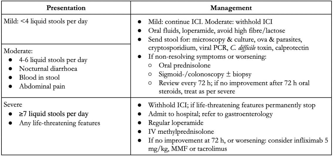
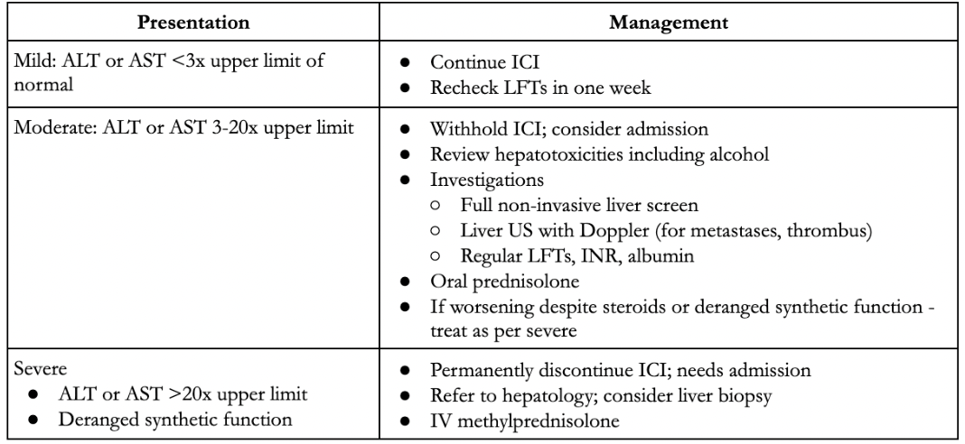
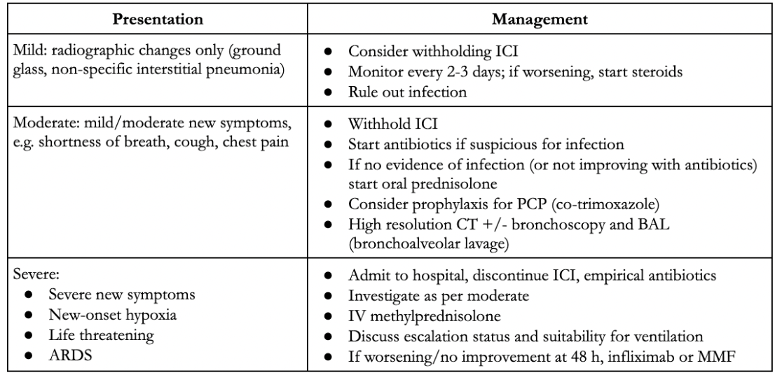
The European Society for Medical Oncology (ESMO) has released comprehensive guidelines to aid in the detection, investigation and management of immune mediated toxicities. The presentation, pertinent investigations and management are summarised by organ system in figures 16-21 below.
Skin toxicities
Skin toxicity presents with a rash, most commonly maculopapular. It may be itchy or painful.
Rarely, severe reactions such as Steven Johnson Syndrome (SJS), toxic epidermal necrolysis (TEN) or bullous dermatitis may occur. These can be life threatening.
| Presentation | Management |
|---|---|
| Mild: <30% body surface area |
|
| Moderate: >30% body surface area, may limit ADLs |
|
Severe:
|
Endocrine toxicities
Endocrinopathies resulting from immune-mediated toxicity can be varied. It is essential to detect and treat hypophysitis early, as otherwise the resultant hypoadrenalism can result in an Addisonian crisis. Thyroid disturbance is common and may occur in 5-10% of patients.
Baseline TSH, T3 are and free T4, pituitary axis bloods (9 am cortisol, ACTH, TSH [thyroid stimulating hormone], T4, LH [luteinising hormone], FSH [follicle-stimulating hormone], oestradiol/testosterone, IGF [insulin-like growth factor]-1, prolactin) and glucose are checked prior to commencing an ICI. TFTs are then monitored regularly throughout treatment and, in some instances, monitored even after cessation of ICI (delayed hypothyroidism/hypophysitis may occur).
| Presentation | Management |
|---|---|
Hypothyroidism
|
If TSH >10 or symptomatic hypothyroidism, start levothyroxine, even with normal T4 |
Thyroiditis
|
|
Hypophysitis
|
|
Diabetes
|
|
Gastrointestinal toxicities (colitis)
Gastrointestinal toxicity is common, with up to 30% of patients experiencing diarrhoea. Severe, high-grade colitis may occur in as many as 1% of patients. This can be life threatening, resulting in perforation or death. It is, therefore, essential to ask about diarrhoea with each cycle and monitor symptoms closely if they occur. Always rule out infection in patients presenting with diarrhoea.
Hepatic toxicities (hepatitis)
Hepatic toxicity or hepatitis is common; 5-10% of patients may present with ICI-induced hepatitis, rising to 30% in patients receiving combined therapy. LFTs are, therefore, measured with each treatment cycle. Severity is determined by the degree of transaminase derangement and presence of derangement in INR or albumin.
Respiratory toxicities (pneumonitis)
Respiratory toxicity, or pneumonitis, is common and can be life threatening. ICIs may be withheld even in mild immune-mediated pneumonitis.
Rare immune-mediated toxicities
It is important to be aware that immune-mediated toxicity can occur in any organ system and to consider immune-mediated toxicity in any patient with almost any symptom! Generally, management is still based on severity. Most instances of these rare toxicities require referral to the specialist team for ongoing management and advice.
| Neurological |
|
|
| Rheumatological |
|
|
| Renal |
|
|
| Cardiac |
|
|
| Ocular |
|
|
| Haematological |
|
|
References
- Cassidy J, Bissett D, Spence RAJ, Payne M, Morris-Stiff GJ. Oxford Handbook of Oncology. 4th ed. Oxford: Oxford University Press; 2015
- Haanen JBAG, Carbonnel F, Robert C, Kerr KM, Peters S, Larkin J, et al. Management of toxicities from immunotherapy: ESMO Clinical Practice Guidelines for diagnosis, treatment and follow-up. Annals of Oncology. 2017 Jul 1;28(suppl_4):iv119–42
- Thompson JA, Schneider BJ, Brahmer J, Andrews S, Armand P, Bhatia S, et al. Management of Immunotherapy-Related Toxicities, Version 1.2019. Journal of the National Comprehensive Cancer Network: JNCCN [Internet]. 2019 Mar 1 [cited 2022 Oct 1];17(3):255–89. Available from: https://pubmed.ncbi.nlm.nih.gov/30865922/
Complete Guide to Clinical Genetics ST3 Application and Recruitment (2027)
Written by Medibuddy. Last updated: October 2026. Clinical Genetics is

Complete Guide to Clinical Radiology ST3 Application and Recruitment (2027)
Recruitment paused: NHS England is not running Clinical Radiology ST3+ recru

Complete Guide to Paediatric Surgery ST3 Application and Recruitment in United Kingdom (2026)
Written by Medibuddy. Last updated: October 2026. If you’re planning


Take your subscriptions with you
Our mobile app allows you to access your interview and exam question banks wherever you are.

