


 Close
Close
 21st February 2025
Pleural and Malignant Lung Disease
Lung cancer
Pathology
Lung cancer is one of the most common cancers in the world and one of the leading causes of cancer deaths.
The most important clinical distinction is between small cell and non-small cell lung cancers, as this defines the management approach.
Table 1 Histological subtypes of lung cancer
| Histological subtype | Key facts | |||
| Small cell lung cancer (SCLC) | Aggressive, fast-growing; most strongly associated with smoking | |||
| Non-small cell lung cancer (NSCLC) | Adenocarcinoma | Most common; arises peripherally | ||
| Squamous cell carcinoma (SCC) | Involves central airways | |||
| Large cell carcinoma | Arises centrally; diagnosis of exclusion (no features of other subtypes) | Other | Neuroendocrine tumours (NET) | Including large cell neuroendocrine carcinoma and well-differentiated NETs (carcinoid tumours) |
| Rarer subtypes | E.g. salivary gland type tumours, lung sarcoma, lung lymphoma | |||
Presentation of lung cancer
Lung cancer is often picked up at an asymptomatic stage via imaging; symptoms can indicate advanced disease. The most common presenting symptoms are cough, haemoptysis, dyspnoea and chest pain. Signs on imaging include lung nodules, hilar enlargement, lobar collapse and pleural effusions.
The NICE suspected cancer referral guidelines for lung cancer are as follows:
| Two week wait referral | Urgent CXR | Consider urgent CXR |
|---|---|---|
| CXR suspicious for lung cancer
OR Age ≥40 with unexplained haemoptysis |
Age ≥40 with 2 of these features, or smoking history with 1 feature:
Cough Fatigue Dyspnoea Chest pain Weight loss Appetite loss |
Age ≥40 and any of the following:
Persistent/recurrent chest infection Finger clubbing Supraclavicular or persistent cervical lymphadenopathy Chest signs consistent with lung cancer Thrombocytosis |
Table 2 NICE guidelines for suspected lung cancer
Complications of lung cancer
Metastases
Common sites to which lung cancers metastasise include the brain, bones, liver and adrenals.
Paraneoplastic syndromes
Paraneoplastic syndromes are frequently seen in lung cancer, especially in association with small cell lung cancer.
| Syndrome | Features | Associated cancer |
|---|---|---|
| SIADH | Euvolaemic hyponatraemia. Improves with treatment of the cancer. | SCLC |
| Lambert-Eaton myasthenic syndrome (LEMS) | Neuromuscular junction disorder similar to myasthenia gravis. Limb weakness is predominant, and characteristically improves with exercise. Autonomic involvement is also common. Antibodies against the voltage-gated calcium channel present. | SCLC |
| Other neurological syndromes | E.g. cerebellar ataxia, sensory neuropathy, limbic encephalitis, encephalomyelitis, autonomic neuropathy, opsomyoclonus. | SCLC |
| Hypertrophic pulmonary osteoarthropathy (HPO) | Painful symmetrical arthropathy and long bone pain secondary to periosteal proliferation. Clubbing of fingers and toes. | Adenocarcinoma |
| Hypercalcaemia | Most commonly caused by tumour secretion of parathyroid hormone-related peptide (PHTrP). In PTHrP-driven hypercalcaemia, you would also expect suppressed serum PTH and low phosphate. | SCC > others |
| Carcinoid syndrome | Flushing, shortness of breath, wheeze, tachycardia, diarrhoea. | NET |
Table 3 Paraneoplastic syndromes associated with lung cancer
Horner’s syndrome
Pancoast tumours (tumours in the lung apex) can involve the sympathetic chain, leading to Horner’s syndrome: ipsilateral ptosis, miosis and anhidrosis.
Superior vena cava obstruction (SVCO)
Lung cancer is the most common malignancy to cause SVCO.
Lymphangitis carcinomatosa
Lymphangitis carcinomatosa is the infiltration of cancer cells into the lymphatic system, leading to inflammation. Pulmonary lymphangitis carcinomatosa is rare but important as it can cause severe symptoms and is associated with a poor prognosis (around half of patients will die within two months of first developing symptoms). Symptoms include shortness of breath and a dry cough. Imaging findings may be similar to those of interstitial lung disease. Lymphangitis is most commonly associated with lung, breast and gastrointestinal cancers.
The most effective treatment is treatment of the underlying cancer. Steroids may be trialled for symptom control.
Management of lung cancer
Management of NSCLC
Treatment algorithms for non-small cell lung cancers are becoming increasingly complex, with choice of treatment depending on various factors including tumour stage, patient fitness and tumour molecular characteristics.
| Curative treatment | Palliative treatment |
|---|---|
| Stage I-IIIA lung cancers are potentially curable, and so patients should be assessed for fitness for surgery.
Lung function testing is an important part of this assessment. Radical chemoradiotherapy may be an option for those not suitable for surgery. In rare cases a stage IIIB tumour is downstaged with treatment to become resectable and therefore curable. |
Stage IIIB, IIIC and IV lung cancers are generally considered unsuitable for radical treatment. The presence of distant metastasis makes a tumour stage IV.
Systemic treatment may be offered, such as chemotherapy, immunotherapy and tyrosine kinase inhibitors. Tumour molecular characteristics are important in treatment decisions at this stage. |
Table 4 Overview of treatment of NSCLC
Prognosis varies significantly depending on the stage of disease, with 5 year survival rates of 67% for stage IA disease down to 1% for stage IV disease.
Management of SCLC
Small cell lung cancer can be broadly divided into limited and extensive stages, which define treatment options.
| Limited-stage SCLC | Extensive-stage SCLC | |
|---|---|---|
| Definition | Confined to ipsilateral haemothorax | Disease beyond ipsilateral haemothorax |
| Approach | Curative treatment may be considered | Treatment is palliative |
| Treatment options | Generally treated with concurrent chemoradiotherapy (typically etoposide + cisplatin/carboplatin).
Surgical resection with adjuvant chemotherapy ± radiotherapy is possible but infrequent. |
Generally treated with chemotherapy alone (again using etoposide + cisplatin/carboplatin).
Radiotherapy may be used for symptom management. |
Table 5 Overview of treatment of SCLC
SCLC often responds well to chemotherapy initially, but overall prognosis is poor, with a 5 year survival of <5% for extensive-stage and 12-24% for limited-stage disease.
Supportive management
Breathlessness is a common problematic symptom in lung cancer. Options for treating breathlessness include low-dose opiates, breathing control techniques and palliative oxygen therapy.
Chest drains and pleurodesis can be helpful in management of symptoms related to pleural effusions.
Radiotherapy can be effective in management of symptoms such as haemoptysis and chest pain.
References
- National Institute for Health and Care Excellence. Suspected cancer: recognition and referral [Internet]. NICE. 2015 [cited 2022 Jan 31]. Available from: https://www.nice.org.uk/guidance/ng12
- National Institute for Health and Care Excellence. Lung cancer: diagnosis and management [Internet]. NICE. 2019 [cited 2022 Jan 31]. Available from: https://www.nice.org.uk/guidance/ng122
- Heist RS. Small cell lung cancer [Internet]. BMJ Best Practice. 2018 [cited 2022 Jan 31]. Available from: https://bestpractice.bmj.com/topics/en-gb/1081
- Baldwin DR, Popat S. Non-small cell lung cancer [Internet]. BMJ Best Practice. 2020 [cited 2022 Jan 31]. Available from: https://bestpractice.bmj.com/topics/en-gb/1082
- Klimek M. Pulmonary lymphangitis carcinomatosis: systematic review and meta-analysis of case reports, 1970-2018. Postgraduate Medicine. 2019 Apr 5;131(5):309–18.
- Anwar A, Jafri F, Ashraf S, Jafri MAS, Fanucchi M. Paraneoplastic syndromes in lung cancer and their management. Annals of Translational Medicine. 2019 Aug;7(15):359–9.
- Midthun D. Clinical manifestations of lung cancer [Internet]. Lilenbaum R, Vora S, editors. www.uptodate.com. 2021 [cited 2022 Jan 31]. Available from: https://www.uptodate.com/contents/clinical-manifestations-of-lung-cancer
- Tazelaar H. Pathology of lung malignancies [Internet]. Nicholson A, Lilenbaum R, Vora S, editors. www.uptodate.com. 2021 [cited 2022 Jan 31]. Available from: https://www.uptodate.com/contents/pathology-of-lung-malignancies
- Longmore M, Wilkinson I, Baldwin A, Wallin E. Oxford Handbook of Clinical Medicine [Internet]. Oxford Medicine Online. Oxford University Press; 2014 [cited 2022 Jan 31]. Available from: https://oxfordmedicine.com/view/10.1093/med/9780199609628.001.0001/med-9780199609628
Mediastinal masses
Differentials for mediastinal masses
The mediastinum is the space between the lungs within the thorax and is separated into three compartments: anterior, middle and posterior. This separation is helpful in identifying the potential causes of a mediastinal mass.
| Anterior mediastinum | Middle mediastinum | Posterior mediastinum |
|---|---|---|
| Thymic mass: thymoma, thymic cyst, thymic carcinoma | Bronchogenic cyst | Neurogenic tumour, e.g. schwannoma, neurofibroma |
| Lymphoma | Lymphadenopathy: lymphoma, sarcoid, metastatic lung cancer | Meningocoele |
| Germ cell tumour | Oesophageal tumour | Thoracic spine lesion |
| Thyroid: substernal goitre, ectopic thyroid tissue | Vascular mass or enlargement, e.g. thoracic aortic aneurysm | |
| Lipoma or liposarcoma | Pericardial cyst |
Table 6 Differentials for mediastinal mass by compartment
Anterior mediastinal masses
Around 50% of mediastinal masses are in the anterior mediastinum and the most common causes can be remembered by the 4 Ts: Thymus, Thyroid, Teratoma (and other germ cell tumours) and ‘Terrible’ lymphoma.
Thymic mass:
- Around a third of anterior mediastinal masses are thymic neoplasms (thymoma or more rarely, carcinoma)
- Thymomas are associated with paraneoplastic syndromes, particularly myasthenia gravis
Germ cell tumour:
- The mediastinum is the most common place outside the testes and ovaries to find germ cell tumours
- Benign germ cell tumours include teratomas and dermoid cysts
- Malignant tumours can be split into seminomatous (more common) and non-seminomatous germ cell tumours (GCT)
Lymphoma:
- Lymphomas are normally found in the anterior mediastinum and tend to grow quickly compared to other causes of anterior mediastinal masses
Presentation of mediastinal masses
Mediastinal masses are often asymptomatic and are frequently identified incidentally on thoracic imaging.
Symptoms and signs will reflect the nature and location of the mass and the structures it involves or compresses. Involvement of the recurrent laryngeal nerve can lead to hoarseness, and damage to the sympathetic chain may result in Horner’s syndrome. Bulky mediastinal masses may cause dysphagia via oesophageal compression.
Emergency presentations are possible and include airway compression and superior vena cava obstruction.
Investigation of mediastinal masses
Investigations depend on the likely differentials, for example:
- Suspected lymphoma: full blood count, blood film and flow cytometry
- Suspected thoracic thyroid tissue: thyroid function tests
- Suspected germ cell tumour: tumour markers including LDH, alpha fetoprotein, beta-HCG. beta-HCG and LDH may be raised in either seminomatous or non-seminomatous tumours, whereas AFP will only be raised in non-seminomatous tumours
- Suspected thymic mass: anti-acetylcholine receptor (AChR) antibodies to evaluate for myasthenia gravis
CT can give information as to the location and composition of a mediastinal mass and may also be helpful in planning for a biopsy.
Ultrasound of the testes should be considered in men presenting with a mediastinal mass to look for evidence of a germ cell tumour.
References
- Muniappan A. Assessment of mediastinal mass [Internet]. BMJ Best Practice. 2020 [cited 2022 Jan 31]. Available from: https://bestpractice.bmj.com/topics/en-gb/998
- 1.Berry M, Bograd A. Approach to the adult patient with a mediastinal mass [Internet]. Muller N, Midthun D, Vallières E, Collins K, Sadhna Vora, editors. www.uptodate.com. 2020 [cited 2022 Jan 31]. Available from: https://www.uptodate.com/contents/approach-to-the-adult-patient-with-a-mediastinal-mass
Asbestos-related lung conditions
Asbestos is a naturally occurring fibrous material previously used widely in construction and manufacturing. Exposure has now been linked to a range of medical conditions, including asbestosis, pleural disease and malignancy.
Deaths related to occupational asbestos exposure should be referred to the coroner and sufferers of asbestos-related disease may be entitled to compensation.
Asbestosis
Asbestosis is an interstitial lung disease caused by progressive pulmonary fibrosis in response to inhaled asbestos fibres.
Presentation of asbestosis
The initial symptoms of asbestosis are usually progressive dyspnoea and a dry cough. These usually develop decades after asbestos exposure.
Signs may include bilateral fine crackles on auscultation (usually starting at the bases), reduced chest expansion and finger clubbing.
Investigation of asbestosis
Initial screening is with a chest X-ray, which may show bilateral interstitial changes. The presence of asbestos-related pleural changes may support a diagnosis of asbestosis as the cause of interstitial changes. Chest X-ray changes can be subtle in early disease so more sensitive HRCT may be considered.
Pulmonary function testing will usually demonstrate a restrictive pattern with reduced TLCO but also reduced KCO as lung parenchyma is affected. If there has been a significant smoking history, there may also be an obstructive element to the results.
Lung biopsy is not normally required.
Management of asbestosis
There is currently no disease-modifying treatment for asbestosis so focus should be on supportive treatment, including avoiding further asbestos exposure, smoking cessation, pulmonary rehabilitation, long-term oxygen therapy and pneumococcal and influenza vaccines.
Asbestos-associated pleural disease
Asbestos exposure can lead to a range of benign pleural disease.
Pleural plaques
Pleural plaques are focal areas of benign pleural thickening which develop as a result of asbestos exposure. They are normally bilateral, may be calcified, and typically overlie the 6th to 9th ribs. The apices and costophrenic recesses are usually spared.
Pleural plaques are usually asymptomatic and do not require any intervention or follow-up.
Diffuse pleural thickening
More extensive pleural thickening can also occur following exposure to asbestos (although diffuse pleural thickening can also develop in response to other processes such as infection, inflammation or pleural effusions).
Diffuse pleural thickening is differentiated from pleural plaques by dimensions on CT imaging. In addition, it is often present in the costophrenic recesses (unlike pleural plaques).
Diffuse pleural thickening may restrict lung expansion and lead to breathlessness. Pulmonary function testing will usually demonstrate a restrictive pattern with reduced TLCO but normal KCO.
Benign asbestos pleural effusion (BAPE)
Small unilateral benign pleural effusions can occur in patients who have been exposed to asbestos. The effusions are exudative.
BAPE is usually asymptomatic and may resolve spontaneously but should be investigated if identified given the increased risk of lung malignancy in this patient group.
Mesothelioma
The malignancy most strongly associated with asbestos exposure is malignant mesothelioma.
Asbestos exposure also increases the risk of developing other types of lung cancer. The investigation and management of these should be the same as for lung cancer of any other cause (see Lung cancer).
Pathology and risk factors in mesothelioma
Exposure to asbestos is the main risk factor for mesothelioma, and around 80% of patients with mesothelioma have a history of this. The average latency between exposure and development of mesothelioma is 20 to 40 years.
Around 90% of mesothelioma cases are pleural, though mesothelioma can also develop in the peritoneum (5-10%), pericardium and testes (<1% each).
Presentation and investigation of mesothelioma
The most common symptoms of pleural mesothelioma include chest pain and dyspnoea, as well as constitutional symptoms such as fever and weight loss. Pleural effusions are often present at diagnosis.
NICE suspected cancer guidelines for mesothelioma are summarised below:
| Two week wait referral | Urgent CXR | Consider urgent CXR |
|---|---|---|
| X-ray findings suggestive of mesothelioma | 2 of the following symptoms
OR Asbestos exposure or smoking history with 1 of the following: Cough Fatigue Dyspnoea Chest pain Weight loss Appetite loss |
Age >40 years with 1 of the following:
Finger clubbing Chest signs compatible with pleural disease |
Table 7 NICE guidelines for suspected mesothelioma
Chest X-ray is recommended as the initial screening investigation. Following an X-ray, A CT of the chest and upper abdomen should be offered if there is high clinical suspicion of malignancy. CT signs suggestive of mesothelioma include nodular pleural thickening, hilar or mediastinal lymphadenopathy and invasion into surrounding structures such as the chest wall.
Even following CT, it can be difficult to differentiate benign from malignant pleural disease on imaging alone. If present, pleural effusions should be tapped for cytology. Yield can be low, so tissue biopsy is often required in addition, either by VATS (video-assisted thoracoscopic surgery) or transcutaneous CT-guided biopsy.
A PET scan should be considered to assess for regional or distant spread of disease.
Management of mesothelioma
Mesothelioma has a poor progress, with a median survival of 10 to 15 months.
In some cases, surgery with curative intent can be considered, usually combined with chemotherapy or radiotherapy.
In most cases, treatment will be palliative. Surgery, radiotherapy and systemic therapies (including chemotherapy, angiogenesis inhibitors and immunotherapy) can all play a role in palliative management. Platinum chemotherapy (usually cisplatin/carboplatin + pemetrexed) has been the mainstay of systemic treatments.
Pleurodesis and indwelling pleural catheters are useful in managing recurrent effusions.
References
- Rosenman K. Asbestosis [Internet]. BMJ Best Practice. 2020 [cited 2022 Jan 31]. Available from: https://bestpractice.bmj.com/topics/en-gb/650
- Thomas A, del Rivero J, Hesdorffer M. Mesothelioma [Internet]. BMJ Best Practice. 2021 [cited 2022 Jan 31]. Available from: https://bestpractice.bmj.com/topics/en-gb/1184
- Woolhouse I, Bishop L, Darlison L, De Fonseka D, Edey A, Edwards J, et al. British Thoracic Society Guideline for the investigation and management of malignant pleural mesothelioma. Thorax [Internet]. 2018 Feb 14 [cited 2019 Dec 18];73(Suppl 1):i1–30. Available from: https://thorax.bmj.com/content/73/Suppl_1/i1
- Popat S, Baas P, Faivre-Finn C, Girard N, Nicholson AG, Nowak AK, et al. Malignant pleural mesothelioma: ESMO Clinical Practice Guidelines for diagnosis, treatment and follow-up. Annals of Oncology [Internet]. 2022 Feb [cited 2022 Jan 31];33(2):129–42. Available from: https://www.annalsofoncology.org/article/S0923-7534(21)04820-1/fulltext
- King T. Asbestos-related pleuropulmonary disease [Internet]. Flaherty K, Hollingsworth H, editors. www.uptodate.com. 2021 [cited 2022 Jan 31]. Available from: https://www.uptodate.com/contents/asbestos-related-pleuropulmonary-disease
- Asbestos-related conditions [Internet]. British Lung Foundation. 2021 Jan [cited 2022 Jan 31]. Available from: https://www.blf.org.uk/sites/default/files/Abestos_and_your_lungs_V4_downloadable_PDF.pdf
- British Thoracic Society. Pleural Plaques Information for Health Care Professionals [Internet]. NHS Greater Glasgow and Clyde. Department of Health; 2011 [cited 2022 Mar 14]. Available from: http://library.nhsggc.org.uk/mediaAssets/Respiratory%20Medicine/BTS%20Plueral_Plaques299912_acc.pdf
Pneumothorax
Pneumothorax is the presence of air in the pleural space (between the visceral and parietal pleura).
Definitions
Pneumothorax is normally categorised by the underlying cause, as this determines management.
| Spontaneous | Primary spontaneous | No identifiable underlying lung disease or precipitating event |
| Secondary spontaneous | Underlying lung disease present but no identifiable trigger | |
| Traumatic | Iatrogenic | Resulting as complication of medical intervention |
| Non-iatrogenic traumatic | Resulting from non-medical trauma (penetrating > blunt force) | |
Table 8 Categorising pneumothorax
The most common cause of an iatrogenic pneumothorax is central line insertion. Other causes include lung biopsy, thoracocentesis and mechanical ventilation.
Risk factors for pneumothorax
| Primary spontaneous | Secondary spontaneous |
|---|---|
| Smoking (tobacco or cannabis) | COPD (most common), ILD (interstitial lung disease), TB, cystic fibrosis, malignancy, necrotising pneumonia |
| Male gender | Male gender |
| Young adults (peak age 20 for first presentation) | Older age group (usually >55 years) |
| Tall, slender body type | |
| Marfan syndrome | |
| Family history |
Table 9 Risk factors for spontaneous pneumothorax
Presentation of pneumothorax
Pneumothorax frequently develops with minimal symptoms or may even be asymptomatic. Symptoms are usually sudden onset and include pleuritic chest pain and shortness of breath. Symptoms are normally more severe in secondary pneumothorax even with pneumothoraces of the same size.
Signs of pneumothorax on examination include reduced breath sounds and hyperresonant percussion notes on the side of the pneumothorax. Hypoxia is often not present in small primary spontaneous pneumothorax but may be present in secondary or traumatic pneumothorax. In traumatic pneumothorax, examination may reveal evidence of rib fractures or penetrating trauma.
Investigation of pneumothorax
The first-line investigation in suspected pneumothorax is an erect chest X-ray.
CT chest should be considered if the diagnosis remains uncertain or in the context of chest trauma. CT can be helpful in differentiating pneumothorax from bullous lung disease, identifying underlying lung pathology, and accurately determining pneumothorax size.
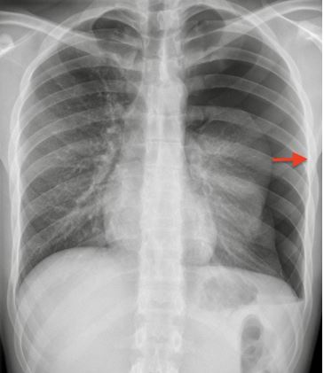
Image Source: Wikimedia Commons (2010). Expiration-left-side-pneumo, Licensed under a creative commons licence. Available for free at: https://commons.wikimedia.org/wiki/File:Expiration-left-side-pneumo.jpg#/media/File:Expiration-left-side-pneumo.jpg (Accessed: 19 January 2024).
Pleural fluid analysis
Often, bilateral pleural effusions can be reliably diagnosed as transudates by history, examination and blood tests. Effusions should be aspirated and analysed if unilateral or if there is not a clear transudative cause.
Recommended for all pleural fluid samples:
- Cell count and differential, cytology
- LDH
- Protein
- Microscopy, culture and sensitivity
Other tests which should be considered include pH (low in infection), glucose, triglycerides and cholesterol (raised in chylothorax), amylase (raised in pancreatitis or oesophageal rupture), NT-proBNP (N-terminal pro b-type natriuretic peptide) (raised in heart failure), tuberculosis culture.
Routine chest X-ray is not recommended by BTS following a pleural aspiration, however a chest X-ray is recommended to check for complications if the procedure was difficult and required multiple attempts, or if air is aspirated during the procedure.
Other investigations
Blood tests should be done to look for evidence of inflammation, bleeding and liver or renal failure. Serum protein and LDH are needed for Light’s criteria. NT-proBNP and transthoracic echo are useful in suspected heart failure.
Management of pneumothorax
Traumatic pneumothorax should be referred to thoracic surgeons and monitored closely, as there is a risk of developing a tension pneumothorax.
Spontaneous pneumothorax management depends on several factors, including:
- Size: rim of air between lung and chest wall at the level of the hilum (small <2 cm, large >2 cm) (Figure 1)
- Symptom severity
- Pneumothorax type (primary spontaneous/secondary spontaneous)
As per British Thoracic Society (BTS) guidelines:
| Primary spontaneous pneumothorax | Secondary spontaneous pneumothorax | ||
|---|---|---|---|
| Small, asymptomatic | Large or symptomatic | Small, asymptomatic | Large or symptomatic |
| Consider discharge with safety netting and outpatient review in 2-4 weeks | Needle aspiration, aiming to improve symptoms and reduce to <2 cm
Successful: consider discharge with safety netting and outpatient review in 2-4 weeks Unsuccessful: chest drain and admission |
If <1 cm, admission for observation
If 1-2 cm, needle aspiration. If it remains >1 cm, proceed to chest drain |
Chest drain and admission |
Table 10 Management of pneumothorax
Bilateral pneumothorax is potentially life-threatening and should proceed straight to bilateral chest drain insertion.
Follow up
Smoking cessation is important to reduce risk of recurrence.
In some cases, surgical intervention via VATS or thoracotomy may be advised to prevent recurrence. Chemical pleurodesis can also be considered.
Flying should be avoided until full resolution of the pneumothorax. Patients should be advised to avoid underwater diving permanently following a pneumothorax unless they have definitive surgical management.
Tension pneumothorax
Tension pneumothorax is a medical emergency. It occurs when a one-way valve develops between the pleural cavity and the external environment, leading to progressive entry of air into the pleural space during inspiration, which is unable to be expelled. The resultant increase in intrathoracic pressure leads to reduced venous return, impaired cardiac output and haemodynamic collapse.
Differences in presentation and management of tension pneumothorax compared to other types of pneumothorax are outlined below:
| Aetiology | More common with traumatic than spontaneous pneumothorax. |
| Symptoms | More severe breathlessness and chest pain; feeling of dread or panic. |
| Signs | Tachycardia, hypotension, sweating, more severe hypoxia and respiratory distress.
Tracheal deviation away from the side of the pneumothorax. |
| Imaging | Management should commence as soon as tension pneumothorax is suspected before imaging.
If an X-ray is done, it may show a mediastinal shift away from pneumothorax. |
| Management | Immediate decompression with a large-bore cannula in the 2nd intercostal space, mid-clavicular line. Chest drain insertion following decompression. |
Table 11 Features, investigations and management of tension pneumothorax
References
- MacDuff A, Arnold A, Harvey J. Management of spontaneous pneumothorax: British Thoracic Society pleural disease guideline 2010. Thorax [Internet]. 2010 Aug 1 [cited 2022 Jan 31];65(Suppl 2):ii18–31. Available from: https://thorax.bmj.com/content/65/Suppl_2/ii18
- Bennett J, Vella C, Umerah O. Pneumothorax [Internet. BMJ Best Practice. 2021. Available from: https://bestpractice.bmj.com/topics/en-gb/3000083
- Lee YG. Pneumothorax in adults: Epidemiology and etiology [Internet]. Broaddus C, Finlay G, editors. Uptodate.com. 2020 [cited 2022 Jan 31]. Available from: https://www.uptodate.com/contents/pneumothorax-in-adults-epidemiology-and-etiology
Pleural effusion
A pleural effusion is a collection of excess fluid in the pleural cavity. In normal circumstances, around 15 ml of fluid is produced by the pleura and reabsorbed by the pleural lymphatics per day, with an average of 20 ml of pleural fluid in each hemithorax at any one time. An imbalance of production and removal will lead to excess fluid.
Causes of pleural effusion
Effusions are traditionally separated into transudates and exudates.
The differential of effusions is wide, but the common underlying pathologies are outlined in the table below, with the most common causes in bold.
| Transudate | Exudate | Other |
|---|---|---|
| Heart failure | Infection (parapneumonic, TB) | Haemothorax |
| Cirrhosis | Malignancy (e.g. lung cancer, mesothelioma, metastatic cancer, primary effusion lymphoma) | Chylothorax (accumulation of chyle due to disruption of lymphatic drainage) |
| Hypoalbuminaemia | Autoimmune pleuritis, e.g. rheumatoid arthritis | |
| Peritoneal dialysis | PE | |
| Hypothyroidism | Benign asbestos pleural effusion | |
| Nephrotic syndrome | Post-myocardial infarction (Dressler syndrome) or post-CABG | |
| Meigs’ syndrome |
Table 12 Causes of pleural effusion
Light’s criteria can be used to differentiate between transudates and exudates. Pleural fluid is thought to be an exudate if any of the following are true:
- Pleural fluid to serum protein ratio is >0.5
- Pleural fluid to serum LDH ratio is >0.6
- Pleural fluid LDH is >⅔ of the upper limit of normal for serum LDH
Presentation of pleural effusion
Symptoms include shortness of breath, cough and pleuritic chest pain. Typical examination findings are ‘stony dull’ percussion, absent breath sounds, reduced chest expansion, and reduced tactile vocal fremitus and vocal resonance.
Other symptoms and signs may point to the underlying pathology.
Investigation of pleural effusion
Imaging
A chest X-ray is the first-line investigation in suspected pleural effusion and can detect effusions from around 200 ml. Ultrasound is more sensitive and specific and should be used to guide any pleural procedure. CT chest is also usually performed, to delineate the effusion and detect loculations, and to look for signs of the underlying pathology such as lung masses or pleural thickening.
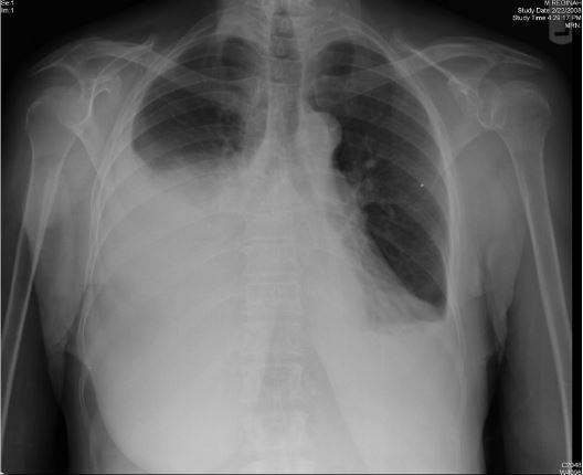
Image Source: Wikimedia Commons (2011). Effusion, Licensed under a creative commons licence. Available for free at: https://commons.wikimedia.org/wiki/File:Effusion.png#/media/File:Effusion.png (Accessed: 19 January 2024).
Pleural fluid analysis
Often, bilateral pleural effusions can be reliably diagnosed as transudates by history, examination and blood tests. Effusions should be aspirated and analysed if unilateral or if there is not a clear transudative cause.
Recommended for all pleural fluid samples:
- Cell count and differential, cytology
- LDH
- Protein
- Microscopy, culture and sensitivity
Other tests which should be considered include pH (low in infection), glucose, triglycerides and cholesterol (raised in chylothorax), amylase (raised in pancreatitis or oesophageal rupture), NT-proBNP (N-terminal pro b-type natriuretic peptide) (raised in heart failure), tuberculosis culture.
Routine chest X-ray is not recommended by BTS following a pleural aspiration, however a chest X-ray is recommended to check for complications if the procedure was difficult and required multiple attempts, or if air is aspirated during the procedure.
Other investigations
Blood tests should be done to look for evidence of inflammation, bleeding and liver or renal failure. Serum protein and LDH are needed for Light’s criteria. NT-proBNP and transthoracic echo are useful in suspected heart failure.
Management of pleural effusion
Management depends on the underlying cause and generally targets the underlying disease, e.g. loop diuretics for an effusion related to heart failure.
Thoracocentesis
Transudates do not generally require drainage, but it can be beneficial for any large-volume symptomatic effusion.
Re-expansion pulmonary oedema is a possible complication of drainage of large effusions, these should be drained at a controlled rate (no more than 1.5 L over the first hour). Other potential complications of thoracocentesis are pneumothorax and trapped lung (incomplete re-expansion of the lung following drainage). A routine chest X-ray is recommended following drainage (this is not normally required after aspiration).
If drainage is required, BTS guidelines suggest that this should be done in working hours, as this minimises the risk of complications. This does not apply to patients in extremis, such as with tension pneumothorax or severe respiratory failure thought to be due to the pleural effusion.
Malignant effusions
Malignant effusions are often recurrent and may require repeat drainage, pleurodesis and indwelling pleural catheters.
Parapneumonic effusions
Parapneumonic effusions exist on a spectrum from a simple parapneumonic effusion to empyema, and differentiating between these is important to define management.
| Simple parapneumonic | Complicated parapneumonic | Empyema | |
|---|---|---|---|
| Definition | No evidence of infection of the pleural fluid on analysis | Evidence of pleural fluid infection, e.g. positive fluid MC&S or pH <7.2 | Frank pus on aspiration |
| Management | Should resolve with appropriate antibiotic management | Urgent chest drain recommended | |
Table 13 Stratification of parapneumonic effusions
Post-myocardial infarction pleural effusion
NSAIDs are the mainstay of treatment for pleural and pericardial effusions following MI. Steroids can be useful if NSAIDs are contraindicated or symptoms are refractory. Colchicine can be used to prevent recurrence.
References
- Bhatnagar R, Maskell N. The modern diagnosis and management of pleural effusions. BMJ [Internet]. 2015 Sep 8;h4520. Available from: https://www.bmj.com/content/351/bmj.h4520
- Hooper C, Lee YCG, Maskell N. Investigation of a unilateral pleural effusion in adults: British Thoracic Society pleural disease guideline 2010. Thorax. 2010 Aug 1;65(Suppl 2):ii4–17.
- Roberts ME, Neville E, Berrisford RG, Antunes G, Ali NJ. Management of a malignant pleural effusion: British Thoracic Society pleural disease guideline 2010. Thorax. 2010 Aug 1;65(Suppl 2):ii32–40.
- Davies HE, Davies RJO, Davies CWH. Management of pleural infection in adults: British Thoracic Society pleural disease guideline 2010. Thorax [Internet]. 2010 Aug 1;65(Suppl 2):ii41–53. Available from: https://thorax.bmj.com/content/65/Suppl_2/ii41#T1
- Orhan O. Pleural effusion [Internet]. BMJ Best Practice. 2019 [cited 2022 Jan 31]. Available from: https://bestpractice.bmj.com/topics/en-gb/287
- Pataka A, Riha RL, Rahman NM. Empyema [Internet]. BMJ Best Practice. 2021. Available from: https://bestpractice.bmj.com/topics/en-gb/1008
- Heffner J. Diagnostic evaluation of a pleural effusion in adults: Initial testing [Internet]. Maldonado F, Finlay G, editors. Uptodate.com. 2021 [cited 2022 Jan 31]. Available from: https://www.uptodate.com/contents/diagnostic-evaluation-of-a-pleural-effusion-in-adults-initial-testing
- Strange C. Management and prognosis of parapneumonic pleural effusion and empyema in adults [Internet]. Broaddus C, Ramirez J, Finlay G, Bond S, editors. www.uptodate.com. 2021 [cited 2022 Jan 31]. Available from: https://www.uptodate.com/contents/management-and-prognosis-of-parapneumonic-pleural-effusion-and-empyema-in-adults
- Sasse T, Eriksson U. Post-cardiac injury syndrome: aetiology, diagnosis, and treatment. e-Journal of Cardiology Practice [Internet]. 2017 Oct 31 [cited 2022 Mar 14];15(21). Available from: https://www.escardio.org/Journals/E-Journal-of-Cardiology-Practice/Volume-15/Post-cardiac-injury-syndrome-aetiology-diagnosis-and-treatment
Complete Guide to Clinical Genetics ST3 Application and Recruitment (2027)
Written by Medibuddy. Last updated: October 2026. Clinical Genetics is

Complete Guide to Clinical Radiology ST3 Application and Recruitment (2027)
Recruitment paused: NHS England is not running Clinical Radiology ST3+ recru

Complete Guide to Paediatric Surgery ST3 Application and Recruitment in United Kingdom (2026)
Written by Medibuddy. Last updated: October 2026. If you’re planning


Take your subscriptions with you
Our mobile app allows you to access your interview and exam question banks wherever you are.

