


 Close
Close
 21st February 2025
Pleural effusion
A pleural effusion is a collection of excess fluid in the pleural cavity. In normal circumstances, around 15 ml of fluid is produced by the pleura and reabsorbed by the pleural lymphatics per day, with an average of 20 ml of pleural fluid in each hemithorax at any one time. An imbalance of production and removal will lead to excess fluid.
Causes of pleural effusion
Effusions are traditionally separated into transudates and exudates.
The differential of effusions is wide, but the common underlying pathologies are outlined in the table below, with the most common causes in bold.
| Transudate | Exudate | Other |
|---|---|---|
| Heart failure | Infection (parapneumonic, TB) | Haemothorax |
| Cirrhosis | Malignancy (e.g. lung cancer, mesothelioma, metastatic cancer, primary effusion lymphoma) | Chylothorax (accumulation of chyle due to disruption of lymphatic drainage) |
| Hypoalbuminaemia | Autoimmune pleuritis, e.g. rheumatoid arthritis | |
| Peritoneal dialysis | PE | |
| Hypothyroidism | Benign asbestos pleural effusion | |
| Nephrotic syndrome | Post-myocardial infarction (Dressler syndrome) or post-CABG | |
| Meigs’ syndrome |
Table 12 Causes of pleural effusion
Light’s criteria can be used to differentiate between transudates and exudates. Pleural fluid is thought to be an exudate if any of the following are true:
- Pleural fluid to serum protein ratio is >0.5
- Pleural fluid to serum LDH ratio is >0.6
- Pleural fluid LDH is >⅔ of the upper limit of normal for serum LDH
Presentation of pleural effusion
Symptoms include shortness of breath, cough and pleuritic chest pain. Typical examination findings are ‘stony dull’ percussion, absent breath sounds, reduced chest expansion, and reduced tactile vocal fremitus and vocal resonance.
Other symptoms and signs may point to the underlying pathology.
Investigation of pleural effusion
Imaging
A chest X-ray is the first-line investigation in suspected pleural effusion and can detect effusions from around 200 ml. Ultrasound is more sensitive and specific and should be used to guide any pleural procedure. CT chest is also usually performed, to delineate the effusion and detect loculations, and to look for signs of the underlying pathology such as lung masses or pleural thickening.
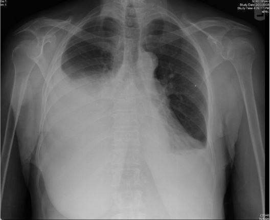
Figure 2 Chest X-ray demonstrating large right-sided pleural effusion
Image Source: Wikimedia Commons (2011). Effusion, Licensed under a creative commons licence. Available for free at: https://commons.wikimedia.org/wiki/File:Effusion.png#/media/File:Effusion.png (Accessed: 19 January 2024).
Pleural fluid analysis
Often, bilateral pleural effusions can be reliably diagnosed as transudates by history, examination and blood tests. Effusions should be aspirated and analysed if unilateral or if there is not a clear transudative cause.
Recommended for all pleural fluid samples:
- Cell count and differential, cytology
- LDH
- Protein
- Microscopy, culture and sensitivity
Other tests which should be considered include pH (low in infection), glucose, triglycerides and cholesterol (raised in chylothorax), amylase (raised in pancreatitis or oesophageal rupture), NT-proBNP (N-terminal pro b-type natriuretic peptide) (raised in heart failure), tuberculosis culture.
Routine chest X-ray is not recommended by BTS following a pleural aspiration, however a chest X-ray is recommended to check for complications if the procedure was difficult and required multiple attempts, or if air is aspirated during the procedure.
Other investigations
Blood tests should be done to look for evidence of inflammation, bleeding and liver or renal failure. Serum protein and LDH are needed for Light’s criteria. NT-proBNP and transthoracic echo are useful in suspected heart failure.
Management of pleural effusion
Management depends on the underlying cause and generally targets the underlying disease, e.g. loop diuretics for an effusion related to heart failure.
Thoracocentesis
Transudates do not generally require drainage, but it can be beneficial for any large-volume symptomatic effusion.
Re-expansion pulmonary oedema is a possible complication of drainage of large effusions, these should be drained at a controlled rate (no more than 1.5 L over the first hour). Other potential complications of thoracocentesis are pneumothorax and trapped lung (incomplete re-expansion of the lung following drainage). A routine chest X-ray is recommended following drainage (this is not normally required after aspiration).
If drainage is required, BTS guidelines suggest that this should be done in working hours, as this minimises the risk of complications. This does not apply to patients in extremis, such as with tension pneumothorax or severe respiratory failure thought to be due to the pleural effusion.
Malignant effusions
Malignant effusions are often recurrent and may require repeat drainage, pleurodesis and indwelling pleural catheters.
Parapneumonic effusions
Parapneumonic effusions exist on a spectrum from a simple parapneumonic effusion to empyema, and differentiating between these is important to define management.
| Simple parapneumonic | Complicated parapneumonic | Empyema | |
|---|---|---|---|
| Definition | No evidence of infection of the pleural fluid on analysis | Evidence of pleural fluid infection, e.g. positive fluid MC&S or pH <7.2 | Frank pus on aspiration |
| Management | Should resolve with appropriate antibiotic management | Urgent chest drain recommended | |
Table 13 Stratification of parapneumonic effusions
Post-myocardial infarction pleural effusion
NSAIDs are the mainstay of treatment for pleural and pericardial effusions following MI. Steroids can be useful if NSAIDs are contraindicated or symptoms are refractory. Colchicine can be used to prevent recurrence.
References
- Bhatnagar R, Maskell N. The modern diagnosis and management of pleural effusions. BMJ [Internet]. 2015 Sep 8;h4520. Available from: https://www.bmj.com/content/351/bmj.h4520
- Hooper C, Lee YCG, Maskell N. Investigation of a unilateral pleural effusion in adults: British Thoracic Society pleural disease guideline 2010. Thorax. 2010 Aug 1;65(Suppl 2):ii4–17.
- Roberts ME, Neville E, Berrisford RG, Antunes G, Ali NJ. Management of a malignant pleural effusion: British Thoracic Society pleural disease guideline 2010. Thorax. 2010 Aug 1;65(Suppl 2):ii32–40.
- Davies HE, Davies RJO, Davies CWH. Management of pleural infection in adults: British Thoracic Society pleural disease guideline 2010. Thorax [Internet]. 2010 Aug 1;65(Suppl 2):ii41–53. Available from: https://thorax.bmj.com/content/65/Suppl_2/ii41#T1
- Orhan O. Pleural effusion [Internet]. BMJ Best Practice. 2019 [cited 2022 Jan 31]. Available from: https://bestpractice.bmj.com/topics/en-gb/287
- Pataka A, Riha RL, Rahman NM. Empyema [Internet]. BMJ Best Practice. 2021. Available from: https://bestpractice.bmj.com/topics/en-gb/1008
- Heffner J. Diagnostic evaluation of a pleural effusion in adults: Initial testing [Internet]. Maldonado F, Finlay G, editors. Uptodate.com. 2021 [cited 2022 Jan 31]. Available from: https://www.uptodate.com/contents/diagnostic-evaluation-of-a-pleural-effusion-in-adults-initial-testing
- Strange C. Management and prognosis of parapneumonic pleural effusion and empyema in adults [Internet]. Broaddus C, Ramirez J, Finlay G, Bond S, editors. www.uptodate.com. 2021 [cited 2022 Jan 31]. Available from: https://www.uptodate.com/contents/management-and-prognosis-of-parapneumonic-pleural-effusion-and-empyema-in-adults
- Sasse T, Eriksson U. Post-cardiac injury syndrome: aetiology, diagnosis, and treatment. e-Journal of Cardiology Practice [Internet]. 2017 Oct 31 [cited 2022 Mar 14];15(21). Available from: https://www.escardio.org/Journals/E-Journal-of-Cardiology-Practice/Volume-15/Post-cardiac-injury-syndrome-aetiology-diagnosis-and-treatment
Complete Guide to Clinical Genetics ST3 Application and Recruitment (2027)
Written by Medibuddy. Last updated: October 2026. Clinical Genetics is

Complete Guide to Clinical Radiology ST3 Application and Recruitment (2027)
Recruitment paused: NHS England is not running Clinical Radiology ST3+ recru

Complete Guide to Paediatric Surgery ST3 Application and Recruitment in United Kingdom (2026)
Written by Medibuddy. Last updated: October 2026. If you’re planning


Take your subscriptions with you
Our mobile app allows you to access your interview and exam question banks wherever you are.

