


 Close
Close
 18th February 2025
Pulmonary Vascular Disease
Pulmonary embolism
Clinical features of PE
Pulmonary embolism (PE) is a condition where one or more dislodged thrombi occlude the pulmonary vasculature, which may cause severe respiratory dysfunction.
Clinical features of acute PE include dyspnoea, pleuritic chest pain, hypoxaemia, tachycardia, cough and haemoptysis. Dizziness, syncope, elevated JVP, gallop rhythm and tricuspid regurgitant murmur are suggestive of right ventricular strain and failure. There may be associated distal limb findings suggestive of a DVT. Hypotension and cardiac arrest are the most serious presentations of acute PE.
Risk factors for PE
Around 30-50% of VTEs are unprovoked, with no identifiable risk factor. However, important risk factors include (from the European Society of Cardiology):
| Strong risk factors
(odds ratio >10) |
Moderate risk factors
(odds ratio 2-9) |
Weak risk factors
(odds ratio <2) |
|---|---|---|
|
|
|
Table 1 Risk factors for PE
Assessing the clinical probability of PE
Various clinical scoring systems are used to assist with objective assessment of the probability of PE and therefore the need (or not) to do further investigations:
- If PE is a differential but with low suspicion: pulmonary embolism rule-out criteria (PERC rule)
- If there is significant suspicion of PE (and/or PERC is positive): Wells’ criteria for PE
| PERC tool for PE: if the suspicion of a PE is low (pre-test probability <15%) AND the total score is 0 then a PE can be ruled out with any further assessment | |
| Age ≥ 50 years old | +1 |
| HR ≥ 100 beats per minute | +1 |
| SaO2 on air < 95% | +1 |
| Unilateral leg swelling | +1 |
| Haemoptysis | +1 |
| Recent surgery or trauma (within past 4 weeks requiring general anaesthesia) | +1 |
| Prior PE or DVT | +1 |
| Hormone use (oral contraceptive, hormone replacement or oestrogen therapy in male/female) | +1 |
Table 2 PERC tool for PE
| Wells’ score for PE: if the total score is ≤4 then PE is unlikely and D-dimer should be checked. If negative, then PE can be ruled out; if positive, CTPA should be performed. If Wells’ score is >4 then proceed to CTPA | |
| Symptoms and signs of a DVT | +3 |
| PE is likely or equally likely diagnosis | +3 |
| Heart rate > 100 | +1.5 |
| Prior PE or DVT | +1.5 |
| Immobilisation for ≥ 3 days or surgery in past 4 weeks | +1.5 |
| Malignancy with active/palliative treatment within past 6 months | +1 |
| Haemoptysis | +1 |
Table 3 Wells’ score for PE
Investigations in PE in a non-pregnant patient
Common investigations in suspected PE include:
- ECG: the most common finding is sinus tachycardia; other abnormalities include non-specific ST-segment and T-wave abnormalities, right axis deviation, right bundle branch block, or the textbook (but unusual) finding of “S1Q3T3” (S wave in lead 1, Q wave and T-wave inversion in lead 3) (figure 3)
- D-dimer: with low clinical suspicion, normal D-dimer can exclude PE
- Chest X-ray: usually normal; may show atelectasis, pleural effusion, or elevation of the hemidiaphragm
- Lower limb venous duplex ultrasound with compression: to investigate suspected concurrent DVT. Proximal DVT in suspected PE is sufficient to start anticoagulation without further investigation
- CTPA (figures 1 and 2): gold standard to confirm PE
- Ventilation-perfusion (VQ) scan: alternative to CTPA in those in whom the latter is contraindicated (e.g. renal impairment, contrast allergy) or in certain patient groups to minimise radiation exposure (e.g. pregnant patients). A VQ scan is of limited use with significant underlying lung disease, abnormal chest radiograph, left ventricular failure or congestive cardiac failure
- Echocardiogram: useful for haemodynamically unstable patients who cannot have CTPA – the presence of right ventricular dysfunction is sufficient to justify urgent thrombolysis. Signs of PE include right ventricle (RV) dilatation and hypokinesis, McConnell’s sign (segmental RV regional wall motion abnormality without apical involvement), tricuspid regurgitation and pulmonary hypertension
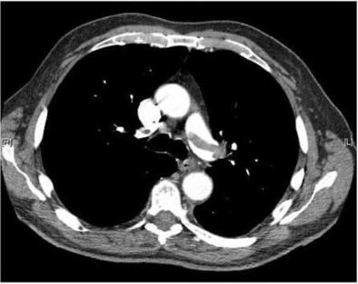
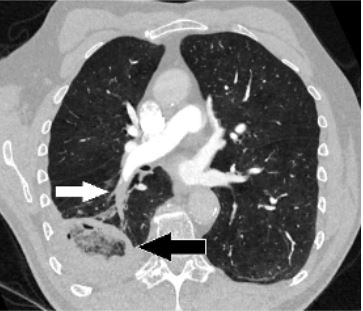
Figure 1: Image Source: Thrombosis Journal (2007). 10.1186, Licensed under a creative commons licence. Available for free at: https://doi.org/10.1186/1477-9560-5-20 (Accessed: 19 January 2024).
Figure 2: Image Source: Wikimedia Commons (2017). Pulmonary_embolism_CTPA, Licensed under a creative commons licence. Available for free at: https://en.wikipedia.org/wiki/Pulmonary_embolism#/media/File:Pulmonary_embolism_CTPA.JPEG (Accessed: 19 January 2024).
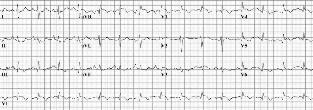
Image Source: Wikimedia Commons (2010). Pulm embolism, Licensed under a creative commons licence. Available for free at: https://en.wikipedia.org/wiki/Pulmonary_embolism#/media/File:Pulm_embolism.jpg (Accessed: 19 January 2024).
Diagnosing PE in pregnancy
Diagnosing acute PE in a pregnant person poses important considerations. D-dimer can be raised in pregnancy due to production by the normal placenta. Furthermore, there is consideration of radiation risks to developing breast tissue as well as the foetus. The approach to diagnosis in this special circumstance is, therefore, tailored to address these. The approach is as follows:
- D-dimer testing: not currently used in pregnancy in the UK, although this may change.
- If concurrent DVT is suspected: lower limb compression venous ultrasound is required. If a DVT is found, then anticoagulation can be started (typically LMWH or UFH; DOACs and warfarin are contraindicated in pregnancy) and no further irradiating tests are needed. Note that in the absence of clinical features of a DVT, a bilateral leg US should not be routinely requested.
- If no concurrent DVT is suspected: anticoagulation (LMWH or UFH) should be started and further investigations ordered. First, a CXR should be requested to ensure there is no underlying lung pathology that may impact the interpretation of a VQ scan. The radiation risk of a CXR to mother and foetus is trivial. VQ scans and CTPA are the next stage of investigation. If the CXR is abnormal, a CTPA is required, as a VQ scan would not be interpretable. If the CXR is normal, either VQ scan or CTPA can be used based on local availability and patient preference, after appropriate counselling regarding radiation risk. Typically, a VQ scan reduces radiation risk to maternal breast tissue and has minimally increased radiation exposure to the foetus; CTPA reduces radiation risk to the foetus but proportionally increases the risk of maternal breast cancer more than the VQ scan increases the risk of childhood cancers to the foetus. If a VQ scan is equivocal, CTPA is indicated, which has a higher specificity and sensitivity.
Management of PE
PE management is usually guided by scoring systems like the PESI (pulmonary embolism severity index) or sPESI (simplified PESI). These have categories of high, intermediate-high, intermediate-low and low risk PEs. High risk PEs (haemodynamically unstable patients) are usually thrombolysed and non-high risk PEs (haemodynamically stable) are usually treated with anticoagulation but with different durations of hospitalisation for monitoring.
Haemodynamically unstable patients
High risk, or “massive” PE is defined as acute PE with haemodynamic instability. There are three main categories for haemodynamic instability. Any single one of the following will count for the definition:
- Cardiac arrest
- Systolic BP <90 mmHg with end-organ hypoperfusion (confusion, skin clamminess, diaphoresis, mottling, oliguria, anuria, raised lactate)
- Systolic BP <90 mmHg for more than 15 minutes, with no additional contributory cause, such as sepsis, hypovolaemia or arrhythmia
For those without contraindications, urgent systemic thrombolysis is given, usually with IV alteplase (bolus then infusion). After thrombolysis, APTTR (APTT ratio) is monitored, and unfractionated heparin started when APTTR is <2.0 (normal: 0.8-1.2). This is continued until the patient is haemodynamically stable, at which point they can be switched to LMWH. If a patient is particularly frail or has relative contraindications to thrombolysis then senior input is needed, with IV UFH infusion being started until a decision about thrombolysis is made.
If there is continued deterioration despite thrombolysis, or if thrombolysis is contraindicated, surgical embolectomy or percutaneous catheter-directed therapy are options.
Haemodynamically stable patients
Anticoagulation is required for haemodynamically stable patients with PE:
- First line: rivaroxaban or apixaban
- Second line: LMWH for at least 5 days followed by dabigatran or edoxaban or warfarin with at least 5 days of LMWH bridging therapy
Mechanical interventions, e.g. IVC (inferior vena cava) filter, can be considered in patients who cannot have anticoagulation or who develop recurrent PE despite adequate anticoagulation. Thrombolytic therapy (systemic or catheter-directed) and open pulmonary embolectomy are alternatives.
Ongoing management of PE
The duration of anticoagulation therapy depends on whether or not the PE was provoked by a transient risk factor:
- Provoked: standard duration of anticoagulation is 3 months
- Unprovoked: longer-term, from 6 months to indefinite anticoagulation may be needed
A full history and examination should be performed, as well as standard blood tests. If no relevant signs or symptoms are found, no further investigations for malignancy screening are required.
If there is a plan to stop anticoagulation treatment in unprovoked PE, it is advised to consider testing for antiphospholipid antibodies and to test for hereditary thrombophilia in those with first-degree relatives who have had DVT or PE. There is no need to test for hereditary thrombophilia in those who are continuing anticoagulation.
Complications of PE
Chronic thromboembolic pulmonary hypertension
Chronic thromboembolic pulmonary hypertension (CTEPH) is a rare complication of PE. Emboli are replaced by fibrous tissue, causing chronic obstruction of the pulmonary vasculature, increased pulmonary artery pressure and pulmonary hypertension. Inadequate anticoagulation, large thrombus mass, residual thrombi and recurrence of VTE may contribute to the development of CTEPH.
Patients typically present with worsening chronic breathlessness after their acute PE and signs of right heart dysfunction including cyanosis, raised JVP, loud P2 heart sound, right ventricular heave, possible tricuspid regurgitation murmur and peripheral oedema. Further testing at a specialist pulmonary hypertension centre would include right heart catheter studies, lung function testing, exercise testing and further imaging (echocardiography, CTPA, MRA, VQ scan and invasive pulmonary angiography).
Treatment options include pulmonary vasculature vasodilators, diuretics for symptom relief, anticoagulation, and more invasive options such as balloon pulmonary angioplasty and pulmonary endarterectomy.
Pulmonary infarction
Pulmonary infarction may complicate PE. Pleuritic chest pain and haemoptysis are more common in these cases. CT thorax is diagnostic – findings include a wedge-shaped focus of consolidation that may not have air bronchograms.
Paradoxical embolism
Deep vein thrombosis in the presence of right-to-left shunts (e.g. patent foramen ovale) may cause paradoxical embolism. Sites of embolisation include the cerebrovasculature causing ischaemic stroke, cardiac vessels causing myocardial infarction, gastrointestinal tract or extremities.
References
- Bennett J, Russell R, Umerah O, Vella C. Pulmonary embolism [Internet]. BMJ Best Practice. 2021 (cited January 10 2022). Available from: https://bestpractice.bmj.com/topics/en-gb/3000115
- National Institute for Health and Care Excellence. Pulmonary Embolism. [Internet]. NICE. 2020 (cited January 10 2022). Available from: https://cks.nice.org.uk/topics/pulmonary-embolism/
- National Institute for Health and Care Excellence. Anticoagulation – oral. [Internet]. NICE. 2021 (cited January 10 2022). Available from: https://cks.nice.org.uk/topics/anticoagulation-oral/
- Aujesky D, Obrosky DS, Stone RA, Auble TE, Perrier A, Cornuz J, Roy PM, Fine MJ. Derivation and validation of a prognostic model for pulmonary embolism. Am J Respir Crit Care Med. 2005 Oct 15;172(8):1041-6. doi: 10.1164/rccm.200506-862OC
- van Belle A, Büller HR, Huisman MV, Huisman PM, Kaasjager K, Kamphuisen PW, Kramer MH, Kruip MJ, Kwakkel-van Erp JM, Leebeek FW, Nijkeuter M, Prins MH, Sohne M, Tick LW; Christopher Study Investigators. Effectiveness of managing suspected pulmonary embolism using an algorithm combining clinical probability, D-dimer testing, and computed tomography. JAMA. 2006 Jan 11;295(2):172-9. doi: 10.1001/jama.295.2.172
- Stavros V Konstantinides et al. ESC Guidelines for the diagnosis and management of acute pulmonary embolism developed in collaboration with the European Respiratory Society (ERS): The Task Force for the diagnosis and management of acute pulmonary embolism of the European Society of Cardiology (ESC), European Heart Journal. 2020. 41;4:543-603. https://doi.org/10.1093/eurheartj/ehz405
Pulmonary hypertension
Overview of pulmonary hypertension
Pulmonary hypertension (PH) is an umbrella term for the pathological condition of raised pressures in the pulmonary vasculature and encompasses numerous aetiological origins. The diagnosis and identification of the underlying cause is vital as it determines the available treatment options.
Classification of PH is into five categories, as per the World Health Organisation (WHO).
Additional categorisation is based on haemodynamic profile, namely whether the disease of the pulmonary vasculature is primarily pre-capillary, post-capillary or combined pre- and post-capillary in origin. Irrespective of the origin, the overarching diagnostic criteria for PH is mean pulmonary artery pressure (mPAP) of >20 mmHg at rest.
Classification of pulmonary hypertension
| Group | Causes |
|---|---|
| I – Pulmonary arterial hypertension (PAH) | Idiopathic
Inherited Drug or toxin induced Associated with other conditions, e.g. connective tissue disease, portal hypertension Pulmonary veno-occlusive disease Persistent PH of the newborn |
| II – PH due to left heart disease | Heart failure with reduced or preserved ejection fraction
Valvular heart disease, e.g. mitral stenosis, mitral regurgitation Congenital/acquired cardiovascular conditions leading to post-capillary PH |
| III – PH due to lung disease and/or hypoxia | Chronic obstructive lung disease
Interstitial lung disease Other lung disease with a mixed obstructive/restrictive pattern Hypoxia without lung disease, e.g. sleep disordered breathing, chronic exposure to high altitudes Developmental lung disorders |
| IV – PH due to pulmonary artery obstruction | Chronic thromboembolic PH (CTEPH)
Other causes of arterial obstruction, e.g. tumours, parasites |
| V – PH with unclear and/or multifactorial mechanisms | Haematological disorders, e.g. chronic haemolytic anaemias, myeloproliferative neoplasms
Systemic or metabolic disorders, e.g. sarcoidosis, neurofibromatosis, glycogen storage disease Complex congenital heart disease |
Table 4 WHO classification of pulmonary hypertension
Cor pulmonale
Cor pulmonale describes the PH-induced structural alteration of the right ventricle (e.g. hypertrophy) associated with chronic lung disease and/or hypoxaemia (i.e. group III PH). Elevation of pulmonary vascular resistance increases pulmonary arterial pressure, which increases right ventricular afterload, resulting in right ventricular dysfunction and failure.
Diagnosis of pulmonary hypertension
Signs and symptoms of pulmonary hypertension
Symptoms are often non-specific and attributed to other conditions, which may themselves have caused the PH (e.g. chronic lung disease or left-sided heart disease). As a result, there is often a delay in diagnosis until the disease becomes severe.
The most common symptoms of PH are:
- Progressive exertional dyspnoea
- Fatigue and weakness
- Substernal chest pain
- Syncope or near syncope on exertion
- Symptoms of right-sided heart failure, e.g. peripheral oedema, abdominal swelling and bloating from ascites
Symptoms such as cough and palpitations can occur but are rarer. A hoarse voice may be a presenting feature of PH from compression of the left recurrent laryngeal nerve by a dilated main pulmonary artery; this is called Ortner’s syndrome.
Early-stage PH may manifest no physical signs. As the disease progresses, signs may develop, such as:
- Elevated JVP with prominent ‘a’ waves and prominent ‘v’ waves in significant tricuspid regurgitation
- Right ventricular heave
- Loud P2 on auscultation, which may be palpable
- Pansystolic murmur of tricuspid regurgitation
- Diastolic murmur of pulmonary regurgitation
- Signs suggestive of tricuspid regurgitation and right heart failure: pulsatile hepatomegaly, peripheral oedema, ascites
- Cyanosis
- Signs of underlying causative disease, e.g. clubbing in ILD, sclerodactyly in systemic sclerosis
Investigations in pulmonary hypertension
The work up for PH is intended to confirm the diagnosis, assess severity and identify the underlying cause.
Echocardiography is recommended as the first-line non-invasive diagnostic investigation in suspected PH and right heart catheterisation (RHC) is the gold standard for diagnosis. However, there may be suggestive features on other investigations that raise suspicion of PH, and there are numerous other recommended investigations in the work up of PH to identify the cause.
Examples of these include:
- ECG – P pulmonale, right axis deviation, right ventricular hypertrophy, right bundle branch block
- Chest X-ray – right heart enlargement, pulmonary artery enlargement, pruning of peripheral vessels; there may also be signs of left heart disease or underlying lung disease
- Lung function testing – may be normal in early disease or show mild restrictive or obstructive disease in later disease stages; it may be abnormal if underlying lung disease is the cause of the PH
- Arterial blood gas – may show hypoxia and hypocapnia
- High-resolution CT chest – identify any underlying lung disease
- Overnight oximetry or polysomnography – to identify sleep-disordered breathing
- Ventilation-perfusion (VQ) scan – to exclude CTEPH as the cause of PH; a CT pulmonary angiogram may also be used, but VQ scanning is preferred as a normal VQ scan essentially excludes CTEPH with high sensitivity and specificity
- 6-minute walk test (6MWT) – helps establish a baseline and is useful for prognosis; those with a walk distance >440 m have a better prognosis
- Blood tests – there are no specific diagnostic blood tests; bloods may be done to screen for underlying disease such as liver function tests or a connective tissue disease screen; elevated NT-proBNP levels are associated with worse prognosis
- Abdominal/liver ultrasound – if liver cirrhosis/portal hypertension is suspected as the underlying cause
As mentioned, transthoracic echocardiography is the first-line investigation in suspected PH. It helps establish the probability of PH and can also help determine the subsequent required tests. Probability of PH is determined by the peak tricuspid regurgitation velocity (TRV) and presence of other features of PH on echocardiogram, e.g dilated right heart chambers. A peak TRV ≤2.8 m/s with no other features of PH suggests a low probability of PH, and a peak TRV of >3.4 m/s suggests a high probability of PH.
Right heart catheterisation (RHC) is the gold standard for diagnosing PH. RHC also allows for determining the contribution of left heart disease to PH and allows assessment of baseline severity and vasoreactivity testing, which is important for choosing treatment and monitoring the subsequent response. However, it is not always necessary to carry out RHC to confirm a diagnosis of PH: for example, in patients with severe left heart disease or severe lung disease with a high probability of PH on echocardiogram, a clinical diagnosis would suffice. However, RHC would be indicated if there is a discrepancy between echocardiogram and clinic suspicion of PH, where the contribution of left heart disease to PH is uncertain if the aetiology of PH remains undetermined after extensive non-invasive testing or where mixed aetiologies are suspected.
Management of pulmonary hypertension
All patients with PH should be referred to a specialist centre with expertise in managing the condition. There are supportive and disease-specific treatments available in PH.
Supportive therapies are non specific and can be employed in the management of all cases of PH.
Disease-specific medical treatments are available for group I PH (PAH) and there are specific interventional treatments for group IV PH. The treatments used in PAH have been shown to have variable and sometimes negative effects on patients with groups II and III PH. As a result, the primary medical management of groups II and III PH focuses on optimisation of the underlying left heart (group II) or pulmonary disease (group III). Given the heterogeneous aetiology of group V PH, PAH-specific therapy is also not utilised in this.
Supportive therapies are similar to those utilised in any patient with a chronic respiratory condition:
- Smoking cessation advice and support
- Pulmonary rehabilitation
- Vaccinations (influenza, coronavirus, pneumococcal)
- Optimising nutrition
- Oxygen therapy if hypoxic
- Diuretics in treating fluid overload from right ventricular failure from PH
- Treating underlying comorbidities
- Psychosocial support
Management of group I PH (PAH)
Patients with suspected PAH will generally have undergone RHC in order to confirm the diagnosis. Vasoreactivity testing at the time of RHC is recommended in those with suspected idiopathic PAH, hereditary PAH and PAH associated with drugs and toxins. PAH associated with other conditions is often not vasoreactive and so testing is not indicated.
Vasoreactivity testing involves administration of a short-acting vasodilator (e.g. inhaled nitric oxide or intravenous epoprostenol) and assessment of haemodynamic response. A test is positive (‘vasoreactive’) if there is a ≥10 mmHg reduction in mPAP to a value ≤40 mmHg, with increased/unchanged cardiac output and minimally reduced/unchanged systolic blood pressure.
Initial treatment is guided by whether vasoreactivity testing is positive or not:
- For those that are vasoreactive, a trial of calcium channel blockers is indicated
- Options include amlodipine, nifedipine, felodipine and diltiazem
- Response to treatment is assessed after three to six months with repeat RHC
- For those who are not vasoreactive or where there is an inadequate response to calcium channel blockers there are other targeted therapies available
- Endothelin receptor antagonists (ERA): ambrisentan (oral), bosentan (oral), macitentan (oral)
- Phosphodiesterase 5 inhibitors (PDE5I): sildenafil (oral), tadalafil (oral)
- Soluble guanylate cyclase stimulators (SGCS): riociguat (oral)
- Prostacyclin receptor agonists (PRA): selexipag (oral)
- Prostacyclin analogues: beraprost (oral), treprostinil (oral, inhaled, subcutaneous or intravenous), iloprost (inhaled), epoprostenol (intravenous)
Treatment decisions are based on WHO functional class (WHO-FC) grade and patient risk (determined by numerous factors including 6MWT distance and NT-proBNP level). WHO-FC ranges from grade I (no limitation in physical activity) to grade IV, where patients are unable to carry out physical activity without symptoms.
Oral monotherapy, often with ERAs, may be appropriate for patients with WHO-FC class I/early class II and low-risk PH.
WHO-FC class II and III disease with low or intermediate risk is usually treated with a combination of ERA and PDE5I, most commonly ambrisentan and tadalafil.
Rapid or progressive disease or WHO-FC class IV or high-risk PH should be treated with a regime containing a parenteral prostacyclin analogue; intravenous epoprostenol is the preferred agent. This is often in addition to the ERA and PDE5I combination.
Inadequate response to combination medical therapy should prompt referral for consideration and work up for lung transplantation.
Management of CTEPH (group IV PH)
All patients with CTEPH should be on lifelong therapeutic anticoagulation, most commonly warfarin.
First-line management is consideration of pulmonary endarterectomy if the patient is an operative candidate.
If the patient is not an operative candidate or has persistent/recurrent PH following endarterectomy, medical therapies used in PAH can be used. Riociguat and parenteral prostacyclin analogues are used based on WHO-FC class at the time of initiation of treatment. ERAs and PDE5Is are also regularly used in clinical practice, albeit off-label currently. An alternative to medical therapy is balloon pulmonary angioplasty in patients who are not operative candidates or have persistent/recurrent PH following endarterectomy.
Prognosis in pulmonary hypertension
Prognosis is variable depending on PH group, WHO-FC class, aetiological origin, sex, age and other factors. In the absence of therapy, group I PH shows worse survival than the rest of the groups. However, with the advent of disease-specific treatment, survival in group I PH is improving and comparable to other groups. Group III PH now appears to have the least favourable prognosis, with a 1-year survival rate of 80% compared to 89% in group I PH and a 5-year survival rate of 38% compared to 59% in group I PH.
References
- Chapman S, Robinson G, Shrimanker R, Turnbull C, Wrightson J (eds.). Oxford Handbook of Respiratory Medicine. 4th ed. Oxford: Oxford University Press; 2021
- Rubin LJ, Hopkins W. Clinical features and diagnosis of pulmonary hypertension of unclear etiology in adults [Internet]. Mandel J, Finlay G, editors. UpToDate. 2023 [cited 2023 May 7]. Available from: https://www.uptodate.com/contents/clinical-features-and-diagnosis-of-pulmonary-hypertension-of-unclear-etiology-in-adults
- Condon DF, Nickel NP, Anderson R, Mirza S, de Jesus Perez VA. The 6th World Symposium on Pulmonary Hypertension: what’s old is new. F1000Res. 2019;8:F1000 Faculty Rev-888. Available from: doi: 10.12688/f1000research.18811.1
- Simonneau G, Montani D, Celermajer DS, Denton CP, Gatzoulis MA, Krowka M et al. Haemodynamic definitions and updated clinic classification of pulmonary hypertension. European Respiratory Journal. 2019;53(1); 180193. Available from: doi: 10.1183/13993003.01913-2018
- Klings ES. Pulmonary hypertension due to lung disease and/or hypoxemia (group 3 pulmonary hypertension): Epidemiology, pathogenesis, and diagnostic evaluation in adults [Internet]. Mandel J, Finlay G, editors. UpToDate. 2022 [cited 2023 May 7] Available from: https://www.uptodate.com/contents/pulmonary-hypertension-due-to-lung-disease-and-or-hypoxemia-group-3-pulmonary-hypertension-epidemiology-pathogenesis-and-diagnostic-evaluation-in-adults
- Chin K, Channick RN. Pulmonary Hypertension. In: Broaddus C, Mason RJ, Ernst JD, King Jr. TE, Lazarus SC, Murray JF (eds.) et al. Murray & Nadel’s Textbook of Respiratory Medicine. 6th ed. Philadelphia: Saunders; 2016. pp. 1031-1049
- Humbert M, Kovacs G, Hoeper MM, Badagliacca R, Berger RMF, Brida M et al. 2022 ESC/ERS Guidelines for the diagnosis and treatment of pulmonary hypertension: Developed by the task force for the diagnosis and treatment of pulmonary hypertension of the European Society of Cardiology (ESC) and the European Respiratory Society (ERS). Endorsed by the International Society for Heart and Lung Transplantation (ISHLT) and the European Reference Network on rare respiratory diseases (ERN-LUNG). European Heart Journal. 2022;43(38); 3618-3731. Available from: doi: 10.1093/eurheartj/ehac237
- Thomas CA, Anderson RJ, Condon DF, de Jesus Perez VA. Diagnosis and Management of Pulmonary Hypertension in the Modern Era: Insights from the 6th World Symposium. Pulmonary Therapy. 2020;6(1); 9-22. Available from: doi: 10.1007/s41030-019-00105-5
- Hopkins W. Treatment and prognosis of pulmonary arterial hypertension in adults (group 1) [Internet]. Mandel J, Finlay G, editors. UpToDate. 2023 [cited 2023 May 8]. Available from: https://www.uptodate.com/contents/treatment-and-prognosis-of-pulmonary-arterial-hypertension-in-adults-group-1
- Fedullo PF. Chronic thromboembolic pulmonary hypertension: Pulmonary hypertension-specific therapy [Internet]. Mandel J, Finlay G, editors. UpToDate. 2021 [cited 2023 May 8]. Available from: https://www.uptodate.com/contents/chronic-thromboembolic-pulmonary-hypertension-pulmonary-hypertension-specific-therapy
- Gall H, Felix JF, Schneck FK, Milger K, Sommer N, Voswinckel R et al. The Giessen Pulmonary Hypertension Registry: Survival in pulmonary hypertension subgroups. The Journal of Heart and Lung Transplantation. 2017;36(9); 957-967. Available from: doi: 10.1016/j.healun.2017.02.016
Cardiogenic pulmonary oedema
Overview of pulmonary oedema
Pulmonary oedema is the accumulation of excess fluid in the lung’s interstitial and/or alveolar spaces, occurring when fluid is filtered into the lungs faster than it can be removed. This fluid reduces the surface area available for gas exchange, leading to hypoxaemia and acute respiratory failure.
Cardiogenic pulmonary oedema occurs when the left heart is unable to pump effectively, resulting in raised left cardiac filling pressures and back pressure in the lungs manifesting as pulmonary oedema. The most common cause of this is acute decompensated heart failure which will be the focus of this section.
Predisposing and precipitating factors of cardiogenic pulmonary oedema
Predisposing factors are the chronic underlying primary conditions that lead to the development of heart failure. Precipitating events are the triggers that cause decompensation and result in episodes of pulmonary oedema.
This section will not cover all the details and causes of heart failure, but examples of predisposing factors include:
- Left ventricular systolic dysfunction, e.g. from coronary artery disease, valvular heart disease, hypertension or a dilated cardiomyopathy
- Left ventricular diastolic dysfunction, e.g. from left ventricular hypertrophy, hypertrophic or restrictive cardiomyopathies or acute hypertensive crisis
- Left ventricular outflow obstruction, e.g. in aortic stenosis or hypertrophic cardiomyopathy with obstruction
- Valvular disease, e.g. mitral stenosis
Precipitating events can be split into those that increase preload, decrease cardiac contractility or increase afterload:
- Increased preload
- Volume overload: chronic liver disease, chronic kidney disease, nephrotic syndrome, high dietary sodium load, iatrogenic intravenous fluid, blood transfusion
- Acute aortic regurgitation: infective endocarditis, aortic dissection
- Acute mitral regurgitation: chordal rupture, papillary muscle rupture from ischaemia
- Decreased contractility
- Myocardial ischaemia or infarction
- Arrhythmias: atrial fibrillation/flutter, ventricular tachyarrhythmias
- Non-ischaemic: cardiomyopathy dilated cardiomyopathy, Takotsubo cardiomyopathy
- High-output cardiac failure: anaemia, sepsis
- Increased afterload
- Hypertensive crisis
- Left ventricular outflow tract obstruction: progressive aortic stenosis, atrial myxoma or thrombus
These precipitating factors lead to decreased ventricular emptying and cardiac output resulting in increased end-diastolic volume and pressure, increased left atrial pressure and increased pulmonary vascular pressure. This, in turn, causes hydrostatic pressure to exceed oncotic pressure in the pulmonary vasculature, resulting in fluid transudating into the pulmonary interstitium and alveoli. Fluid accumulates once the rate of leakage surpasses the speed of lymphatic drainage. Fluid accumulation leads to impaired gas exchange and hypoxaemia.
These downstream effects precipitate further physiological responses that feed back into a vicious cycle and worsen pulmonary oedema. Hypoxaemia triggers release of catecholamines, increasing systemic vascular resistance and increasing afterload. There is also activation of the renin-angiotensin-aldosterone system, leading to increased salt and water retention, causing increased overall fluid and an increase in pre- and afterload and increases cardiac strain and demand. All these lead back to decreased ventricular emptying, ventricular relaxation and cardiac output, the factors that started the cycle.
Clinical features of cardiogenic pulmonary oedema
Common symptoms include:
- Progressive dyspnoea
- Fatigue
- Orthopnoea
- Paroxysmal nocturnal dyspnoea
- Cough productive of pink frothy sputum
- Abdominal fullness and tenderness
- Ankle swelling
- Sweating and palpitations (from excess catecholamines)
- Other signs from underlying causes, e.g. chest pain from myocardial ischaemia
Common examination findings include:
- Tachypnoea and tachycardia
- Cool, pale and clammy peripherally
- Elevated jugular venous pressure
- Lung auscultation: bibasal crepitations, reduced basal air entry secondary to effusions, cardiac wheeze
- Murmurs if there is underlying valvular heart disease
- Pulsatile hepatomegaly
- Peripheral oedema
Investigations in cardiogenic pulmonary oedema
Acute decompensated heart failure is mainly a clinical diagnosis based on signs and symptoms. Subsequent investigations are used to confirm the suspected diagnosis and investigate underlying causes. Investigations include:
- Electrocardiogram – will often show tachycardia and possible LVH (left ventricular hypertrophy); may show precipitating causes, e.g. acute ischaemic changes or arrhythmia
- Chest x-ray – can rule out other causes of dyspnoea; in pulmonary oedema can show alveolar oedema, cardiomegaly, upper lobe diversion and pleural effusions
- Arterial blood gas – type 1 respiratory failure and assess lactate
- Biochemical tests – full blood count (identify anaemia or infection), renal function and liver function (identify causes of fluid overload), troponin (rule out acute coronary syndrome)
- NT-proBNP – typically raised but may be normal in hyperacute or flash pulmonary oedema or acute MR (mitral regurgitation)
- Echocardiography – can identify ventricular systolic and diastolic function, valvular abnormalities and regional wall motion abnormalities from ischaemia; urgent if a mechanical problem is suspected, e.g. valvular dysfunction, ventricular wall rupture, tamponade
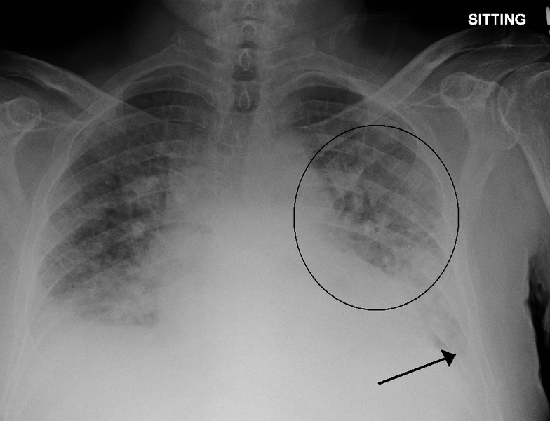
Image Source: Wikimedia Commons (2011). PulmEdema, Licensed under a creative commons licence. Available for free at: https://en.wikipedia.org/wiki/File:PulmEdema.PNG#/media/File:PulmEdema (Accessed: 19 January 2024).
Management of cardiogenic pulmonary oedema
The aims of management are to relieve symptoms and restore haemodynamic stability and tissue perfusion. Cardiogenic pulmonary oedema is a marker of poor long-term outcome, but effective early management can improve short-term survival.
Aspects of acute management include:
- Sitting the patient upright
- Oxygen if hypoxia is present aiming for appropriate target saturations (94-98% or 88-92% in chronic CO2 retainers) – this can be delivered by any method of oxygen delivery
- IV diuretics – this will need monitoring of urine output, patient weight and electrolytes. IV furosemide is often the first choice. A trial bolus of 40 mg would be appropriate initial management in someone who is diuretic naive if the blood pressure will tolerate this. If the patient is normally treated with diuretics then the initial dose should be equal to or greater than their normal dose. Diuretic boluses can be used twice or three times a day. If the patient’s blood pressure is low and they are not tolerating diuretic boluses then a 24-hour infusion can be used, such as 120-240 mg/day. However, infusions should not be used if the patient is not responsive to boluses and there is no evidence that infusions are more effective than boluses at achieving diuresis.
- Vasodilators – The choice of vasodilator depends on the aim. Sodium nitroprusside can be used to reduce arterial tone and reduce cardiac afterload; glyceryl trinitrate can be used to reduce venous tone as an adjunct to diuretics. Routine use of vasodilators is not recommended, only if there is a specific aim as above.
- Continuous positive airway pressure (CPAP) – if there is severe oedema or they are failing to respond to optimal medical therapy then CPAP should be initiated. CPAP can facilitate the delivery of oxygen, splinting open airways and raising intrapulmonary pressure and, therefore, reduce pulmonary oedema. Pressures are normally started at around 5 cmH2O and uptitrated to around 10 cmH2O. If there is associated respiratory acidosis and hypercapnia, the patient may require non-invasive ventilation (NIV) rather than CPAP. There is a risk of hypotension with CPAP as the venous return is reduced.
- Mechanical ventilation – patients who fail to respond to CPAP/NIV or those who have contraindications to CPAP/NIV should be intubated if appropriate and mechanically ventilated
- Additional support – in the presence of shock preventing optimisation of medical therapy and severe systolic dysfunction, the patient may require inotropes (e.g. dobutamine or milrinone) or mechanical support (e.g. intra-aortic balloon pump, left ventricular assist device or extracorporeal membrane oxygenation)
References
- Garan AR. Pathophysiology of cardiogenic pulmonary edema [Internet]. Colucci WS, Dardas TF, editors. UpToDate. 2022 [cited 2023 May 8]. Available from: https://www.uptodate.com/contents/pathophysiology-of-cardiogenic-pulmonary-edema
- Meyer TE. Approach to diagnosis and evaluation of acute decompensated heart failure in adults [Internet]. Colucci WS, Dardas TF, editors. UpToDate. 2023 [cited 2023 May 8]. Available from: https://www.uptodate.com/contents/approach-to-diagnosis-and-evaluation-of-acute-decompensated-heart-failure-in-adults
- Henson VL. Cardiogenic Pulmonary Oedema. Kendall JM, editor. RCEM Learning. 2021 [cited 2023 May 8]. Available from: https://www.rcemlearning.co.uk/reference/cardiogenic-pulmonary-oedema
- Matthay MA, Murray JF. Pulmonary Edema. In: Broaddus C, Mason RJ, Ernst JD, King Jr. TE, Lazarus SC, Murray JF (eds.) et al. Murray & Nadel’s Textbook of Respiratory Medicine. 6th ed. Philadelphia: Saunders; 2016. p. 1096-1117
- Oxford Medical Education. Acute Pulmonary Oedema. Oxford Medical Education. 2015 [cited 2023 May 8]. Available from: https://oxfordmedicaleducation.com/cardiology/acute-pulmonary-oedema/
- Colucci WS. Treatment of acute decompensated heart failure: Specific therapies [Internet]. Gottlieb SS, HoekstraJ, Dardas TF, editors. UpToDate. 2023 [cited 2023 May 8]. Available from: https://www.uptodate.com/contents/treatment-of-acute-decompensated-heart-failure-specific-therapies
- McDonagh TA, Metra M, Adamo M, Gardner RS, Baumbach A, Böhm M et al. 2021 ESC Guidelines for the diagnosis and treatment of acute and chronic heart failure: Developed by the Task Force for the diagnosis and treatment of acute and chronic heart failure of the European Society of Cardiology (ESC) With the special contribution of the Heart Failure Association (HFA) of the ESC. European Heart Journal. 2021;42(36); 3599-3726. Available from: doi: 10.1093/eurheartj/ehab368
Non-cardiogenic pulmonary oedema
Overview of non-cardiogenic pulmonary oedema
This describes alveolar fluid accumulation which is not secondary to increased pulmonary capillary pressure (pulmonary artery wedge pressure ≤18 mmHg in pulmonary artery or Swan-Ganz right heart catheter study).
Acute respiratory distress syndrome
Acute respiratory distress syndrome (ARDS) is an important cause of non-cardiogenic pulmonary oedema. It can result from sepsis, pneumonia, massive trauma with shock, DIC, acute pancreatitis, and inhalation of vomit, smoke or toxic chemicals.
Diagnostic (Berlin) criteria are as follows:
- Timing: onset of lung injury within 1 week of insult or new/worsening respiratory symptoms over a week
- Imaging: bilateral opacities on CXR not fully explained by effusions, collapse or nodules
- Origin of oedema: respiratory failure not fully explained by cardiac failure or fluid overload
- Oxygenation: hypoxaemia (PaO2/FiO2 ratio ≤ 300 requiring a minimum PEEP/CPAP of 5 cmH2O to maintain target sats)
Management is with respiratory support (often in ITU) and treatment of underlying causes. CPAP may be adequate in the first instance but a majority of patients will need intubation and ventilation.
High altitude pulmonary oedema
High altitude pulmonary oedema (HAPE) occurs after rapid ascent to altitudes above 2500-3000 m and typically occurs 2-5 days after the ascent. It is the most common cause of death in high altitude sickness, with a mortality rate of more than 10% when treated and 50% when untreated. This is thought to occur due to reduced environmental FiO2 at higher altitudes, which leads to uneven hypoxia-driven vasoconstriction to physiologically address ventilation-perfusion mismatch. This increases vascular resistance in the pulmonary arteries, causing pulmonary hypertension. Increased hydrostatic pressure leads to capillary leak and pulmonary oedema.
Symptoms include breathlessness (particularly reduced exercise tolerance), cough and chest tightness. Acute altitude sickness and cerebral oedema may occur at the same time.
Risk factors include: male sex, prior high altitude pulmonary oedema, fast ascent rate, high peak altitude, cold exposure, high intensity of exertion, pre-existing pulmonary hypertension and genetic factors.
The most important management strategy is to descend until there is resolution of symptoms. Oxygen may be a temporising measure if descent is delayed. Nifedipine and phosphodiesterase inhibitors may be used as adjuncts.
Other causes of non-cardiogenic pulmonary oedema
- Transfusion-related acute lung injury (TRALI) – pulmonary oedema and hypoxia within 6 hours of a blood transfusion. It can be associated with fever and usually causes hypotension compared to transfusion-associated circulatory overload (TACO), with which there is often hypertension. Troponin and NT-pro-BNP are not usually raised, but the latter may be.
- Re-expansion pulmonary oedema – occurs after rapid expansion of a collapsed lung, e.g. after large volume pleural tap or drainage (greater than 1-1.5 L) with acute shortness of breath usually occurring within hours. It is self-limiting and usually requires only supportive management.
- Neurogenic pulmonary oedema – may be precipitated by epileptic seizures, traumatic brain injury or intracranial haemorrhage. Symptom onset is usually within minutes to hours and resolves after 48-72 h.
- Post-obstructive (e.g. after upper airway obstruction due to foreign object or hanging) pulmonary oedema – occurs due to increasing negative intrathoracic pressure whilst trying to inspire despite upper airway obstruction, which substantially increases venous return and may cause vascular damage
- Salicylate toxicity and opioid overdose
- Hypoalbuminemia is not a cause of pulmonary oedema but carries a worse prognosis when present
References
- Colucci WS. Treatment of acute decompensated heart failure: Specific therapies. [Internet]. Gottlieb SS, Hoekstra J, Dardas TF, editors. UpToDate. 2022 [cited January 30 2022]. Available from: https://www.uptodate.com/contents/treatment-of-acute-decompensated-heart-failure-specific-therapies
- Garan AR. Pathophysiology of cardiogenic pulmonary edema. [Internet]. Colucci WS, Dardas TF, editors. UpToDate. 2022 [cited January 30 2022]. Available from: https://www.uptodate.com/contents/pathophysiology-of-cardiogenic-pulmonary-edema
- Tidy C, Jackson C. Acute Pulmonary Oedema. [Internet]. Patient.info. 2018 [cited January 30 2022]. Available from: https://patient.info/doctor/acute-pulmonary-oedema
- Henson VL, Kendall JM, Burrows L, Kilgour P. Cardiogenic Pulmonary Oedema. [Internet]. Royal College of Emergency Medicine. 2021 [cited January 30 2022]. Available from: https://www.rcemlearning.co.uk/reference/cardiogenic-pulmonary-oedema
- Knott L, Bonsall A. Acute (Adult) Respiratory Distress Syndrome. [Internet]. Patient.info. 2019 [cited June 01 2022]. Available from: https://patient.info/doctor/acute-adult-respiratory-distress-syndrome
- Givertz M. Noncardiogenic pulmonary edema [Internet]. Gottlieb S, Finlay G, editors. UpToDate. 2021 [cited June 01 2022]. Available from: https://www.uptodate.com/contents/noncardiogenic-pulmonary-edema
- Auerbach, Paul S. (2017). Wilderness Medicine. Elsevier. pp. 20–25
Vasculitis affecting the lungs
Overview of vasculitic lung disease
Pulmonary vasculitides are a rare set of conditions that may affect the lung or pulmonary vessels. The most drastic presentation is large volume haemoptysis and hypoxaemia. The respiratory system may be involved in all systemic vasculitides, to a variable degree, and VTE is also associated. Eliciting the underlying diagnosis is based heavily on clinical phenotype and autoimmune serological testing.
Granulomatosis with polyangiitis
Granulomatosis with polyangiitis (GPA; previously Wegener’s granulomatosis) is an ANCA (antineutrophilic cytoplasmic antibody)-associated vasculitis causing small vessel necrosis affecting the upper and lower respiratory tract, kidneys, skin and nervous system. Typically, both upper airway and pulmonary involvement are present.
Clinical features of GPA
Nasal, sinus and ear disease are found in 90%. They may be present without other manifestations and precede generalised disease by weeks to months. Patients often present with nasal congestion and discharge suggestive of a sinus infection that has failed to resolve with antibiotic therapy.
Specific features include:
- Nasal crusting and obstruction
- Chronic rhinosinusitis – can lead to septal perforation, saddle nose deformity or nasocutaneous fistula, but not nasal polyps
- Purulent or bloody nasal discharge
- Excessive tearing (epiphora)
- Sinus mucoceles
- Conductive and/or sensorineural hearing loss
Pulmonary features include:
- Tracheobronchial disease – subglottic stenosis is most common and tracheostomy may be required
- Lung nodules +/- cavitation (usually asymptomatic) – most common radiological manifestation
- Interstitial lung disease – in around 25% of patients (less common in GPA than microscopic polyangiitis)
- Alveolar haemorrhage (often seen as ground glass changes on CT) – a life-threatening pulmonary manifestation causing rapid-onset dyspnoea, cough, haemoptysis, anaemia and potentially hypoxaemia. Untreated, it can progress quickly to respiratory failure
- Pulmonary artery stenosis is reported rarely and is diagnosed with CTPA
Investigations in GPA
Findings on blood tests include:
- Positive c-ANCA in up to 75% of cases and, less classically, p-ANCA in 25% of cases
- Normochromic, normocytic anaemia
- Raised WCC and platelets (>400 x 109/L)
- Raised ESR and CRP are common, but negative ESR and CRP do not exclude GPA
- Acute creatinine/urea rise (acute kidney injury)
Chest radiograph is typically obtained as an initial investigation. Features include pulmonary nodules that may cavitate, reticular markings, diffuse, hazy opacities, lobar or segmental atelectasis, pleural opacities, and hilar adenopathy.
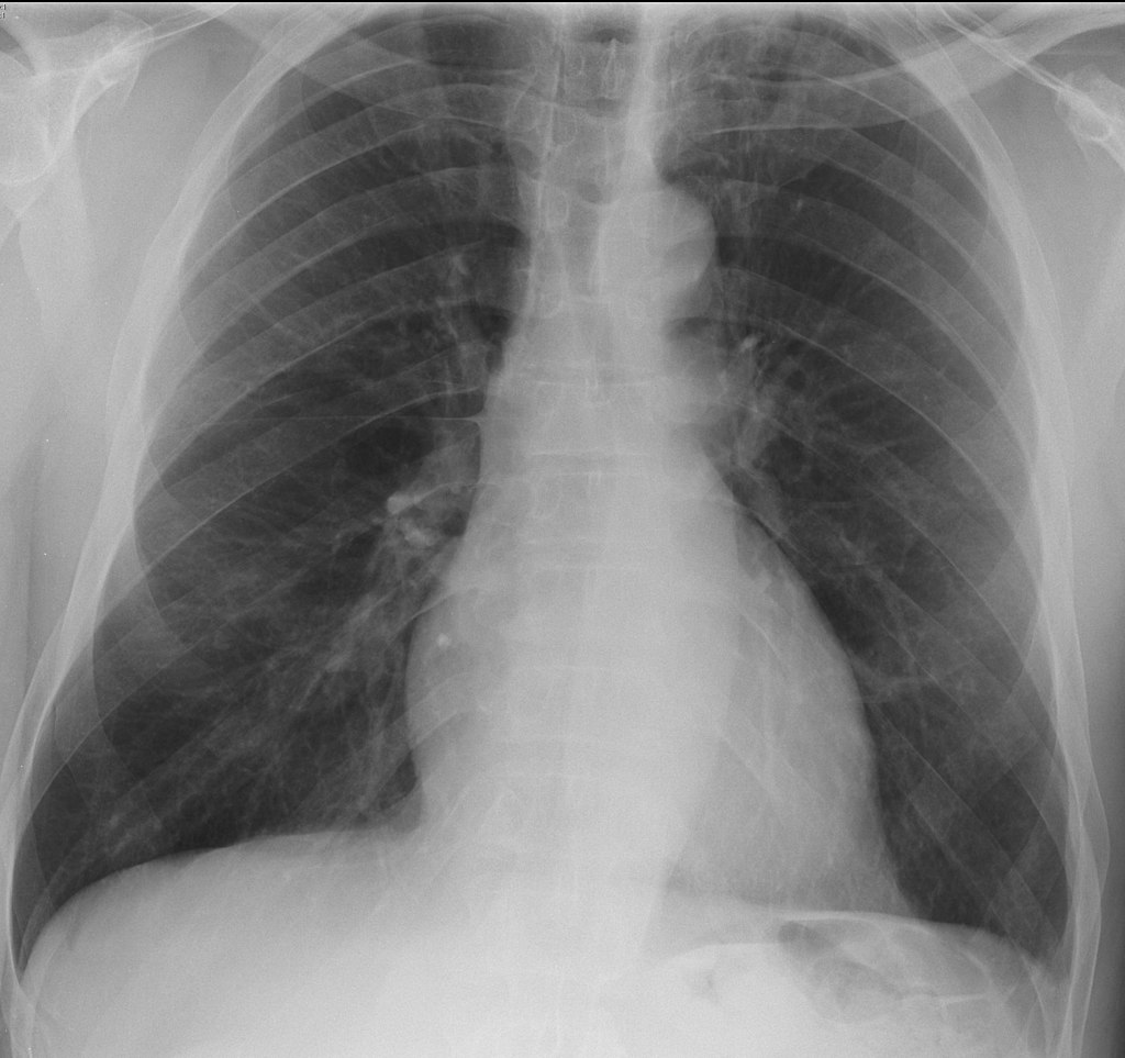
Figure 5: Image Source: Wikimedia Commons (2012). Granulomatosis with polyangiitis, Licensed under a creative commons licence. Available for free at: https://en.wikipedia.org/wiki/Granulomatosis_with_polyangiitis#/media/File:Granulomatosis_with_polyangiitis.jpg (Accessed: 19 January 2024).
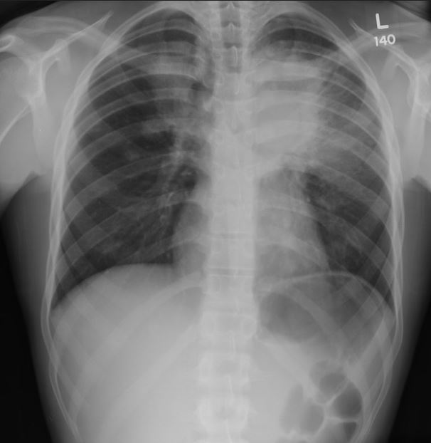
Figure 6: Image Source: Flickr (2011). Pulmonary pathology Licensed under a creative commons licence. Available for free at: https://www.flickr.com/photos/pulmonary_pathology/5958079204/in/photostream/ (Accessed: 19 January 2024).
High-resolution CT should be obtained in patients undergoing evaluation for vasculitis with abnormal chest X-ray. Common findings include:
- Multiple pulmonary nodules
- Patchy or diffuse ground-glass or consolidative opacities (pulmonary haemorrhage or active vasculitis)
- Small, peripheral, wedge-shaped densities (pulmonary microinfarction)
- Enlarged, irregular, stellate-shaped, peripheral pulmonary arteries (‘vasculitis’ sign)
- Tracheal or bronchial stenosis
Other investigations may include:
- CT of the sinuses – to evaluate chronic rhinosinusitis in suspected GPA.
- Bronchoscopy – to assess and take biopsies of endobronchial disease and perform bronchoalveolar lavage (BAL). BAL can identify pulmonary haemorrhage when imaging shows diffuse parenchymal opacities. Subsequent lavage aliquots become increasingly haemorrhagic. Transbronchial biopsy is often too small to make a diagnosis of GPA but can identify alternative diagnoses.
- Lung biopsy – when there are single or multiple pulmonary nodules and extrapulmonary disease is absent or not accessible to biopsy. Transthoracic needle biopsies are usually too small for a definitive diagnosis, so lung tissue must be obtained via video-assisted thoracoscopic surgery (VATS) or thoracotomy.
Management of GPA
Treatment involves an induction regimen of glucocorticoids and rituximab/cyclophosphamide. Plasma exchange may be useful in patients with pulmonary haemorrhage not readily responding to other therapies and optimal supportive care.
In patients with less severe disease without any organ- or life-threatening features (i.e. those with only ENT features, arthritis and/or pulmonary nodules), initial induction therapy with glucocorticoids and weekly methotrexate or, alternatively, mycophenolate mofetil (MMF) monotherapy is suggested. Note that methotrexate is associated with pulmonary fibrosis.
Maintenance therapy typically involves tapering the glucocorticoids and using rituximab or azathioprine as maintenance therapy, usually after a few months.
Microscopic polyangiitis
Microscopic polyangiitis (MPA) is an ANCA-associated necrotising vasculitis without granulomatous inflammation. It predominantly affects small vessels including the lungs and kidneys. Features and presentation are similar to GPA, but it typically lacks upper respiratory tract involvement.
MPA is generally associated with p-ANCA (versus c-ANCA in GPA) but either can be positive. MPA may still be the preferred diagnosis even with a positive c-ANCA over GPA if there are no ear, nose or throat nor upper respiratory features, although they can be difficult to distinguish.
The most common pulmonary manifestation is pulmonary haemorrhage, which can be seen in up to 40% of cases. Other clinical features, as well as investigation and management, are similar to GPA. Some classify the term of idiopathic pauci-immune pulmonary capillaritis (IPIPC) as a lung-specific subtype of MPA as it is also associated with p-ANCA.
Treatment is with IV methylprednisolone and cyclophosphamide/rituximab.
Eosinophilic granulomatosis with polyangiitis
Eosinophilic granulomatosis with polyangiitis (EGPA) (previously Churg-Strauss syndrome) is an ANCA-positive, small- and medium-sized vasculitis characterised by chronic rhinosinusitis, asthma and eosinophilia. The most commonly involved organ is the lung.
EGPA is typically associated with p-ANCA (versus c-ANCA in GPA), although the antibodies are not found in all patients. It is rare to find an isolated positive c-ANCA in EGPA and this would put the diagnosis of EGPA in serious doubt. Leucocytosis with eosinophilia is also a feature, as are raised CRP and ESR. Similarly to GPA, renal impairment can also be seen in EGPA.
Clinical features of EGPA
The clinical features of EGPA typically develop in three phases:
- Prodromal phase: atopy, asthma and allergic rhinitis, sinusitis, nasal polyps
- Eosinophilic phase: eosinophilic organ infiltration (e.g. in lung, heart, GI system)
- Vasculitic phase: purpura, peripheral neuropathy and constitutional symptoms
Asthma is the cardinal feature of EGPA. It should be suspected in patients with poorly controlled asthma on moderate doses of inhaled corticosteroids. EGPA-associated asthma often requires long-term courses of systemic glucocorticoids. Commonly, asthma exacerbations increase during the vasculitic phase but the initial asthmatic presentation may predate the vasculitic phase by up to 10 years. Haemoptysis is unusual, unlike in GPA and MPA.
Other respiratory manifestations include:
- ENT involvement in 70-85% – otitis media, allergic rhinitis, nasal obstruction, sinusitis, nasal polyps
- Chronic rhinosinusitis can lead to chronic serous otitis media and sensorineural hearing loss
- Imaging findings of bilateral peripheral/random consolidation or ground-glass changes and eosinophilic pleural effusions
Investigation and management of EGPA
Investigation and treatment are similar to other ANCA-associated vasculitides, such as GPA and MPA.
Induction treatment is with cyclophosphamide and IV methylprednisolone, especially if there are organ- or life-threatening features. Rituximab can be a substitute for cyclophosphamide as per clinician preference.
Anti-GBM (Goodpasture) disease
Anti-glomerular basement membrane (GBM) disease is a small-vessel vasculitis associated with anti-GBM antibodies (against type IV collagen). It is rare, with peak incidences in the 3rd and 6-7th decades. The classic presentation is with large volume haemoptysis and rapidly progressive glomerulonephritis, although younger patients are more likely to present with pulmonary haemorrhage and older patients with isolated glomerulonephritis.
It is thought that a triggering stimulus is needed on a background of genetic predisposition (HLA-DR15 and HLA-DR4 are associated). Stimuli include drugs such as alemtuzumab, cocaine use, infections including influenza, smoking and extracorporeal shockwave lithotripsy for ureteric stones.
Clinical features of anti-GBM disease
The most common pulmonary feature is alveolar haemorrhage. Other features include:
- Systemic symptoms such as malaise, weight loss, fever or arthralgia for typically only a few weeks
- Iron-deficiency anaemia due to prolonged pulmonary bleeding
- Rapidly progressive glomerulonephritis causing nephritic syndrome and proteinuria (not usually in the nephrotic range)
Investigations in anti-GBM disease
Diagnosis of anti-GBM disease requires demonstration of anti-GBM antibodies either in the serum or the kidney.
ANCA testing should be performed in all patients suspected of anti-GBM disease, as the presence of both anti-GBM and ANCA antibodies supports a diagnosis of double-positive anti-GBM and ANCA-associated disease. This requires aggressive early treatment of anti-GBM disease and maintenance of immunosuppression for ANCA-associated vasculitis.
Other investigations include:
- Urine dipstick and microscopy to assess for nephritic syndrome
- Full blood count, liver function tests, urea and electrolytes, clotting +/- group and save depending on presence of pulmonary haemorrhage
- Kidney biopsy should be performed without delay unless contraindicated
- Serum assay for anti-GBM antibodies if kidney biopsy cannot be performed or will be delayed
- Bronchoalveolar lavage is the gold standard to diagnose pulmonary haemorrhage
- Alveolar haemorrhage may be seen on chest X-ray (patchy or diffuse opacities) or CT (ground-glass or consolidative opacities)
- Pulmonary function testing is not routinely performed but may show increased carbon monoxide diffusing capacity (DLCO)
Management of anti-GBM disease
The standard treatment is urgent plasmapheresis to rapidly remove culprit antibodies and subsequent cyclophosphamide and glucocorticoid to inhibit autoantibody production and resolve inflammation. Plasmapheresis can be initiated before antibody testing comes back if clinical suspicion of the disease is high. Rituximab and mycophenolate have emerging data as agents for induction of remission.
A conservative approach may be appropriate in patients without alveolar haemorrhage (i.e. only renal involvement) but needing dialysis at presentation. This is because their kidneys are likely fully and permanently damaged already (as evidenced by their need for dialysis) and there is a low probability of recovery of kidney function despite plasmapheresis with immunosuppression. If subsequent renal biopsy shows 100% crescents, this is even more reason to continue a conservative approach as the chance of renal recovery is low. A short trial of treatment could be considered if patients are particularly young and able to tolerate aggressive treatment, if the presentation is very acute and thus irreversible damage is less predictable, or if renal biopsy shows only focal crescents or evidence of concurrent ANCA-associated vasculitis.
Pulmonary manifestations of other vasculitides
Rheumatoid arthritis
If longstanding, rheumatoid arthritis is associated with interstitial lung disease, inflammation of the pleura (pleuritis and pleural effusions) and, rarely, pulmonary vasculitis leading to pulmonary hypertension.
Takayasu arteritis
Pulmonary arteritis may cause pulmonary hypertension.
Giant cell arteritis
Pulmonary involvement is rare, but interstitial lung disease has been reported.
Behçet’s disease
Behçet’s may affect the pulmonary artery (the second most commonly affected site after the aorta), generally causing aneurysms rather than thrombosis. Haemoptysis is the most prevalent pulmonary presenting symptom. Behçet’s disease is also typically associated with orogenital ulceration, arthralgia, uveitis and pericarditis.
Epidemiologically, it is classically seen in people with Middle Eastern, Southeast Asian and Mediterranean ethnic origin. It is classically seronegative but can be investigated with a pathergy test.
Hughes-Stovin syndrome
This is thought to be a cardiovascular variant of Behçet’s disease, characterised by widespread pulmonary or bronchial artery aneurysms and deep vein thrombosis.
Idiopathic pauci-immune pulmonary capillaritis
IPIPC is a rare cause of pulmonary vasculitis. It doesn’t present with the systemic features of other vasculitides and is largely lung specific. Although pauci-immune, it may be associated with a weakly positive p-ANCA. Some consider this to be a lung-specific form of MPA. Treatment is with glucocorticoids and cyclophosphamide.
ANCA-associated vasculitides
It may be helpful to group some of the below conditions as investigation and management are similar. The primary ANCA-associated vasculitides are GPA, MPA, EGPA and IPIPC. Treatment, where there is life-/organ-threatening disease, is usually IV methylprednisolone and cyclophosphamide/rituximab.
| Upper airway and ENT features | Asthma | Nodules, cavities and infiltrates | Alveolar haemorrhage | c-ANCA positivity (%) | p-ANCA positivity (%) | |
|---|---|---|---|---|---|---|
| GPA | Very common | Rare | Very common | 5-10% | 70 | 25 |
| MPA | Rare | Rare | Uncommon | 10-30% | 25 | 60 |
| EGPA | Very common | Very common | Very common | Rare | < 5 | 35 |
| Anti-GBM | Rare | Rare | Very common | 25-60% | 5 | 25 |
Table 5 Typical pulmonary manifestations of several small-vessel vasculitides
References
- King TE Jr. Granulomatosis with polyangiitis and microscopic polyangiitis: Respiratory tract involvement [Internet]. Flaherty KR, Glassock RJ, Dieffenbach P, editors. UpToDate. 2023 [cited August 14 2023]. Available from: https://www.uptodate.com/contents/granulomatosis-with-polyangiitis-and-microscopic-polyangiitis-respiratory-tract-involvement
- Merkel PA, Kaplan AA, Falk RJ. Granulomatosis with polyangiitis and microscopic polyangiitis: Induction and maintenance therapy [Internet]. Appel GB, Fervenza FC, Lam AQ, editors. UpToDate. 2022 [cited January 19 2022]. Available from: https://www.uptodate.com/contents/granulomatosis-with-polyangiitis-and-microscopic-polyangiitis-induction-and-maintenance-therapy
- Falk RJ, Merkel PA, King TE Jr. Granulomatosis with polyangiitis and microscopic polyangiitis: Clinical manifestations and diagnosis. [Internet]. Glassock RJ, Appel GB, Lam QA, Seo P, editors. UpToDate. 2022 [cited January 19 2022]. Available from: https://www.uptodate.com/contents/granulomatosis-with-polyangiitis-and-microscopic-polyangiitis-clinical-manifestations-and-diagnosis
- King TE Jr. Clinical features and diagnosis of eosinophilic granulomatosis with polyangiitis (Churg-Strauss) [Internet]. Flaherty KR, Glassock RJ, Bochner BS, Dieffenbach P, editors. UpToDate. 2020 [cited January 19 2022]. Available from: https://www.uptodate.com/contents/clinical-features-and-diagnosis-of-eosinophilic-granulomatosis-with-polyangiitis-churg-strauss
- Kaplan AA, Appel GB, Pusey CD. Anti-GBM (Goodpasture) disease: Treatment and prognosis [Internet]. Glassock RJ, Fervena FC, Lam AQ, editors. UpToDate. 2022 [cited August 14 2023]. Available from: https://www.uptodate.com/contents/anti-gbm-goodpasture-disease-treatment-and-prognosis
- Frankel SK, Schwarz MI. The pulmonary vasculitides. Am J Respir Crit Care Med. 2012 Aug 1;186(3):216-24. doi: 10.1164/rccm.201203-0539CI. Epub 2012 Jun 7. PMID: 22679011
- Pusey CD. Anti-GBM (Goodpasture) disease: Pathogenesis, clinical manifestations, and diagnosis. [Internet]. Segelmark M, Glassock RJ, Fervenza FC, Lam AQ, editors. UpToDate. 2021 [cited January 30 2022]. Available from: https://www.uptodate.com/contents/anti-gbm-goodpasture-disease-pathogenesis-clinical-manifestations-and-diagnosis
- Khalid U, Saleem T. Hughes-Stovin syndrome. Orphanet J Rare Dis. 2011;6:15. doi:10.1186/1750-1172-6-15
- McAdoo SP, Tanna A, Hrušková Z, et al. Patients double-seropositive for ANCA and anti-GBM antibodies have varied renal survival, frequency of relapse, and outcomes compared to single-seropositive patients. Kidney Int. 2017;92(3):693-702. doi:10.1016/j.kint.2017.03.014
- Weerakkody Y, Idiopathic pauci immune pulmonary capillaritis. Reference article, Radiopaedia.org. https://doi.org/10.53347/rID-19687
- Mehrotra AK, Gupta PR, Khublani TK, Anupam, Soni S, Feroz A. Isolated pauci-immune pulmonary capillaritis. Lung India. 2015 Jan-Feb; 32(1):56-9. doi: 10.4103/0970-2113.148453. PMID: 25624599; PMCID: PMC4298921
- DeVrieze BW, Hurley JA. Goodpasture Syndrome. [Updated 2022 Sep 26]. In: StatPearls [Internet]. Treasure Island (FL): StatPearls Publishing; 2023 Jan-. Available from: https://www.ncbi.nlm.nih.gov/books/NBK459291/
- Ronco P et al. KDIGO 2021 Clinical Practice guidelines for management of glomerular disease. Journal of the international society of nephrology. 2021; 100(4s):s231
- Kitching, A.R., Anders, HJ., Basu, N. et al. ANCA-associated vasculitis. Nat Rev Dis Primers. 2020; 6(1):71. https://doi.org/10.1038/s41572-020-0204-y
- Feragalli B, Mantini C, Sperandeo M, Galluzzo M, Belcaro G, Tartaro A, Cotroneo AR. The lung in systemic vasculitis: radiological patterns and differential diagnosis. Br J Radiol. 2016;89(1061):20150992. doi: 10.1259/bjr.20150992. Epub 2016 Feb 15. PMID: 26876879; PMCID: PMC4985478
- Levy J, Hammad T, Coulthart A et al. Clinical features and outcome of patients with both ANCA and anti-GBM antibodies. Kidney International. 2004; 66(4):1535-1540. https://doi.org/10.1111/j.1523-1755.2004.00917
Pulmonary complications of sickle cell disease
Overview of pulmonary complications of sickle cell disease
Sickle cell disease (SCD) refers to a group of haemoglobinopathies where the sickle mutation is co-inherited with another mutation affecting the other beta globin subunit in haemoglobin (Hb), causing reduced or absent normal beta globin production. Sickle cell anaemia (SCA) is the term used when someone is homozygous for the sickle mutation, resulting in the formation of the HbS (sickle haemoglobin) molecule. The HbS molecule is much less soluble than normal HbA when deoxygenated, causing red cells to deform (sickle). The complications seen in SCD occur as a result of this sickling, such as vaso-occlusive events, haemolysis and pulmonary complications such as acute chest syndrome and pulmonary hypertension (PH).
The pulmonary complications of SCD are particularly important because they are a major cause of morbidity and mortality, with acute chest syndrome and pulmonary hypertension having the highest associated mortality in these populations.
There are numerous pulmonary complications seen in SCD:
- Acute chest syndrome (discussed in detail later)
- Asthma – asthma is much more common in people with SCD, with up to 70% of children with SCD having airway hypersensitivity. Asthma is more common in children with a history of acute chest syndrome and children with asthma have a higher incidence of acute chest syndrome.
- Sleep disordered breathing – increased incidence in people with SCD, and is commonly due to obstructive sleep apnoea, associated with nocturnal hypoxaemia. These are linked with increased risk of vaso-occlusive events, cardiovascular complications and neurological disease.
- Pneumonia – more common in people with SCD. Common causative organisms include Chlamydophila pneumoniae and Mycoplasma pneumoniae. They’re also at increased risk of infection from encapsulated organisms such as Streptococcus pneumoniae and Haemophilus influenzae as they are functionally asplenic.
- Pulmonary venous thromboembolism – SCD is a hypercoagulable state, leading to increased risk of thromboembolisms
- Pulmonary hypertension – occurs in 6-11% of people with SCD and is an independent risk factor for mortality
- Chronic dyspnoea – likely multifactorial and often results in a reduction in exercise capacity in early to late adolescence; contributing factors include the pulmonary complications listed above, anaemia and myocardial dysfunction
- Pulmonary fibrosis – occasionally seen in people with recurrent acute chest syndrome episodes
Acute chest syndrome
Overview of acute chest syndrome
Acute chest syndrome is defined as a new pulmonary infiltrate on chest imaging associated with fever and/or respiratory symptoms in someone with SCD. Acute chest syndrome is the second most common cause of hospitalisation in people with SCD after vaso-occlusive events and is a leading cause of morbidity and mortality with a lifetime incidence of around 50% in those with SCD.
Clinically, acute chest syndrome is indistinguishable from pneumonia, but acute management of both is generally the same.
Increased incidence of acute chest syndrome occurs with:
- More severe genotypes of SCD, e.g. HbS or HbS-beta0-thalassaemia
- Lower HbF (foetal haemoglobin) concentrations
- Acute pain (vaso-occlusive events)
- Other respiratory problems: asthma, chronic hypoxaemia
- Environmental factors: active smoking, environmental smoke exposure, winter months
Common triggers for acute episodes of acute chest syndrome include:
- Respiratory infection or asthma
- Pulmonary infarction
- Fat embolism
- Vaso-occlusive pain
Multiple triggers may coexist in the same person, whereas in some cases, an acute trigger is not identified. Infection and asthma are more common triggers in children, while vaso-occlusive pain and fat embolism are more common causes in adults.
Clinical features and investigation of acute chest syndrome
The presentation and severity is variable. Some people present with an acute chest syndrome while others will develop it a few days into admission for another reason, mainly vaso-occlusive events. The most frequent presenting symptoms include:
- Fever
- Cough
- Chest pain
- Shortness of breath
- Pain preceding the acute chest syndrome episode (pain in arms, legs, rib and sternal pain)
Acute chest syndrome can be very severe, with around 10% of cases having respiratory failure so severe they require invasive mechanical ventilation.
These are pertinent investigations to perform in a person with suspected acute chest syndrome:
- Full blood count including reticulocytes
- Haemoglobinopathy screen with HbS percentage – HbS percentage needs to be monitored before and after transfusions (simple or exchange)
- Group and save
- Metabolic profile – acute chest syndrome can be associated with acute multiorgan failure syndrome
- Renal profile
- Liver function tests
- Cultures: blood and sputum
- Respiratory viral screen and atypical pneumonia screen
- Arterial blood gas – not routine unless there is significant respiratory compromise
- Chest x-ray – to identify if there are any new pulmonary infiltrates
- CT pulmonary angiogram – if a pulmonary embolism is suspected
Management of acute chest syndrome
The main aims of treatment are early intervention to prevent deterioration and early escalation if required.
Acute interventions include:
- Analgesia – to reduce hypoventilation and atelectasis
- Incentive spirometry and chest physiotherapy – to reduce atelectasis
- Empirical broad-spectrum antibiotics – as per local protocols covering for community-acquired pneumonia
- IV fluids – care should be taken to avoid hypervolaemia as this can precipitate pulmonary oedema
- Bronchodilators – if wheezing is present or there is a history of asthma
- Venous thromboembolism prophylaxis
- Respiratory support – supplementary oxygen should be given to maintain saturations ≥95% or at their baseline; may require continuous positive airway pressure, non-invasive ventilation or invasive ventilation
- Simple or exchange transfusion – prompt transfusion decreases progression of disease; the aim of simple transfusion is to increase haematocrit to ≥30% or Hb >100 g/L and the aim of exchange transfusion is to reduce HbS to <30%; Hb should not rise above 110 g/L due to risks of hyperviscosity.
| Indications for simple transfusion | Indications for exchange transfusion |
|---|---|
|
|
Table 6 Indications for simple versus exchange transfusion in acute chest syndrome
Exchange transfusion allows for large transfusion volumes (6-8 units) whilst not risking hyperviscosity.
There are also numerous preventative measures that can be put in place to reduce the incidence of acute chest syndrome:
- Regular incentive spirometry if admitted to hospital for other reasons such as vaso-occlusive events
- Prevent infections: prophylactic penicillin and up-to-date vaccinations
- Optimise underlying lung conditions, e.g. asthma
- Reduce vaso-occlusive events
- Hydroxyurea – increases HbF concentration
- Chronic transfusions or exchange transfusion programme (generally for patients with severe disease and recurrent episodes)
References
- Chapman S, Robinson G, Shrimanker R, Turnbull C, Wrightson J (eds.). Oxford Handbook of Respiratory Medicine. 4th ed. Oxford: Oxford University Press; 2021
- Machado RF, Gladwin MT. Pulmonary Complications of Hematological Diseases. In: Broaddus C, Mason RJ, Ernst JD, King Jr. TE, Lazarus SC, Murray JF (eds.) et al. Murray & Nadel’s Textbook of Respiratory Medicine. 6th ed. Philadelphia: Saunders; 2016. p. 1653-1670
- Klings ES, Willen S. Overview of the pulmonary complications of sickle cell disease [Internet]. Mandel J, Vichinsky EP, Dieffenback P, Tirnauer JS, editors. UpToDate. 2022 [cited 2023 May 9]. Available from: https://www.uptodate.com/contents/overview-of-the-pulmonary-complications-of-sickle-cell-disease
- Miller AC, Gladwin MT. Pulmonary Complications of Sickle Cell Disease. American Journal of Respiratory and Critical Care Medicine. 2012;185(11); 1154-1165. Available from: doi: 10.1164/rccm.201111-2082CI
- Vichinsky EP. Overview of the clinical manifestations of sickle cell disease. DeBaun MR, Tirnauer JS, editors. UpToDate. 2023 [cited 2023 May 13]. Available from: https://www.uptodate.com/contents/overview-of-the-clinical-manifestations-of-sickle-cell-disease
- Howard J, Hart N, Roberts-Harewood M, Cummins M, Awogbae M, Davis B. Guidelines on the management of acute chest syndrome in sickle cell disease. British Journal of Haematology. 2015;169(4); 492-505. Available from: doi: 10.1111/bjh.13348
Complete Guide to Clinical Genetics ST3 Application and Recruitment (2027)
Written by Medibuddy. Last updated: October 2026. Clinical Genetics is

Complete Guide to Clinical Radiology ST3 Application and Recruitment (2027)
Recruitment paused: NHS England is not running Clinical Radiology ST3+ recru

Complete Guide to Paediatric Surgery ST3 Application and Recruitment in United Kingdom (2026)
Written by Medibuddy. Last updated: October 2026. If you’re planning


Take your subscriptions with you
Our mobile app allows you to access your interview and exam question banks wherever you are.

