


 Close
Close
 18th February 2025
Clinical features of PE
Pulmonary embolism (PE) is a condition where one or more dislodged thrombi occlude the pulmonary vasculature, which may cause severe respiratory dysfunction.
Clinical features of acute PE include dyspnoea, pleuritic chest pain, hypoxaemia, tachycardia, cough and haemoptysis. Dizziness, syncope, elevated JVP, gallop rhythm and tricuspid regurgitant murmur are suggestive of right ventricular strain and failure. There may be associated distal limb findings suggestive of a DVT. Hypotension and cardiac arrest are the most serious presentations of acute PE.
Risk factors for PE
Around 30-50% of VTEs are unprovoked, with no identifiable risk factor. However, important risk factors include (from the European Society of Cardiology):
| Strong risk factors
(odds ratio >10) |
Moderate risk factors
(odds ratio 2-9) |
Weak risk factors
(odds ratio <2) |
|---|---|---|
|
|
|
Table 1 Risk factors for PE
Assessing the clinical probability of PE
Various clinical scoring systems are used to assist with objective assessment of the probability of PE and therefore the need (or not) to do further investigations:
- If PE is a differential but with low suspicion: pulmonary embolism rule-out criteria (PERC rule)
- If there is significant suspicion of PE (and/or PERC is positive): Wells’ criteria for PE
| PERC tool for PE: if the suspicion of a PE is low (pre-test probability <15%) AND the total score is 0 then a PE can be ruled out with any further assessment | |
| Age ≥ 50 years old | +1 |
| HR ≥ 100 beats per minute | +1 |
| SaO2 on air < 95% | +1 |
| Unilateral leg swelling | +1 |
| Haemoptysis | +1 |
| Recent surgery or trauma (within past 4 weeks requiring general anaesthesia) | +1 |
| Prior PE or DVT | +1 |
| Hormone use (oral contraceptive, hormone replacement or oestrogen therapy in male/female) | +1 |
Table 2 PERC tool for PE
| Wells’ score for PE: if the total score is ≤4 then PE is unlikely and D-dimer should be checked. If negative, then PE can be ruled out; if positive, CTPA should be performed. If Wells’ score is >4 then proceed to CTPA | |
| Symptoms and signs of a DVT | +3 |
| PE is likely or equally likely diagnosis | +3 |
| Heart rate > 100 | +1.5 |
| Prior PE or DVT | +1.5 |
| Immobilisation for ≥ 3 days or surgery in past 4 weeks | +1.5 |
| Malignancy with active/palliative treatment within past 6 months | +1 |
| Haemoptysis | +1 |
Table 3 Wells’ score for PE
Investigations in PE in a non-pregnant patient
Common investigations in suspected PE include:
- ECG: the most common finding is sinus tachycardia; other abnormalities include non-specific ST-segment and T-wave abnormalities, right axis deviation, right bundle branch block, or the textbook (but unusual) finding of “S1Q3T3” (S wave in lead 1, Q wave and T-wave inversion in lead 3) (figure 3)
- D-dimer: with low clinical suspicion, normal D-dimer can exclude PE
- Chest X-ray: usually normal; may show atelectasis, pleural effusion, or elevation of the hemidiaphragm
- Lower limb venous duplex ultrasound with compression: to investigate suspected concurrent DVT. Proximal DVT in suspected PE is sufficient to start anticoagulation without further investigation
- CTPA (figures 1 and 2): gold standard to confirm PE
- Ventilation-perfusion (VQ) scan: alternative to CTPA in those in whom the latter is contraindicated (e.g. renal impairment, contrast allergy) or in certain patient groups to minimise radiation exposure (e.g. pregnant patients). A VQ scan is of limited use with significant underlying lung disease, abnormal chest radiograph, left ventricular failure or congestive cardiac failure
- Echocardiogram: useful for haemodynamically unstable patients who cannot have CTPA – the presence of right ventricular dysfunction is sufficient to justify urgent thrombolysis. Signs of PE include right ventricle (RV) dilatation and hypokinesis, McConnell’s sign (segmental RV regional wall motion abnormality without apical involvement), tricuspid regurgitation and pulmonary hypertension
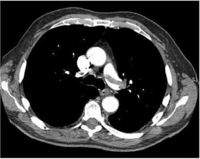
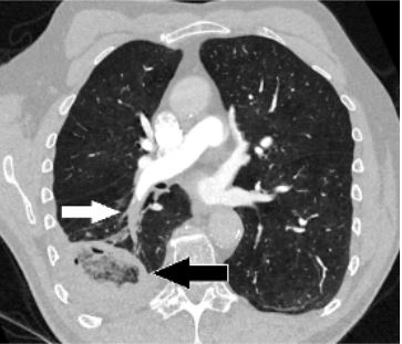
Figure 1: Image Source: Thrombosis Journal (2007). 10.1186, Licensed under a creative commons licence. Available for free at: https://doi.org/10.1186/1477-9560-5-20 (Accessed: 19 January 2024).
Figure 2: Image Source: Wikimedia Commons (2017). Pulmonary_embolism_CTPA, Licensed under a creative commons licence. Available for free at: https://en.wikipedia.org/wiki/Pulmonary_embolism#/media/File:Pulmonary_embolism_CTPA.JPEG (Accessed: 19 January 2024).
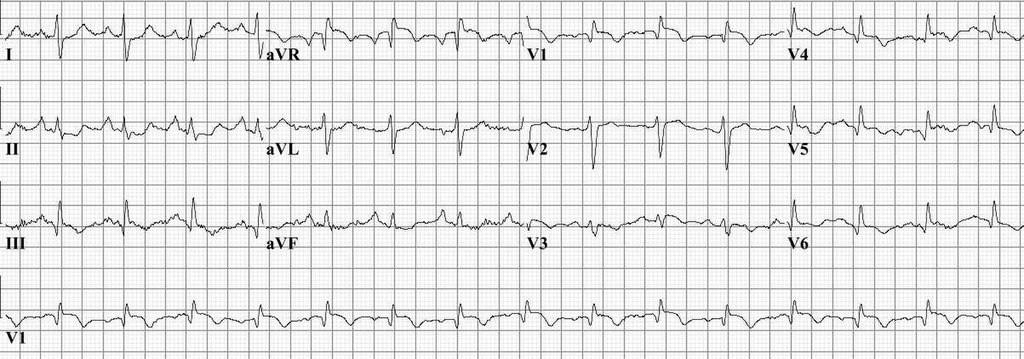
Image Source: Wikimedia Commons (2010). Pulm embolism, Licensed under a creative commons licence. Available for free at: https://en.wikipedia.org/wiki/Pulmonary_embolism#/media/File:Pulm_embolism.jpg (Accessed: 19 January 2024).
Diagnosing PE in pregnancy
Diagnosing acute PE in a pregnant person poses important considerations. D-dimer can be raised in pregnancy due to production by the normal placenta. Furthermore, there is consideration of radiation risks to developing breast tissue as well as the foetus. The approach to diagnosis in this special circumstance is, therefore, tailored to address these. The approach is as follows:
- D-dimer testing: not currently used in pregnancy in the UK, although this may change.
- If concurrent DVT is suspected: lower limb compression venous ultrasound is required. If a DVT is found, then anticoagulation can be started (typically LMWH or UFH; DOACs and warfarin are contraindicated in pregnancy) and no further irradiating tests are needed. Note that in the absence of clinical features of a DVT, a bilateral leg US should not be routinely requested.
- If no concurrent DVT is suspected: anticoagulation (LMWH or UFH) should be started and further investigations ordered. First, a CXR should be requested to ensure there is no underlying lung pathology that may impact the interpretation of a VQ scan. The radiation risk of a CXR to mother and foetus is trivial. VQ scans and CTPA are the next stage of investigation. If the CXR is abnormal, a CTPA is required, as a VQ scan would not be interpretable. If the CXR is normal, either VQ scan or CTPA can be used based on local availability and patient preference, after appropriate counselling regarding radiation risk. Typically, a VQ scan reduces radiation risk to maternal breast tissue and has minimally increased radiation exposure to the foetus; CTPA reduces radiation risk to the foetus but proportionally increases the risk of maternal breast cancer more than the VQ scan increases the risk of childhood cancers to the foetus. If a VQ scan is equivocal, CTPA is indicated, which has a higher specificity and sensitivity.
Complete Guide to Clinical Genetics ST3 Application and Recruitment (2027)
Written by Medibuddy. Last updated: October 2026. Clinical Genetics is

Complete Guide to Clinical Radiology ST3 Application and Recruitment (2027)
Recruitment paused: NHS England is not running Clinical Radiology ST3+ recru

Complete Guide to Paediatric Surgery ST3 Application and Recruitment in United Kingdom (2026)
Written by Medibuddy. Last updated: October 2026. If you’re planning


Take your subscriptions with you
Our mobile app allows you to access your interview and exam question banks wherever you are.

