


 Close
Close
 18th February 2025
Vasculitis affecting the lungs
Overview of vasculitic lung disease
Pulmonary vasculitides are a rare set of conditions that may affect the lung or pulmonary vessels. The most drastic presentation is large volume haemoptysis and hypoxaemia. The respiratory system may be involved in all systemic vasculitides, to a variable degree, and VTE is also associated. Eliciting the underlying diagnosis is based heavily on clinical phenotype and autoimmune serological testing.
Granulomatosis with polyangiitis
Granulomatosis with polyangiitis (GPA; previously Wegener’s granulomatosis) is an ANCA (antineutrophilic cytoplasmic antibody)-associated vasculitis causing small vessel necrosis affecting the upper and lower respiratory tract, kidneys, skin and nervous system. Typically, both upper airway and pulmonary involvement are present.
Clinical features of GPA
Nasal, sinus and ear disease are found in 90%. They may be present without other manifestations and precede generalised disease by weeks to months. Patients often present with nasal congestion and discharge suggestive of a sinus infection that has failed to resolve with antibiotic therapy.
Specific features include:
- Nasal crusting and obstruction
- Chronic rhinosinusitis – can lead to septal perforation, saddle nose deformity or nasocutaneous fistula, but not nasal polyps
- Purulent or bloody nasal discharge
- Excessive tearing (epiphora)
- Sinus mucoceles
- Conductive and/or sensorineural hearing loss
Pulmonary features include:
- Tracheobronchial disease – subglottic stenosis is most common and tracheostomy may be required
- Lung nodules +/- cavitation (usually asymptomatic) – most common radiological manifestation
- Interstitial lung disease – in around 25% of patients (less common in GPA than microscopic polyangiitis)
- Alveolar haemorrhage (often seen as ground glass changes on CT) – a life-threatening pulmonary manifestation causing rapid-onset dyspnoea, cough, haemoptysis, anaemia and potentially hypoxaemia. Untreated, it can progress quickly to respiratory failure
- Pulmonary artery stenosis is reported rarely and is diagnosed with CTPA
Investigations in GPA
Findings on blood tests include:
- Positive c-ANCA in up to 75% of cases and, less classically, p-ANCA in 25% of cases
- Normochromic, normocytic anaemia
- Raised WCC and platelets (>400 x 109/L)
- Raised ESR and CRP are common, but negative ESR and CRP do not exclude GPA
- Acute creatinine/urea rise (acute kidney injury)
Chest radiograph is typically obtained as an initial investigation. Features include pulmonary nodules that may cavitate, reticular markings, diffuse, hazy opacities, lobar or segmental atelectasis, pleural opacities, and hilar adenopathy.
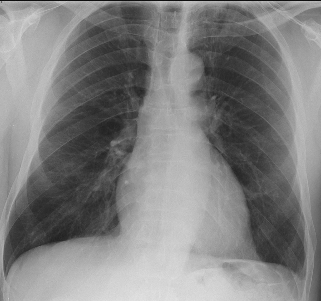
Figure 5: Image Source: Wikimedia Commons (2012). Granulomatosis with polyangiitis, Licensed under a creative commons licence. Available for free at: https://en.wikipedia.org/wiki/Granulomatosis_with_polyangiitis#/media/File:Granulomatosis_with_polyangiitis.jpg (Accessed: 19 January 2024).
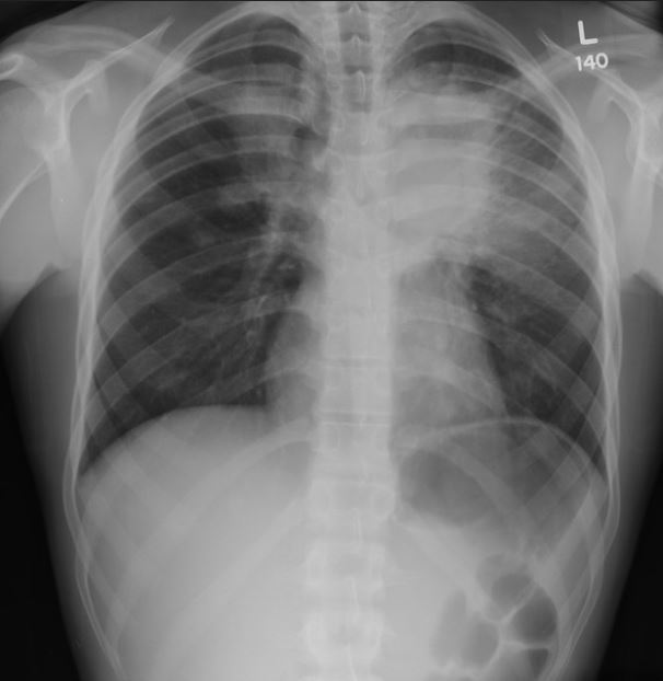
Figure 6: Image Source: Flickr (2011). Pulmonary pathology Licensed under a creative commons licence. Available for free at: https://www.flickr.com/photos/pulmonary_pathology/5958079204/in/photostream/ (Accessed: 19 January 2024).
High-resolution CT should be obtained in patients undergoing evaluation for vasculitis with abnormal chest X-ray. Common findings include:
- Multiple pulmonary nodules
- Patchy or diffuse ground-glass or consolidative opacities (pulmonary haemorrhage or active vasculitis)
- Small, peripheral, wedge-shaped densities (pulmonary microinfarction)
- Enlarged, irregular, stellate-shaped, peripheral pulmonary arteries (‘vasculitis’ sign)
- Tracheal or bronchial stenosis
Other investigations may include:
- CT of the sinuses – to evaluate chronic rhinosinusitis in suspected GPA.
- Bronchoscopy – to assess and take biopsies of endobronchial disease and perform bronchoalveolar lavage (BAL). BAL can identify pulmonary haemorrhage when imaging shows diffuse parenchymal opacities. Subsequent lavage aliquots become increasingly haemorrhagic. Transbronchial biopsy is often too small to make a diagnosis of GPA but can identify alternative diagnoses.
- Lung biopsy – when there are single or multiple pulmonary nodules and extrapulmonary disease is absent or not accessible to biopsy. Transthoracic needle biopsies are usually too small for a definitive diagnosis, so lung tissue must be obtained via video-assisted thoracoscopic surgery (VATS) or thoracotomy.
Management of GPA
Treatment involves an induction regimen of glucocorticoids and rituximab/cyclophosphamide. Plasma exchange may be useful in patients with pulmonary haemorrhage not readily responding to other therapies and optimal supportive care.
In patients with less severe disease without any organ- or life-threatening features (i.e. those with only ENT features, arthritis and/or pulmonary nodules), initial induction therapy with glucocorticoids and weekly methotrexate or, alternatively, mycophenolate mofetil (MMF) monotherapy is suggested. Note that methotrexate is associated with pulmonary fibrosis.
Maintenance therapy typically involves tapering the glucocorticoids and using rituximab or azathioprine as maintenance therapy, usually after a few months.
Microscopic polyangiitis
Microscopic polyangiitis (MPA) is an ANCA-associated necrotising vasculitis without granulomatous inflammation. It predominantly affects small vessels including the lungs and kidneys. Features and presentation are similar to GPA, but it typically lacks upper respiratory tract involvement.
MPA is generally associated with p-ANCA (versus c-ANCA in GPA) but either can be positive. MPA may still be the preferred diagnosis even with a positive c-ANCA over GPA if there are no ear, nose or throat nor upper respiratory features, although they can be difficult to distinguish.
The most common pulmonary manifestation is pulmonary haemorrhage, which can be seen in up to 40% of cases. Other clinical features, as well as investigation and management, are similar to GPA. Some classify the term of idiopathic pauci-immune pulmonary capillaritis (IPIPC) as a lung-specific subtype of MPA as it is also associated with p-ANCA.
Treatment is with IV methylprednisolone and cyclophosphamide/rituximab.
Eosinophilic granulomatosis with polyangiitis
Eosinophilic granulomatosis with polyangiitis (EGPA) (previously Churg-Strauss syndrome) is an ANCA-positive, small- and medium-sized vasculitis characterised by chronic rhinosinusitis, asthma and eosinophilia. The most commonly involved organ is the lung.
EGPA is typically associated with p-ANCA (versus c-ANCA in GPA), although the antibodies are not found in all patients. It is rare to find an isolated positive c-ANCA in EGPA and this would put the diagnosis of EGPA in serious doubt. Leucocytosis with eosinophilia is also a feature, as are raised CRP and ESR. Similarly to GPA, renal impairment can also be seen in EGPA.
Clinical features of EGPA
The clinical features of EGPA typically develop in three phases:
- Prodromal phase: atopy, asthma and allergic rhinitis, sinusitis, nasal polyps
- Eosinophilic phase: eosinophilic organ infiltration (e.g. in lung, heart, GI system)
- Vasculitic phase: purpura, peripheral neuropathy and constitutional symptoms
Asthma is the cardinal feature of EGPA. It should be suspected in patients with poorly controlled asthma on moderate doses of inhaled corticosteroids. EGPA-associated asthma often requires long-term courses of systemic glucocorticoids. Commonly, asthma exacerbations increase during the vasculitic phase but the initial asthmatic presentation may predate the vasculitic phase by up to 10 years. Haemoptysis is unusual, unlike in GPA and MPA.
Other respiratory manifestations include:
- ENT involvement in 70-85% – otitis media, allergic rhinitis, nasal obstruction, sinusitis, nasal polyps
- Chronic rhinosinusitis can lead to chronic serous otitis media and sensorineural hearing loss
- Imaging findings of bilateral peripheral/random consolidation or ground-glass changes and eosinophilic pleural effusions
Investigation and management of EGPA
Investigation and treatment are similar to other ANCA-associated vasculitides, such as GPA and MPA.
Induction treatment is with cyclophosphamide and IV methylprednisolone, especially if there are organ- or life-threatening features. Rituximab can be a substitute for cyclophosphamide as per clinician preference.
Anti-GBM (Goodpasture) disease
Anti-glomerular basement membrane (GBM) disease is a small-vessel vasculitis associated with anti-GBM antibodies (against type IV collagen). It is rare, with peak incidences in the 3rd and 6-7th decades. The classic presentation is with large volume haemoptysis and rapidly progressive glomerulonephritis, although younger patients are more likely to present with pulmonary haemorrhage and older patients with isolated glomerulonephritis.
It is thought that a triggering stimulus is needed on a background of genetic predisposition (HLA-DR15 and HLA-DR4 are associated). Stimuli include drugs such as alemtuzumab, cocaine use, infections including influenza, smoking and extracorporeal shockwave lithotripsy for ureteric stones.
Clinical features of anti-GBM disease
The most common pulmonary feature is alveolar haemorrhage. Other features include:
- Systemic symptoms such as malaise, weight loss, fever or arthralgia for typically only a few weeks
- Iron-deficiency anaemia due to prolonged pulmonary bleeding
- Rapidly progressive glomerulonephritis causing nephritic syndrome and proteinuria (not usually in the nephrotic range)
Investigations in anti-GBM disease
Diagnosis of anti-GBM disease requires demonstration of anti-GBM antibodies either in the serum or the kidney.
ANCA testing should be performed in all patients suspected of anti-GBM disease, as the presence of both anti-GBM and ANCA antibodies supports a diagnosis of double-positive anti-GBM and ANCA-associated disease. This requires aggressive early treatment of anti-GBM disease and maintenance of immunosuppression for ANCA-associated vasculitis.
Other investigations include:
- Urine dipstick and microscopy to assess for nephritic syndrome
- Full blood count, liver function tests, urea and electrolytes, clotting +/- group and save depending on presence of pulmonary haemorrhage
- Kidney biopsy should be performed without delay unless contraindicated
- Serum assay for anti-GBM antibodies if kidney biopsy cannot be performed or will be delayed
- Bronchoalveolar lavage is the gold standard to diagnose pulmonary haemorrhage
- Alveolar haemorrhage may be seen on chest X-ray (patchy or diffuse opacities) or CT (ground-glass or consolidative opacities)
- Pulmonary function testing is not routinely performed but may show increased carbon monoxide diffusing capacity (DLCO)
Management of anti-GBM disease
The standard treatment is urgent plasmapheresis to rapidly remove culprit antibodies and subsequent cyclophosphamide and glucocorticoid to inhibit autoantibody production and resolve inflammation. Plasmapheresis can be initiated before antibody testing comes back if clinical suspicion of the disease is high. Rituximab and mycophenolate have emerging data as agents for induction of remission.
A conservative approach may be appropriate in patients without alveolar haemorrhage (i.e. only renal involvement) but needing dialysis at presentation. This is because their kidneys are likely fully and permanently damaged already (as evidenced by their need for dialysis) and there is a low probability of recovery of kidney function despite plasmapheresis with immunosuppression. If subsequent renal biopsy shows 100% crescents, this is even more reason to continue a conservative approach as the chance of renal recovery is low. A short trial of treatment could be considered if patients are particularly young and able to tolerate aggressive treatment, if the presentation is very acute and thus irreversible damage is less predictable, or if renal biopsy shows only focal crescents or evidence of concurrent ANCA-associated vasculitis.
Pulmonary manifestations of other vasculitides
Rheumatoid arthritis
If longstanding, rheumatoid arthritis is associated with interstitial lung disease, inflammation of the pleura (pleuritis and pleural effusions) and, rarely, pulmonary vasculitis leading to pulmonary hypertension.
Takayasu arteritis
Pulmonary arteritis may cause pulmonary hypertension.
Giant cell arteritis
Pulmonary involvement is rare, but interstitial lung disease has been reported.
Behçet’s disease
Behçet’s may affect the pulmonary artery (the second most commonly affected site after the aorta), generally causing aneurysms rather than thrombosis. Haemoptysis is the most prevalent pulmonary presenting symptom. Behçet’s disease is also typically associated with orogenital ulceration, arthralgia, uveitis and pericarditis.
Epidemiologically, it is classically seen in people with Middle Eastern, Southeast Asian and Mediterranean ethnic origin. It is classically seronegative but can be investigated with a pathergy test.
Hughes-Stovin syndrome
This is thought to be a cardiovascular variant of Behçet’s disease, characterised by widespread pulmonary or bronchial artery aneurysms and deep vein thrombosis.
Idiopathic pauci-immune pulmonary capillaritis
IPIPC is a rare cause of pulmonary vasculitis. It doesn’t present with the systemic features of other vasculitides and is largely lung specific. Although pauci-immune, it may be associated with a weakly positive p-ANCA. Some consider this to be a lung-specific form of MPA. Treatment is with glucocorticoids and cyclophosphamide.
ANCA-associated vasculitides
It may be helpful to group some of the below conditions as investigation and management are similar. The primary ANCA-associated vasculitides are GPA, MPA, EGPA and IPIPC. Treatment, where there is life-/organ-threatening disease, is usually IV methylprednisolone and cyclophosphamide/rituximab.
| Upper airway and ENT features | Asthma | Nodules, cavities and infiltrates | Alveolar haemorrhage | c-ANCA positivity (%) | p-ANCA positivity (%) | |
|---|---|---|---|---|---|---|
| GPA | Very common | Rare | Very common | 5-10% | 70 | 25 |
| MPA | Rare | Rare | Uncommon | 10-30% | 25 | 60 |
| EGPA | Very common | Very common | Very common | Rare | < 5 | 35 |
| Anti-GBM | Rare | Rare | Very common | 25-60% | 5 | 25 |
Table 5 Typical pulmonary manifestations of several small-vessel vasculitides
References
- King TE Jr. Granulomatosis with polyangiitis and microscopic polyangiitis: Respiratory tract involvement [Internet]. Flaherty KR, Glassock RJ, Dieffenbach P, editors. UpToDate. 2023 [cited August 14 2023]. Available from: https://www.uptodate.com/contents/granulomatosis-with-polyangiitis-and-microscopic-polyangiitis-respiratory-tract-involvement
- Merkel PA, Kaplan AA, Falk RJ. Granulomatosis with polyangiitis and microscopic polyangiitis: Induction and maintenance therapy [Internet]. Appel GB, Fervenza FC, Lam AQ, editors. UpToDate. 2022 [cited January 19 2022]. Available from: https://www.uptodate.com/contents/granulomatosis-with-polyangiitis-and-microscopic-polyangiitis-induction-and-maintenance-therapy
- Falk RJ, Merkel PA, King TE Jr. Granulomatosis with polyangiitis and microscopic polyangiitis: Clinical manifestations and diagnosis. [Internet]. Glassock RJ, Appel GB, Lam QA, Seo P, editors. UpToDate. 2022 [cited January 19 2022]. Available from: https://www.uptodate.com/contents/granulomatosis-with-polyangiitis-and-microscopic-polyangiitis-clinical-manifestations-and-diagnosis
- King TE Jr. Clinical features and diagnosis of eosinophilic granulomatosis with polyangiitis (Churg-Strauss) [Internet]. Flaherty KR, Glassock RJ, Bochner BS, Dieffenbach P, editors. UpToDate. 2020 [cited January 19 2022]. Available from: https://www.uptodate.com/contents/clinical-features-and-diagnosis-of-eosinophilic-granulomatosis-with-polyangiitis-churg-strauss
- Kaplan AA, Appel GB, Pusey CD. Anti-GBM (Goodpasture) disease: Treatment and prognosis [Internet]. Glassock RJ, Fervena FC, Lam AQ, editors. UpToDate. 2022 [cited August 14 2023]. Available from: https://www.uptodate.com/contents/anti-gbm-goodpasture-disease-treatment-and-prognosis
- Frankel SK, Schwarz MI. The pulmonary vasculitides. Am J Respir Crit Care Med. 2012 Aug 1;186(3):216-24. doi: 10.1164/rccm.201203-0539CI. Epub 2012 Jun 7. PMID: 22679011
- Pusey CD. Anti-GBM (Goodpasture) disease: Pathogenesis, clinical manifestations, and diagnosis. [Internet]. Segelmark M, Glassock RJ, Fervenza FC, Lam AQ, editors. UpToDate. 2021 [cited January 30 2022]. Available from: https://www.uptodate.com/contents/anti-gbm-goodpasture-disease-pathogenesis-clinical-manifestations-and-diagnosis
- Khalid U, Saleem T. Hughes-Stovin syndrome. Orphanet J Rare Dis. 2011;6:15. doi:10.1186/1750-1172-6-15
- McAdoo SP, Tanna A, Hrušková Z, et al. Patients double-seropositive for ANCA and anti-GBM antibodies have varied renal survival, frequency of relapse, and outcomes compared to single-seropositive patients. Kidney Int. 2017;92(3):693-702. doi:10.1016/j.kint.2017.03.014
- Weerakkody Y, Idiopathic pauci immune pulmonary capillaritis. Reference article, Radiopaedia.org. https://doi.org/10.53347/rID-19687
- Mehrotra AK, Gupta PR, Khublani TK, Anupam, Soni S, Feroz A. Isolated pauci-immune pulmonary capillaritis. Lung India. 2015 Jan-Feb; 32(1):56-9. doi: 10.4103/0970-2113.148453. PMID: 25624599; PMCID: PMC4298921
- DeVrieze BW, Hurley JA. Goodpasture Syndrome. [Updated 2022 Sep 26]. In: StatPearls [Internet]. Treasure Island (FL): StatPearls Publishing; 2023 Jan-. Available from: https://www.ncbi.nlm.nih.gov/books/NBK459291/
- Ronco P et al. KDIGO 2021 Clinical Practice guidelines for management of glomerular disease. Journal of the international society of nephrology. 2021; 100(4s):s231
- Kitching, A.R., Anders, HJ., Basu, N. et al. ANCA-associated vasculitis. Nat Rev Dis Primers. 2020; 6(1):71. https://doi.org/10.1038/s41572-020-0204-y
- Feragalli B, Mantini C, Sperandeo M, Galluzzo M, Belcaro G, Tartaro A, Cotroneo AR. The lung in systemic vasculitis: radiological patterns and differential diagnosis. Br J Radiol. 2016;89(1061):20150992. doi: 10.1259/bjr.20150992. Epub 2016 Feb 15. PMID: 26876879; PMCID: PMC4985478
- Levy J, Hammad T, Coulthart A et al. Clinical features and outcome of patients with both ANCA and anti-GBM antibodies. Kidney International. 2004; 66(4):1535-1540. https://doi.org/10.1111/j.1523-1755.2004.00917
Complete Guide to Clinical Genetics ST3 Application and Recruitment (2027)
Written by Medibuddy. Last updated: October 2026. Clinical Genetics is

Complete Guide to Clinical Radiology ST3 Application and Recruitment (2027)
Recruitment paused: NHS England is not running Clinical Radiology ST3+ recru

Complete Guide to Paediatric Surgery ST3 Application and Recruitment in United Kingdom (2026)
Written by Medibuddy. Last updated: October 2026. If you’re planning


Take your subscriptions with you
Our mobile app allows you to access your interview and exam question banks wherever you are.

