


 Close
Close
 8th August 2024
Chronic neurological disability
Following a stroke or other intracranial event many patients are left with high levels of morbidity or disability. Key to managing patients with chronic neurological disability is the joined-up working of a multidisciplinary team (MDT).
Cognitive impairment
Chronic neurological conditions such as stroke can be life changing. Up to 75% of patients are left with some degree of cognitive impairment following a stroke. This may be global or in a specific domain. Two thirds of patients suffer from post-stroke fatigue at six months; no therapy has been proven beneficial for this.
Mood disorders
Up to a third of patients suffer from depression at some point following a stroke and a quarter from anxiety. Mood disturbance and emotional lability may be transient and related to the event, resolving as function and life return to normal. However, depression can negatively impact rehabilitation, reduce independence and functional outcomes and increase mortality.
- Detection: patients should routinely be assessed for mood and cognition disorders throughout follow up, with access to psychological services as part of the MDT.
- Pharmacotherapy: SSRIs are often first line pharmacological treatment in the management of depression but treatment should follow current guidance and be guided by patient discussion.
Communication disorders
Following a stroke, patients may be left with a degree of aphasia, alexia, acalculia or agraphia, impacting independence and quality of life. Key types of aphasia are:
| Aphasia | Speech pattern | Comprehension | Repetition |
|---|---|---|---|
| Expressive (Broca’s) | Non-fluent | Intact | Impaired |
| Global | Non-fluent | Impaired | Impaired |
| Conductive | Fluent | Intact | Impaired |
| Receptive (Wernicke’s) | Fluent | Impaired | Impaired |
| Anomic | Fluent | Intact | Intact |
Capacity
Following a stroke, patients may lose capacity to make certain decisions in the short or long-term. It is important that disorders of communication are not confused with lack of capacity. The MDT, particularly speech and language therapists (SLT), must be involved to assist with assessing capacity and help with decision making. This is to ensure all efforts are taken to provide the information in a way understandable to the patient and to understand responses which may not be verbal.
Nutrition and hydration
Difficulty with nutrition and hydration may stem from reduced functional ability affecting cooking and feeding or due to impaired coordination of muscles used in swallowing. Dysphagia affects up to 50% of people in the acute phase following a stroke. It is associated with significant morbidity, including from aspiration pneumonia, malnutrition and prolonged hospitalisation and as a consequence increased mortality.
All patients with suspected stroke should be assessed with a swallow screen within four hours. If swallowing is deemed to be impaired at this point or if concerns develop during the admission, involve SLT to assess the dysphagia and dieticians to advise on nutrition.
Interventions include:
- 1ˢᵗ line: nasogastric tube (NGT) insertion if swallow deemed to be unsafe >24 hours
- 2ⁿᵈ line: if persisting dysphagia >4 weeks (or in anticipation of dysphagia in some neurological conditions such as motor neurone disease), consider percutaneous endoscopic gastrostomy (PEG) insertion, which confers reduced risk of treatment failure compared to long-term NGT use. Main side effects of PEG insertion include aspiration and infection; pneumoperitoneum is a common, benign finding and, in the absence of peritonitis, should resolve spontaneously in days

Image Source: Wikimedia Commons (2014). Diagram showing the position of a nasogastric tube CRUK 340, Licensed under a creative commons licence. Available for free at: https://commons.wikimedia.org/wiki/File:Diagram_showing_the_position_of_a_nasogastric_tube_CRUK_340.svg (Accessed: 17 January 2024)
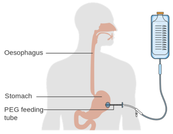 Image Source: Wikimedia Commons (2014). Diagram showing the position of a percutaneous endoscopic gastrostomy (PEG) feeding tube CRUK 341, Licensed under a creative commons licence. Available for free at: https://commons.wikimedia.org/wiki/File:Diagram_showing_the_position_of_a_percutaneous_endoscopic_gastrostomy_(PEG)_feeding_tube_CRUK_341.svg (Accessed: 17 January 2024)
Reduced mobility and pressure ulcers
In chronic neurological conditions, mobility may be reduced due to flaccid or spastic paralysis. Functional limitation can result in disuse atrophy, increased dependence, falls and fractures, increased risk of venous thromboembolism, predisposition to infection such as pneumonia, mood disturbance and pressure ulcers. Manage with comprehensive MDT and family input.
Pressure ulcers
People with chronic neurological disability may be at increased risk of pressure sores through various factors: reduced mobility and inability to reposition themselves, reduced sensation, incontinence, nutritional deficiency and cognitive impairment.
- Prevention:
- On admission, assess risk of pressure sores using a validated assessment tool (e.g. Waterlow);
- Frequent skin assessment, repositioning, nutritional supplementation if inadequate intake, barrier creams and pressure redistributing devices.
- Management:
- Document pre-existing sores and raise safeguarding concerns if required;
- Assess severity (see classification system below);
- Choose dressings based on the degree of maceration, exudate and signs of infection: hydrocolloid dressings promote healing and alginate dressings are considered if significant exudate;
- Consider Tissue Viability Nurse involvement;
- Debridement if significant necrosis;
- Consider systemic antibiotics if evidence of sepsis, spreading cellulitis or underlying osteomyelitis.
| Pressure ulcer grade by the National Pressure Ulcer Advisory Panel classification system | |
|---|---|
| Grade | Description |
| I | Non-blanchable erythema of intact skin |
| II | Partial thickness skin loss involving the epidermis, dermis or both |
| III | Full thickness skin loss involving damage to or necrosis of subcutaneous tissue that may extend down to, but not through, underlying fascia |
| IV | Full thickness skin loss with extensive destruction, necrosis, or damage to muscle, bone or supporting structures |
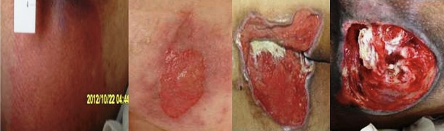
Image Source: Wikimedia Commons (2012). Wound stage, Licensed under a creative commons licence. Available for free at: https://commons.wikimedia.org/wiki/File:Wound_stage.jpg (Accessed: 17 January 2024)
Pain
Pain is often encountered by patients with chronic neurological disability. Following a stroke, this is most commonly musculoskeletal and is related to paralysis and immobility.
Headache
Common following a stroke or due to medications used, such as GTN (glyceryl trinitrate). Manage with simple analgesia.
Hemiplegic shoulder pain
Hemiplegic shoulder pain or subluxation is related to muscular flaccidity and poor handling. Management is with prevention, physiotherapy, positioning and simple analgesia.
Central post-stroke pain
A neuropathic pain syndrome occurring following stroke. Characterised by pain in the areas affected by the stroke and can cause allodynia, hyperalgesia or abnormal sensations. Managed with neuropathic agents; may need Pain Team input.
Spasticity
Common due to upper motor neurone damage. Caused by hyperexcitability of the stretch reflex, resulting in functional impairment, risk of infection related to skin breakdown, pain and the development of contractures.
- Prevention: physical rehabilitation using a variety of techniques
- Management:
- Physical rehabilitation
- IM botulinum toxin (type A) for focal spasticity – well tolerated with minimal side effects
- Trial of oral baclofen or tizanidine – use is limited to generalised spasticity or progressive neurological conditions due to side effects of drowsiness, fatigue and weakness
Continence
Urinary incontinence affects half of patients admitted with an acute stroke, with a significant minority remaining incontinent one year post stroke. Functional urinary incontinence is related to physical limitations, aphasia and cognitive impairment. Short-term urinary catheterisation may be required for symptomatic retention while investigations are done to ascertain the cause. Faecal incontinence is less common and also often related to function, although faecal loading and infection must be ruled out.
References
- Berlowitz DR. Pressure ulcer. In: BMJ Best Practice. [Internet]. London: BMJ Publishing Group, 2021 [August 3 2021; cited January 21 2022]. Available from: https://bestpractice.bmj.com/topics/en-gb/378
- Clark DG. Approach to the patient with aphasia. In: Mendez MF, Wilterdink JL, editors. UpToDate. [Internet]. Waltham (MA): UpToDate Inc; 2021. [July 14 2020; cited January 21 2022]. Available from: https://www.uptodate.com/contents/approach-to-the-patient-with-aphasia
- Creutzfeldt CJ, Robinson MT. Neuropalliative care of stroke. In: Biller J, Morrison RS, Dashe JF, editors. UpToDate. [Internet]. Waltham (MA): UpToDate Inc; 2021. [December 6 2021; cited January 21 2022]. Available from: https://www.uptodate.com/contents/neuropalliative-care-of-stroke
- Improvement, NHS. Psychological care after stroke. Leicester: NHSI, 2011. [cited January 21 2022]. Available from: https://www.england.nhs.uk/improvement-hub/publication/psychological-care-after-stroke-improving-stroke-services-for-people-with-cognitive-and-mood-disorders/
- National Institute for Health and Care Excellence. Stroke and TIA. [Internet] London: NICE, 2021 [September 2021; cited January 21 2022]. Available from: https://cks.nice.org.uk/topics/stroke-tia/
- National Institute for Health and Care Excellence. Stroke and transient ischaemic attack in over 16s: diagnosis and initial management. London: NICE, 2019 [May 1 2019; cited January 21 2022]. Available from: https://www.nice.org.uk/guidance/ng128
- Stroke.org.uk. [Internet] London: Stroke Association, 2022 [cited January 21 2022]. Available from: https://www.stroke.org.uk/
Complete Guide to Clinical Genetics ST3 Application and Recruitment (2027)
Written by Medibuddy. Last updated: October 2026. Clinical Genetics is

Complete Guide to Clinical Radiology ST3 Application and Recruitment (2027)
Recruitment paused: NHS England is not running Clinical Radiology ST3+ recru

Complete Guide to Paediatric Surgery ST3 Application and Recruitment in United Kingdom (2026)
Written by Medibuddy. Last updated: October 2026. If you’re planning


Take your subscriptions with you
Our mobile app allows you to access your interview and exam question banks wherever you are.

