


 Close
Close
 10th May 2025
Syncope and Sudden Cardiac Death
Syncope
Syncope is defined as transient loss of consciousness due to cerebral hypoperfusion, characterized by rapid onset, short duration and spontaneous complete recovery.
Table 1: Classification of Syncope
| Reflex (neurally mediated) syncope |
|---|
|
| Orthostatic hypotension |
|
| Cardiac syncope |
|
Table 1: Syncope
Orthostatic hypotension
Orthostatic hypotension is a condition in which a person’s blood pressure (BP) drops abnormally when they stand up after sitting or lying down.
Diagnostic criteria
Abnormal BP fall is defined as a progressive and sustained fall in systolic BP from baseline value ≥ 20mmHg or diastolic BP ≥ 10 mmHg, or a decrease in systolic BP to < 90 mmHg.
|
Drugs: |
|---|
|
| Volume depletion: |
|
| Primary autonomic failure: |
|
| Secondary autonomic failure: |
|
Table 2: Causes of orthostatic hypotension
Management
- Explanation of the diagnosis, reassurance, and avoidance of triggers
- Adequate hydration and salt intake
- Modification and discontinuation of hypotensive drug regimens
- Isometric physical counterpressure maneuvers if symptoms persist
- Abdominal binders/ support stockings to reduce venous pooling if symptoms persist
- Midodrine (2.5-10mg t.i.d) if symptoms persist
- Fludrocortisone (0.1-0.3mg once daily) if symptoms persist
- Head-up tilt sleeping (>10 degrees) to increase fluid volume if symptoms persist.
Long QT Syndrome
Long QT syndrome (LQTS) is a disease of ventricular repolarization identified by the prolongation of the QT interval on ECG. Normal QT interval is <440 msec in male and <460 msec in female when corrected for heart rate of 60/min.
Diagnostic criteria
- Corrected QT interval (QTc) ≥ 480ms
- LQTS score >3 (See modified long QT syndrome diagnostic score)
- In the presence of cardiac arrest or syncope, a QTc ≥ 460 ms
- Pathogenic genetic mutation irrespective of QT interval
Classification
- Congenital:
- Autosomal recessive: Jervell and Lange-Nielsen syndrome (associated with deafness)
- Autosomal dominant: Also known as Romano Ward Syndrome (has 3 main phenotypes: LQT1, LQT2 and LQT3)
- Acquired: Caused by drugs or electrolyte imbalance
| Drugs |
|---|
|
| Electrolyte disturbance |
|
Table 3: Acquired causes of LQTS
Clinical features
- Asymptomatic
- Sudden cardiac death
- Syncope
- Aborted ventricular arrhythmia including Torsades de pointes (Figure-2)
- Patients with LQT 1 are triggered by physical exercise, specially swimming
- Patients with LQT2 are triggered by noise
- Patients with LQT3 are triggered during sleep
ECG findings
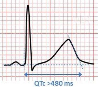
Image Source: Wikimedia Commons (2020). ECG, Licensed under a creative commons licence. Available for free at: https://commons.wikimedia.org/w/index.php?search=long+qt+ecg&title=Special:MediaSearch&go=Go&type=image (Accessed 7 May 2025).
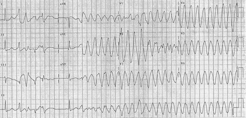
Image Source: Wikimedia Commons (2010). Torsade ECG, Licensed under a creative commons licence. Available for free at: https://commons.wikimedia.org/w/index.php?search=torsade+pointes+ecg&title=Special:MediaSearch&go=Go&type=image (Accessed 7 May 2025).
Management
- Torsades de pointes should be terminated by intravenous Magnesium sulfate. Alternatives are, overdrive pacing and intravenous isoprenaline. Although, isoprenaline should be avoided in congenital long QT syndrome.
- Avoidance of QT prolonging drugs, electrolyte imbalance and genotype specific triggers in all patients
- Asymptomatic congenital LQTS are advised to be treated with beta blockers
- Implantable cardioverted defibrillator (ICD) implantation should be considered in patients with previous cardiac arrest, syncope despite beta blockers and in asymptomatic cases with QTc >500 msec
- Left cardiac sympathetic denervation can be considered in patients who remain symptomatic.
Brugada syndrome
Brugada syndrome is characterized by right bundle branch block (RBBB) and ST-segment elevation in the anterior precordial leads of ECG, often without evidence of structural heart disease
Classification
- Type 1: Coved ST segment elevation
- Type 2: Saddleback ST elevation ≥ 1 mm
- Type 3: Saddleback ST elevation < 1mm
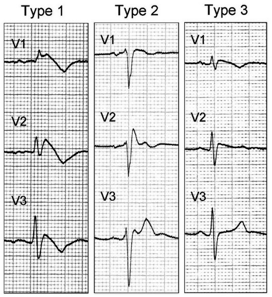
Image Source: Wikimedia Commons (2008). Brugada ECG, Licensed under a creative commons licence. Available for free at: https://commons.wikimedia.org/w/index.php?search=brugada+ecg&title=Special:MediaSearch&go=Go&type=image (Accessed 7 May 2025).
Diagnosis
Diagnostic criteria
- Type 1 ECG pattern occurring spontaneously
- Type 1 ECG provoked by class I anti-arrhythmic drugs with one additional clinical feature: documented ventricular tachycardia (VT) or ventricular fibrillation (VF), syncope of probable arrhythmic cause, family history of sudden cardiac death (SCD) before age 45, family history of brugada type 1 or nocturnal agonal respiration.
Clinical features
- Asymptomatic
- Syncope
- Aborted cardiac arrest due to VT/VF
Management
- Fever should be aggressively treated (can precipitate VT/VF)
- Avoid drugs that provoke Brugada syndrome (e.g. flecainide)
- ICD is indicated in patients with aborted SCD or spontaneous VT/VF or with a history of syncope
- Ablation may be considered in patients with frequent ICD shocks
- Quinidine may be considered in patients with frequent ICD shocks or who refuse ICD or in whom ICD is contraindicated
Catecholaminergic Polymorphic Ventricular Tachycardia
Catecholaminergic polymorphic ventricular tachycardia (CPVT) is a heritable disorder characterized by catecholamine induced bidirectional VT and polymorphic VT in the absence of structural heart disease or ischaemia.
Clinical feature
- Bidirectional VT or polymorphic VT induced by physical activity or emotional stress
- Most patients have normal ECG and structurally normal heart
- Two main genetic types: dominant disease due to mutation of gene encoding RYR2 receptor and recessive disease due to mutation of gene encoding CASQ2
Investigations
- Exercise stress test: Elicits bidirectional or polymorphic VT (Figure-4)
- Genetic testing
- Epinephrine or isoprenaline challenge may be considered when exercise testing is not possible
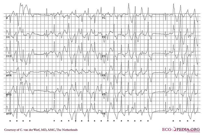
Image Source: Wikimedia Commons (2010). Catecholaminergic polymorphic ventricular tachycardia, Licensed under a creative commons licence. Available for free at: https://commons.wikimedia.org/wiki/Category:Catecholaminergic_polymorphic_ventricular_tachycardia#/media/File:ECG000033_(CardioNetworks_ECGpedia).jpg (Accessed 7 May 2025).
Management
- Avoidance of competitive sports, strenuous exercise and exposure to stressful environments is recommended to all patients
- Non-selective beta-blockers (nadolol or propranolol) are recommended in all patients.
- ICD implantation is indicated in patients with aborted cardiac arrest in combination with beta-blockers and flecainide
- Left cardiac sympathetic denervation or Flecainide can be considered when above measures fail.
References
- Brignole M, Moya A, Lange FJ de, et al. 2018 ESC Guidelines for the diagnosis and management of syncope. Eur Heart J 2018; 39: 1883–1948.
- Zeppenfeld K, Tfelt-Hansen J, Riva M de, et al. 2022 ESC Guidelines for the management of patients with ventricular arrhythmias and the prevention of sudden cardiac death. Eur Heart J 2022; 43: 3997–4126.
- Penman ID, Ralston SH, Strachan MWJ, et al. (eds). Davidson’s principles and practice of medicine. 24th edition, international edition. London, New York, Oxford, Philadelphia, St. Louis, Sydney: Elsevier, 2023.
- Mizusawa Y and Wilde AAM. Brugada syndrome. Circ Arrhythm Electrophysiol 2012; 5: 606–616.
- Brugada J, Katritsis DG, Arbelo E, et al. 2019 ESC Guidelines for the management of patients with supraventricular tachycardiaThe Task Force for the management of patients with supraventricular tachycardia of the European Society of Cardiology (ESC). Eur Heart J 2020; 41: 655–720.
Complete Guide to Clinical Genetics ST3 Application and Recruitment (2027)
Written by Medibuddy. Last updated: October 2026. Clinical Genetics is

Complete Guide to Clinical Radiology ST3 Application and Recruitment (2027)
Recruitment paused: NHS England is not running Clinical Radiology ST3+ recru

Complete Guide to Paediatric Surgery ST3 Application and Recruitment in United Kingdom (2026)
Written by Medibuddy. Last updated: October 2026. If you’re planning


Take your subscriptions with you
Our mobile app allows you to access your interview and exam question banks wherever you are.

