


 Close
Close
 23rd May 2025
Disorders of Hormonal Deficiencies
Diabetes Insipidus
Diabetes insipidus (DI) is a disorder that leads to an increased production of dilute urine, polydipsia and can include nocturia. It is rare, with a global prevalence of 1 per 25,000 of the population.
Types of DI:
- Cranial/Central: Impaired production/transportation/release of ADH. Caused by disorders in areas responsible for ADH secretion, such as hypothalamic osmoreceptors, supraoptic/paraventricular nuclei or the superior part of the supraoptic hypophyseal tract. Pathology in the neurohypophysis alone often does not cause DI as ADH produced in the hypothalamus can enter the systemic circulation.
- Nephrogenic: Renal collecting ducts are resistant to ADH
- Gestational: Occurs during pregnancy
Aetiology
Cranial DI
- Transsphenoidal surgery for pituitary adenoma
- Craniopharyngioma
- Lesions to the pituitary stalk
- Traumatic brain injury
- Subarachnoid haemorrhage affecting the anterior communicating artery supplying the anterior hypothalamus
- Congenital hypothalamic-pituitary defects
- Wolfram syndrome: Autosomal recessive progressive neurodegenerative disorder leading to diabetes mellitus, optic atrophy, sensorineural deafness and DI
- Associated with autoimmune disorders e.g., Hashimoto’s thyroiditis
- Medications e.g., phenytoin
- Late complication of meningitis/encephalitis
Nephrogenic DI
- Lithium therapy
- Chronic kidney disease
- Renal amyloidosis
- Renal sarcoidosis
- Associated with chronic hypercalcaemia/ hypokalaemia
- Genetics: mutations AVPR2 receptor (X linked recessive) or AQP2 gene (autosomal recessive)
Initial Investigations
- Paired urine serum osmolality: Low urine (<300 mmol/kg) and high serum osmolality
- Urea and electrolytes, calcium: Assesses renal function and electrolyte abnormalities
- 24 hr urine collection to measure volume: >3L in 24 hrs
- To rule out diabetes mellitus: urine dipstick to test for glucose and serum glucose levels
Diagnostic Tests
- 1st Water deprivation test
- Patient deprived of fluids for 8 hours. Every hour serum osmolality, urine osmolality and urine volume are measured
- Contraindications: Renal insufficiency, uncontrolled diabetes mellitus, uncorrected adrenal or thyroid hormone deficiency
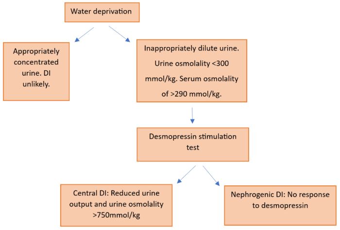
- DI patients are unable to concentrate their urine in response to dehydration so results show inappropriately dilute urine with osmolality <300 mmol/kg and a serum osmolality of >290 mmol/kg
- 2nd Desmopressin stimulation test
- Done after positive water deprivation test
- 2 micrograms desmopressin administered subcutaneously. Every hour for 4 hours serum osmolality, urine osmolality and urine volume are measured
- Patients with central DI will have a reduced urine output and increased urine osmolality (>750 mmol/kg) in response to desmopressin
- Patients with nephrogenic DI show no response to desmopressin
- Hypertonic saline stimulated test can be considered
- Baseline copeptin level >24.1 picomol/L diagnoses nephrogenic DI
- After infusing 3% sodium chloride, a copeptin level >4.9 picomol/L diagnoses central DI
Further investigations to be considered
- MRI brain with contrast
- Genetic testing
- Pituitary hormone levels
Diagram for Diagnosis of Diabetes Insipidus
Management
- Hypernatremia: treat with IVF e.g., 5% dextrose
- Central DI: Desmopressin
- Nephrogenic DI: Adequate fluid intake to match losses, treatment of underlying cause. Can consider low sodium diet (<500mg/day), thiazide diuretics or indomethacin to reduce urine output
Growth Hormone Deficiency in Adults
GH deficiency is a rare disorder caused by inadequate secretion of GH. It can be idiopathic, congenital or acquired. GH deficiency in adults is commonly due to pituitary tumour (78%), extra-pituitary tumour such as craniopharyngioma (13%) and unknown (8%). Therefore, it is important to test adults with known hypothalamic or pituitary disease, such as panhypopituitarism, for GH deficiency.
Clinical manifestations of GH deficiency
- Reduced energy levels
- Reduced lean body mass
- Increase in proportion of fat
- Reduced bone mineral density leading to osteoporosis
- Cardiovascular: increased LDL cholesterol, impaired cardiac function
- Insulin resistance,
- Poor quality of life and increased mortality
Diagnosis
- History of pituitary/hypothalamic disease or panhypopituitarism
- Low serum IGF-1
- If IGF-1 levels are normal, a GHRH-Arginine simulation test can be done to diagnose. This involves taking a blood sample then injecting GHRH and then arginine over 30 minutes. Further blood samples are taken over the next two hours. Facial flushing is a common side effect of GHRH injection. A diagnosis is made if GH remains suppressed. The exact level depends on peak GH levels relative to patient’s BMI
- For further investigations, can consider pituitary imaging if not already completed
Management
Treatment involves injections of recombinant human growth hormone (rHGH). Please note a positive simulation test is required to initiate treatment with rHGH in the National Health Service (NHS), UK. Although patients taking this treatment are unlikely to have marked increases in their energy levels, it can improve muscle strength, bone mineral density, CVS risk factors and has some impact on quality of life
Panhypopituitarism
This is a disorder characterised by the reduced/absent secretion of most or all of the hormones by the pituitary gland. Panhypopituitarism can be due to hypothalamic disease, leading to reduced secretions of releasing hormones, or pituitary disease.
Aetiology
Hypothalamic disease
- Tumour
- Radiation therapy for nasopharyngeal cancer or brain tumours
- Post meningitis
- Traumatic brain injury
- Stroke
Pituitary disease
- Tumours and lesions, e.g., Pituitary adenoma (most common association with panhypopituitarism)
- Pituitary surgery
- Radiotherapy to the pituitary gland
- Hemochromatosis of pituitary gland
- Immunotherapy side effect, e.g., ipilimumab
- Sheehan syndrome
- Pituitary apoplexy: haemorrhage into infarction or existing adenoma
- Pituitary abscess or infection
- Pituitary stalk interruption syndrome- congenital hypopituitarism
- Empty sella
Clinical Features
- ACTH and cortisol deficiency: postural hypotension, tachycardia, anorexia, reduced libido, hypoglycaemia
- TSH deficiency: symptoms of hypothyroidism
- Low levels of gonadotropins lead to hypogonadotropic hypogonadism
- GH deficiency
- PRL deficiency
Investigations
- U&Es
- Serum and urine osmolality
- Morning cortisol, ACTH, testosterone/oestradiol, FSH, LH
- PRL, IGF-1 levels
- Synthetic ACTH stimulation test: serum cortisol levels taken 30- and 60-mins post injection. Suppression of cortisol (<500 nanomol/L) suggests adrenal insufficiency
- Can consider tests for Diabetes insipidus and diabetes mellitus
- Consider neuroimaging
Management
- Treat underlying cause
- Deficient hormones are replaced
- Pituitary apoplexy can lead to acute severe hypopituitarism which is a medical emergency. This is treated with IV hydrocortisone and some cases require emergency surgery.
Complete Guide to Clinical Genetics ST3 Application and Recruitment (2027)
Written by Medibuddy. Last updated: October 2026. Clinical Genetics is

Complete Guide to Clinical Radiology ST3 Application and Recruitment (2027)
Recruitment paused: NHS England is not running Clinical Radiology ST3+ recru

Complete Guide to Paediatric Surgery ST3 Application and Recruitment in United Kingdom (2026)
Written by Medibuddy. Last updated: October 2026. If you’re planning


Take your subscriptions with you
Our mobile app allows you to access your interview and exam question banks wherever you are.

