


 Close
Close
 23rd May 2025
Thyroid Disorders
The Thyroid Gland
Anatomy
The thyroid gland consists of a midline isthmus horizontally (below the cricoid cartilage) and two lateral lobes extending upwards over the lower half of the thyroid cartilage . The gland lies deep to the strap muscles, enclosed in the pretracheal fascia, which anchors it to the trachea and means the thyroid moves upwards on swallowing.
Anatomical relations
- Anteriorly: infrahyoid muscles
- Laterally: carotid sheath, comprising of the common carotid artery, internal jugular vein, and vagus nerve
Medially: larynx, pharynx, trachea, and oesophagus, and the external and recurrent laryngeal nerves (the left and right recurrent laryngeal nerves are in close proximity to the thyroid gland, and may be damaged by compression (e.g. by a nearby mass) or during thyroidectomy)
Physiology
Iodine is a substrate for thyroid hormone biosynthesis. It is actively transported into the follicular layer and produces mono-iodotyrosine (MIT) and diiodotyrosine (DIT). Thyroid peroxidase links two DITs to form T4, and a DIT and MIT to form T3 and reverse T3. The thyroid’s main product is T4 (inactive form) and it also secretes 20% of circulating T3 (active form); the remainder is generated in extra-glandular tissues by the conversion of T4 to T3 (mainly in the liver and kidneys). In the blood, T3 and T4 are bound to the plasma protein. Production of the thyroxine hormones and its function can be affected by the following:
- Thioureas (e.g., carbimazole and propylthiouracil) and mercaptoimidazole inhibit the initial oxidation of iodide and coupling of iodothyronines.
- In large doses, iodine itself blocks organic binding and coupling reactions.
- Lithium has several inhibitory effects on intrathyroidal iodine metabolism.
Thyroid hormones function
In general, they increase metabolic rate where oxygen, energy consumption and thermogenesis increase. Effects on target organs are as follows:
- Cardiac:
- Increase the expression and activity of cardiogenic B receptors
- Results in increased heart rate, stroke volume, cardiac output, and contractility.
- Respiratory:
- Increase respiratory centre stimulation and lung perfusion
- Increases respiratory rate and oxygenation.
- Thyroxine also stimulates surfactant production in new-borns.
- Neurological:
- Stimulate the maturation of the nervous system.
- Musculoskeletal:
- Stimulate bone growth and the development of type II muscle fibres.
- Endocrine:
- Promote fertility, ovulation, and menstruation.
- Metabolism:
- increase the basal metabolic rate (BMR) leading to an increase in oxygen consumption and body temperature, with an additional thermoregulation function.
- stimulate protein anabolism.
- increase glucose reabsorption, gluconeogenesis, glycogenolysis, and glucose oxidation, leading to increased carbohydrate metabolism
- stimulate lipolysis or lipogenesis depending on metabolic status.
Hyperthyroidism
It is important to differentiate between thyrotoxicosis and hyperthyroidism, as these terms are not synonymous:
- Thyrotoxicosis is a referral to hypermetabolic state or symptoms caused by excessive thyroid hormone levels which is usually due to gland hyperactivity (i.e., hyperthyroidism).
- Overt hyperthyroidism is usually the experience of thyrotoxicosis symptoms with the presence of low serum TSH level and high T3 and T4.
- Subclinical hyperthyroidism is notation of asymptomatic patients who will show low serum levels of TSH but normal T3 and T4 levels.
Any condition which results in increased levels of circulating T3/T4 may result in the following clinical signs and symptoms:
- Ocular signs:
- Lid lag
- Lid retraction
- Exophthalmos (Graves’ disease)
- Extraocular signs:
- Cardiovascular:
- ↑ Heart rate (HR)
- Palpitations and irregular pulse, with abnormal heart beats (e.g., atrial fibrillation (AF))
- Hypertension with widened pulse pressure
- Chest pain
- Neuromuscular and psychiatric signs:
- Anxiety and restlessness
- Insomnia
- Fine tremors
- Hyperreflexia
- Myopathy (proximal muscle weakness with normal creatinine kinase (CK) levels
- Osteoporosis
- Reproductive:
- Oligo/amenorrhoea
- Gynaecomastia, infertility problems and decreased libido (50% of men may have sexual dysfunction)
- Metabolic and dermatological:
- Heat intolerance
- Weight loss with increased appetite
- Fatigue
- Warm moist skin
- Excessive sweating
- Pretibial myxoedema (infiltrative dermopathy)
- Fine hair with loss
- Onycholysis
- Palmar erythema
- Gastrointestinal:
- Increased bowel movements
- Cardiovascular:
Thyrotoxicosis
As mentioned above, thyrotoxicosis is the description of the hypermetabolic state caused by increased levels of circulating thyroid hormones. This can occur as a result of hyperthyroidism (i.e. over-activity of the thyroid gland), or due to other causes, detailed below:
| Associated with hyperthyroidism | Excessive thyroid stimulation:
|
Endogenous thyroid-hormone secretion:
|
|
| Other causes | Thyroiditis (see section below):
|
Exogenous thyroid hormone excess:
|
|
Ectopic thyroid tissue:
|
|
Table 1: Causes of thyrotoxicosis
Graves’ disease
- Grave’s disease is an autoimmune thyroid condition that is associated with hyperthyroidism. It is due to TSH receptor autoantibodies that lead to overstimulation of thyroid gland, and excessive thyroid hormone production. It is the most common cause for hyperthyroidism in iodine sufficient parts of the world, affecting females more and of peak age between 20-40 years old.
- 50% of the patients will report having a family history of other autoimmune disorders such as T1DM or pernicious anaemia. There is also association with human leukocytic antigen (HLA-DR3, HLA-B8) alleles.
- Grave’s disease occurs due to B and T cell autoimmunity, and an increase in the production of stimulating IgG against TSH receptors, leading to an increase in the thyroid gland size and function with manifest hyperthyroidism and goitre.
Associated conditions
- Endocrine: T1DM, Addison’s disease,
- Dermatological: Vitiligo, Alopecia areata
- Haematological: Pernicious anaemia
- Neurological: Myasthenia gravis
- Gastrointestinal: Coeliac disease
- Other autoimmune conditions occur with HLA-DR3
Risk factors
- Female sex
- Smoking
- Drug-induced (alemtuzumab used for multiple sclerosis)
- Infection with Yersinia enterocolitica and Borrelia burgdorferi
- Pregnancy (due to overstimulation by high levels of hCG and TSH receptor antibodies (TRAb) during 1st trimester)
- Stress (due to cortisol mediated immune suppression followed by rebound immunologic hyperactivity)
Clinical picture
In addition to the thyrotoxic picture described above, patients also present with triad of: Diffuse goitre, Graves ophthalmopathy and dermopathy.
- Diffuse goitre which is smooth, uniformly enlarged, and non-tender. Sometimes bruit can be heard over the superior poles due to increased vasculature.
- Ophthalmopathy occurs due to activated B and T cells lymphocytic infiltration retro-orbitally, that leads to a cascade of immunological reactions occurring and cytokine release (TNF-α and INF-γ). Accordingly, fibroblast proliferation leads to an increase in the hyaluronic acid, glycosaminoglycans (GAGs) and adipocytes causing an increase in intra-orbital fat and muscle tissue volume. These changes are manifested as exophthalmos, ophthalmoplegia, lid retraction and exposure conjunctivitis and keratopathy.
- Other manifestations of Grave’s disease can be pretibial myxoedema which is a non-pitting oedema on both lateral and anterior aspects of the leg.
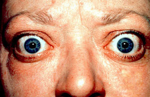
Image Source: Wikimedia Commons (2022). Graves Opthalmopathy, Licensed under a Creative Commons licence. Available for free at: https://en.wikipedia.org/w/index.php?title=Graves%27_ophthalmopathy&oldid=1130342091 (Accessed 13 May 2025).
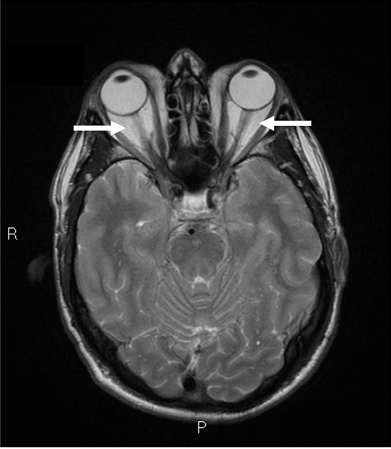
Image Source: Wikimedia Commons (2022). Graves Opthalmopathy, Licensed under a creative commons licence. Available for free at: https://en.wikipedia.org/w/index.php?title=Graves%27_ophthalmopathy&oldid=1130342091 (Accessed 13 May 2025).
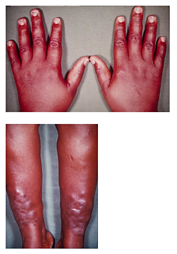 Image Source: Wikimedia Commons (2008). Pretibial Myxedema, Licensed under a creative commons licence. Available for free at: https://commons.wikimedia.org/wiki/Category:Pretibial_myxedema#/media/File:Myxedema.jpg (Accessed 13 May 2025).
Investigations
Diagnosis is based on the clinical picture and is confirmed through thyroid function tests (TFTs) and antibody findings.
- TFTs: low or undetectable TSH with high T3 and T4
- Thyroid antibodies can be specific such as high TRAbs, or non-specific as high Antithyroid peroxidase (anti-TPO) antibodies, and Thyroglobulin (anti-Tg) antibodies.
- Thyroid ultrasound which shows enlarged hypervascular thyroid. Can be used in pregnancy if TRAbs are low.
Management
Goals and principles:
- Induce remission.
- Monitor for any relapse, Monitoring initially is 6-8 weekly for the 1st 6 months, then 6-monthly for 2 years. Afterwards, annually depending on the symptoms.
- For relapse, radiotherapy or surgery are considered as definitive treatment.
- Treatment is usually subdivided into hyperthyroidism state control and symptomatic control.
NICE guidelines recommends the following management:
- Hyperthyroidism state control via Antithyroid drugs (ATDs)
| Medication | Mechanism | Indication | Side effects | Contraindication |
|---|---|---|---|---|
| Carbimazole |
|
|
|
|
| Propylthiouracil (PTU) |
|
|
Table 2: Antithyroid (ATDs) drugs mechanism of action and indications
Two treatment regimens are identified; dose titration and block and replace regimes.
- Dose titration regime:
A relatively high dose of ATDs is started and dose is titrated according to TFTs (TSH particularly) that are performed every 4-8 weeks. It continues for 18 weeks. Some cases of relapses can be seen after discontinuing the therapy. Hence, Long-term therapy at a low dose can be an option. This regime has lower SE compared to block and replace regime.
2. Block and replace regime:
After achieving euthyroid state on carbimazole, Levothyroxine is started. This regime I can continue for 6 months. It allows fewer visits to the hospital for multiple TFTs.
- Hyperthyroidism state control via Radioactive ablation:
Intention is ablation of the gland then commencing thyroxine replacement therapy. It is becoming introduced more to children >10 years old. Sometimes ATDs are started after radioiodine administration and tapered down gradually as per TFTs. There is no risk of thyroid malignancy post-treatment.
Indications for radioactive ablation can be as 1st line after failure of ATDs or surgery. Multinodular goitre or adenoma or relapsed Grave’s disease are other indications. Side Effects of radioactive ablation are radiation induced-thyroiditis, transient hypothyroidism and rarely rebound thyroid storm.
Contraindications for radioactive ablation are pregnancy and lactation. Women of childbearing age should avoid pregnancy for a minimum of 6 months post-treatment. Similarly for men should avoid fathering for minimum of 4 months post-radiation. Other contraindications are Graves ophthalmopathy (due to risk of symptoms worsening), or young children (due to risk of thyroid carcinogenesis).
- Hyperthyroidism state control via Surgery:
Prior to surgery, ATDs, are used to achieve euthyroidism. Rarely, potassium iodide is needed. Β-blockers are needed to prevent an unwanted liberation of thyroid hormones during surgery.
Indications for surgical management are usually preserved for suspicious/malignant thyroid nodules. pregnant women with post-ATDs failure or complications. Surgery is done in the 2nd trimester. Patients who have poor compliance to medical treatment, refuse radiation therapy, have either relapsed Graves’ disease, severe Graves’ ophthalmopathy, or large goitres with have low-radioiodine uptake and with local compressive symptoms are also offered surgical management.
Thyroidectomy can lead to nerve damage, hypoparathyroidism, local haemorrhage or wound infection, hypothyroidism, or keloid formation.
- Symptomatic control
One of the common symptoms that requires treatment in hyperthyroidism is AF. Rate control medications particularly propranolol is commonly used. Propranolol is non-selective β-Blocker which not only has HR control effect but also, inhibits the conversion of T4 to T3 peripherally. Anticoagulation should also be considered.
Nodular thyroid disease
Thyroid nodules are abnormal growths within the thyroid gland that are subdivided into benign ( ~ 95% of the cases) and malignant ones which are less common. Types of thyroid nodules are shown in Table 3.
| Benign nodules | Malignant Nodules |
|---|---|
Thyroid adenomas
Thyroid cyst
|
|
Table 3: Types of nodular thyroid disease
Thyroid nodules are present in 50% of the population but only palpable in 5-10% of the population. They are more predominant in females and are more prevalent in inland regions where iodine content in food and water is low. Less than 5% of these nodules can be malignant.
Clinical picture
The clinical picture depends on the activity and the virulence of the nodule. For example, the follicular type is considered an inactive nodule. whereas toxic adenoma and toxic multinodular goitre are considered active nodules, and patients present with symptoms of hyperthyroidism. These nodules are usually painless.
Thyroid cysts are due to cystic degeneration in the thyroid tissue or involution of adenoma. They usually present with a palpable nodule that can be tender due to haemorrhage inside the cyst or may cause compressive symptoms such as hoarseness and dysphagia. Pemberton’s sign (facial erythema and jugular venous distension on raising the arms) is a sign of Superior Vena Cava (SVC) obstruction by a substernal mass. Thyroid nodules in pregnancy have shown increase in size and number with increase in malignancy risk. As a result, surgical removal can be indicated in the 2nd trimester or post-partum.
Malignancy should be suspected in patients <20 or >60 years old. Other red flags for malignancy can be male gender, history of radiation to the head and neck, rapidly growing nodules with compression symptoms, and firm fixed nodules that may be associated with cervical lymphadenopathy. Some genetic associations increasing risk of malignancy are:
- MEN2 syndrome
- Gardner syndrome
- Cowden syndrome
- Family history of thyroid malignancy.
Investigations
- Fine needle aspiration biopsy (FNAC).
- Serum TSH: High levels are associated with higher risk of malignancy.
- Imagining can be thyroid US or thyroid scintigraphy (which shows cold nodules are associated with malignancy, hot nodules are less likely to be malignant).
- Investigation of complications: e.g., symptoms suggestive of SVC obstruction from a goitre require CT imaging
Management
NICE guidelines recommend management of nodular goitre depending on the activity of the nodules and virulence.
Management of non-toxic multinodular goitre may involve:
- Surgery
- Compressive symptoms
- Cosmetic reasons
- Solitary nodule with FNAC suggestive of malignancy
- Radioiodine
- Indications same as for surgery, in patients not fit enough to undergo surgery
- Medical treatment
- Not generally used, but T4 replacement may be useful if TSH is detectable
Management of toxic multi-nodular goitre:
- Radioiodide management
- 1st line definitive treatment.
- That might be due to the accumulation of 131 I preferentially in the hot nodules , not the normal thyroid tissue causing an euthyroid state as a result
- Surgery
- Aims to remove the nodular tissue.
- For symptomatic relief
- Post-operative monitoring of the TFTs is advised as patients at risk of becoming hypothyroid and may require thyroxine replacement.
- It is considered the 2nd line of management if ATDs fail if a patient is pregnant or fathering a child in 6 months.
- ATDs such as Carbimazole
- Effective for hyperthyroidism, but are not curative treatments
- There is a risk of recurrence when the medications are stopped.
Thyroiditis
Thyroiditis refers to a transient state of thyrotoxicosis followed by hypothyroidism features. It occurs due to gland inflammation. It is more common in women, with a peak incidence of 30-50 years old.
Causes of thyroiditis include:
- Hashimoto’s disease (discussed later on in this chapter)
- Post-partum thyroiditis
- Usually occurs within 6 months of giving birth.
- It occurs in about 30-52% of patients of +ve TPO antibodies.
- Is associated with type 1 diabetes
- Most patients show complete remission with fewer incidence of permanent hypothyroidism as a complication.
- Drug-induced e.g., amiodarone, lithium, interleukin 2 or interferon-α(due to thyroid peroxidase antibodies formation in 15% of the cases).
- Subacute (De Quervain’s thyroiditis)
- May occur due to granuloma formation, usually associated with viral upper respiratory tract infection (mumps, coxsackie, influenza, adenovirus, and echovirus).
- May also occur due to lymphocytic infiltration, which may occur in the post-partum period, in association with autoimmune disease, or as a side effect of a drug.
- Has a triphasic picture:
- The thyrotoxic phase occurs due to the inflammatory damage of the follicular cells, releasing thyroid hormones (pre-formed colloid).
- The hypothyroid phase follows due to the depletion of the thyroid hormones and reduced synthesis as a result of follicular cell damage.
- The euthyroid phase is achieved where, a recovery of the thyroid gland occurs with pathological changes and the thyroid function normalises
- Riedel’s thyroiditis
- A rare condition characterised by extensive thyroid gland fibrosis and the surrounding structure.
- One third of the patients have associated extracervical manifestations for example: pulmonary, sclerosing cholangitis, mediastinal, and retroperitoneal fibrosis. Parathyroid glands fibrosis is rare.
- Patients are usually in euthyroid state. 30% of the patients develop hypothyroidism eventually with extensive fibrosis.
- Malignancy should be excluded.
- Radiation
- Pyogenic
- e.g., Staphylococcus aureus, streptococci, Escherichia coli, tuberculosis, fungal.
- Usually caused by another septic focus.
- Piriform sinus infection should be excluded.
Clinical picture
The clinical picture depends on the cause of thyroiditis:
- Patients may present with features consistent with hypothyroidism or hyperthyroidism
- In suppurative and subacute causes, painful tender glands and fever is common.
- With subacute thyroiditis, there is usually a prior history of upper respiratory tract infection, generalised malaise, and arthralgia.
- Hashimoto’s disease, as will be described later, is usually associated with goitrous appearance and a clinical picture of hypothyroidism.
- With Riedel’s thyroiditis, a hard woody consistency of the thyroid may be noted. Patients are usually euthyroid in Riedel’s thyroiditis.
Investigations
- Erythrocyte sedimentation rate (ESR) or C-reactive protein (CRP) are raised in a state of inflammation.
- TFTs will show either a picture of hyper or hypothyroidism depending on the cause of the thyroiditis.
- Anti-TPO might be elevated.
- Thyroid scintigraphy
- Radionuclide uptake is reduced in subacute thyroiditis (due to follicular cell damage).
- Inconclusive in acute and chronic conditions.
- Thyroid US
- Some nodular pattern and cobblestone appearance are seen in subacute thyroiditis (due to reduced vascularity and hypoechoic regions)
- FNAC
- Can show lymphocytic or granulomatous picture in subacute thyroiditis. Aspiration culture should be sent in acute cases.
Management
- When it comes to treatment of thyroiditis, two aspects are considered: generic management of symptoms and acute thyroid function change; and management of the underlying cause of the thyroiditis. Patients should have their thyroid function monitored regularly, with the frequency of this depending on the underlying cause.
- In terms of general management, as was discussed previously, the management of hyperthyroidism entails treatment of the hyperactivity as well as treating thyrotoxic symptoms. If hypothyroidism is prevalent, T4 replacement is required.
Management of the different causes of the thyroiditis is summarised in Table 4.
| Type of thyroiditis | Management |
|---|---|
| Riedel’s thyroiditis | Surgery to relieve obstruction, and in suspected malignancy.
Tamoxifen has shown some benefit. Corticosteroids are of limited benefit. |
| Pyogenic thyroiditis | Antibiotic therapy is usually required
Incision and drainage/excision in case of presence of abscess, sinus, or tract formation respectively. |
| Subacute thyroiditis | Mild cases can benefit from non-steroidal anti-inflammatory drugs (NSAIDs) and paracetamol for analgesia.
Severe cases are treated with glucocorticoids (prednisolone 20-40mg/day). Treatment is given till T4 returns to normal. Once a patient is in a hypothyroid state, they are given T4 replacement. ATDs are not indicated. |
| Drug induced thyroiditis | Stopping of the offending agent. |
Table 4: Thyroiditis management
Thyrotoxic crisis (storm)
- A thyrotoxic crisis is a rare but life-threatening exacerbation of thyrotoxicosis. Mortality can reach 30-50%. Some data shows higher predominance in females especially in pubertal children.
- It occurs as a response to stressful conditions e.g., infection, DKA, gland trauma, pregnancy, surgical treatments or during radioiodine treatment. Other causes can be excessive thyroid hormone ingestion or non-compliance with ATDs.
Clinical picture
Patients with thyroid storms are generally unwell, presenting with fever, altered mental status with tachycardia or tachyarrhythmias. Multisystem decompensation can occur, which can be fatal. This entails cardiac failure/arrhythmias, respiratory distress, congestive hepatomegaly (causing jaundice), dehydration and pre-renal failure (due to vomiting and diarrhoea). Patients may also exhibit neurological dysfunction in the form of severe agitation, delirium, seizures, and coma.
Investigations
The diagnosis of a thyroid crisis is based on clinical features, not on laboratory findings. So, in suspicion of a thyroid storm, treatment should not be delayed whilst awaiting blood results.
- ECG: will show AF which is the commonest form of arrhythmia occurring in thyroid storms.
- Full blood count (FBC): Can show signs of infection.
- Biochemical screen: Electrolyte disturbance and renal failure may be seen. Raised liver transaminases and bilirubin and raised Ca+2.
- TFTs: Thyroid hormones are elevated, with suppressed TSH. Thyroid antibodies can be present.
Management
Treating thyroid storm must address two aspects: stabilisation of the patient; and management of the underlying thyroid pathology:
- Resuscitation, supportive therapy, and treatment of the cause which involves:
- Best management is delivered in ITU for better monitoring of the cardiorespiratory status and fluid balance.
- Electrolyte imbalance should be corrected.
- If the patient develops AF, arrhythmia management and anticoagulation should be considered.
- Usually, high dose propranolol 160-480mg/day is given in divided doses as PO or IV.
- Cardio-selective β-blockers can be used as alternatives in patients with reactive airway disease.
- Calcium channel blockers are another alternative.
- In a state of infection, broad spectrum antibiotics should be started.
- Chlorpromazine (50-100mg IM) has shown effect in treating agitation and hyperpyrexia. Hyperthermia can be treated by applying ice packs and cooling blankets.
- Treatment of the thyrotoxic state, which aims to inhibit thyroid hormone synthesis completely. Hence, the following should be given:
- Propylthiouracil 200-300mg 6 hourly via NG.
- It has the ability to block peripheral conversion of T4 to T3.
- High dose carbimazole NG 45-60mg/d can be an alternative.
- Potassium iodide 60mg NG 6 hourly and started after 6 hours from the propylthiouracil.
- It inhibits thyroid hormone release.
- High dose of glucocorticoids (Prednisolone 60mg/day or hydrocortisone 400mg IM QDS)
- Blocks T4 to T3 conversion.
- Cholestyramine (3g TDS)
- Improves thyrotoxicosis by reducing enterohepatic circulation of thyroid hormones.
- Propylthiouracil 200-300mg 6 hourly via NG.
Thyrotoxicosis in pregnancy
Thyrotoxicosis occurs in ~0.2% of pregnancies, where Grave’s disease is the most prevalent clinical thyrotoxic picture. It is considered the 2nd most common endocrine complication to occur in pregnancy after diabetes. Other risk factors for developing gestational hyperthyroidism are hyperemesis gravidarum and trophoblastic neoplasia.
Clinical picture and investigations
- Physiological changes in pregnancy, like increased basal metabolic rate and cardiovascular changes, can mimic hyperthyroidism. The most suggestive symptoms of thyrotoxicosis in pregnancy are failure to gain weight despite adequate intake and tachycardia. Grave’s ophthalmopathy can occur in 1/3 of cases. Symptoms are more severe in the 1st trimester and post-partum periods.
- Diagnosis is confirmed with the lab findings of suppressed TSH and elevated T3 and T4. Normally in pregnancy, T3 and t4 increase in the 1st trimester with low/normal TSH. In the 2nd and 3rd trimester TSH normalises and T3 and T4 are slightly lower.
Complications
Potential complication due to untreated gestational hyperthyroidism can be either affecting the mother or the foetus:
- Maternal complications
- Pregnancy-induced hypertension
- Preterm delivery
- Congestive heart failure
- Thyroid storm
- Miscarriage
- Abruptio placentae
- Accidental haemorrhage
- Foetal complications
- Hyperthyroidism
- Neonatal hyperthyroidism
- Intrauterine growth retardation
- Small-for-gestational age
- Prematurity
- Stillbirth
- Craniosynostosis
Management
- The aim of treatment is alleviation of thyroid symptoms and normalisation of thyroid function. Patients should have their TFTs monitored every 4-8 weeks. The block-and-replace regimen is not used due to the risk of foetal hypothyroidism. ATDs used can be PTU (preferably in the 1st trimester as it can cause child hepatotoxicity). Dosage depends on the TFTs done at 2-4 weeks intervals, aiming to maintain free thyroid hormone in the upper third of the reference range. β-blocker can be used to alleviate the high BMR state, but only prescribed for a few weeks.
- Iodides and radioiodine are contraindicated in pregnancy. Surgical management is reserved for refractory cases and better done in the 2nd trimester. Breastfeeding is preferred to continue while the mother is on the lowest possible dose of carbimazole. As mentioned before, PTU has greater risk of child hepatotoxicity.
Hypothyroidism
Hypothyroidism is a condition where the thyroid gland is underactive, leading to T3 and T4 deficiencies. The commonest cause is autoimmune thyroid diseases.
Causes of hypothyroidism can be classified as thyroid-related or central, and are summarised in table 5 below.
| Thyroid-related causes | Non-goitrous:
Goitrous:
Self-Limiting:
High TSH and low free T4 are usually found in thyroid related conditions. |
| Central causes | Pituitary- related:
Hypothalamic-related:
Low TSH and free T4 are usually associated with central causes. |
Table 5: Hypothyroidism causes
Hypothyroidism may also be classified as primary, secondary, or tertiary:
- Primary hypothyroidism: failure in thyroid hormone production due to disease targeting thyroid gland itself, e.g., Hashimoto’s thyroiditis, postpartum thyroiditis, subacute thyroiditis, iatrogenic, gland dysplasia, and Riedel’s thyroiditis.
- Secondary hyperthyroidism: occurs due to deficiency of TSH which occurs with pituitary disorders.
- Tertiary hyperthyroidism: occurs due to hypothalamic disorders causing Thyroid Releasing Hormone (TRH) deficiency and subsequently thyroid hormone deficiency.
Clinical picture in hypothyroidism disorders:
- Cardiovascular:
- ↓ HR
- Myxoedematous heart disease (bradycardia, dilated cardiomyopathy, dyspnoea)
- ↓ Cardiac output (CO)
- Neuromuscular and psychiatric signs:
- Impaired cognition
- Somnolence
- Depression
- Hyporeflexia
- Woltman sign (delayed relaxation of deep tendon)
- Myopathy (proximal muscle weakness with increased creatinine kinase levels)
- Entrapment syndrome (e.g., carpal tunnel syndrome)
- Myxoedema (localised muscle contraction induced by light muscle percussion)
- Reproductive:
- Abnormal menstrual cycle (either amenorrhoea or menorrhagia)
- Galactorrhoea
- Decreased libido, erectile dysfunction, delayed ejaculation, and infertility
- Metabolic and dermatological signs:
- Cold intolerance
- Weight gain
- Fatigue
- Cold dry skin
- Decreased sweating.
- Pretibial oedema
- Coarse hair with loss especially of the outer 1/3 of eyebrows
- Gastrointestinal:
- Constipation
- Other manifestations:
- Deep hoarse voice
Hashimoto’s disease
- Hashimoto’s’ thyroiditis (also known as chronic autoimmune of atrophic thyroiditis) is the commonest form of hypothyroidism in non-iodine deficient areas. Its peak incidence is mainly between 30–50-year-olds with female predominance.
- It is an autoimmune disease associated with HLA-DR5 genotype. Cytotoxic T cells attack the follicular cells of the gland and associated with unregulated production of autoantibodies against thyroid peroxidase (TPO), and thyroglobulin(Tg). As a result, the gland is destroyed and hypofunction results.
Clinical picture
- It can be either subclinical (75%) or with overt clinical picture of hypothyroidism (20%) (see full description above). It is of insidious onset slowly progressing over months to years.
- In the early stage, patients occasionally may have painless goitre especially which tends to be rubbery and of symmetrical enlargement. Hashitoxicosis (5%) sometimes present with transient hyperthyroidism picture. Upon later stage (end-stage gland extensive fibrosis), the gland tends to be of normal size as seen in atrophic thyroiditis. Hypothyroidism picture is predominant. Patients have increased risk to other autoimmune conditions as T1DM, SLE, Grave’s disease, Addison disease, other HLA-associated conditions, or non-Hodgkin lymphoma.
Investigations
- TFTs: within the early-stage transient hyperthyroidism picture occurs then progress to hypothyroid picture of high TSH and low levels of T3 and T4.
- Thyroid antibodies: may be positive for anti-TPO (>90%) also known as thyroid microsomal antibodies, and anti-Tg (20-25%).
- Other blood tests can show macrocytic anaemia and hypercholesterolemia.
- Thyroid biopsy: It is characterised with extensive infiltration of the thyroid parenchyma by lymphocytes and plasma cells. Germinal centres are formed as a result. Occasionally done in case of malignancy suspicion.
Prognosis and management
- The long-term prognosis of the chronic type is good because it can be easily corrected with T4 replacement. Local manifestations due to goitre size are not common. However, in case of rapidly enlarging goitre and pain, further investigation may be required. A short course of prednisolone may prove helpful.
- The mainstay of management for hypothyroidism is oral thyroxine replacement.
- Levothyroxine has a long half-life which enables the patient to take 2 doses at once if a dose is omitted. Normal metabolic state should be restored gradually to avoid precipitating cardiac arrhythmias. Fasting ingestion of levothyroxine in the morning or the evening is recommended, as concurrent ingestion of calcium, iron, food, and other medications may impair its absorption. NICE guidelines advise the following:
- For adults <65 with primary hypothyroidism and no cardiovascular disease history, start levothyroxine at a dosage of 1.6 mcg/kg of body weight per day (rounded to the nearest 25 mcg).
- Adults ≥ 65 and patients with cardiovascular history, start levothyroxine at a dose of 25 to 50 mcg/ day with titration by 25 mcg at 4-week intervals until normal metabolic state and TSH level is reached.
- In antenatal and perinatal period: Patients who become pregnant and on replacement therapy, recommendation to increase levothyroxine by 25 mcg. Dose requirements can increase by 25-50% in pregnancy due to increased thyroid-binding globulin (TBG), to avoid impaired cognitive function in offspring.
Subclinical hypothyroidism
This condition refers to a clinical picture of raised TSH in the presence of normal free thyroid hormone concentrations in an asymptomatic patient. It is most commonly caused by chronic autoimmune thyroiditis. It occurs more commonly in postmenopausal women.
Clinical picture and investigations
- Many patients will be asymptomatic, and subclinical hypothyroidism will be picked up on blood tests done for other reasons.
- Some patients may have mild nonspecific symptoms such as tiredness or weight gain. It can progress to overt hypothyroidism with a rate 2-5% per annum. There is increased incidence of cardiovascular risk which is greater if <65 years old.
- As mentioned above, TSH will be raised but thyroid hormones are within normal. Patients who show +ve anti-TPO antibodies (TPOABs) are more likely to develop overt hypothyroidism. Thus, monitoring for TSH is required for them for 6-12 months. If TPOAbs are -ve, then TSH monitoring is advised every 3-5 years.
Management
There is some controversy with regards to treatment of subclinical hypothyroidism. Some authorities prefer to start treatment before progression to overt hypothyroidism. NICE guidelines suggest:
- Measuring TPOAbs for adults with TSH above the reference range.
- Considering treatment with levothyroxine for adults when TSH is ≥ 10mIU/L on 2 separate occasions of 3 months apart.
- 6-month trial of levothyroxine for adults <65 with TSH findings above reference range but below 10mIU/L on 2 separate occasions (3-months apart) and symptoms of hypothyroidism. NICE advises to re-adjust dose accordingly if symptoms do not improve. If TSH falls below 10mIU/L, then levothyroxine is advised to stop but monitoring continues.
In case of subclinical hypothyroidism due to non-thyroidal illness or transient thyroiditis from which the patient is recovering, it is advised not to start treatment on the basis of a single raised TSH level with negative TPOAbs.
Congenital hypothyroidism
- Congenital hypothyroidism refers to the deficiency of thyroid hormone from birth. It is the commonest neonatal endocrine disorder. It occurs in about 1 in 4000 live births in the UK, with predominance in females. Most of the cases are familial and inherited as autosomal recessive conditions. Errors in the formation or migration of the thyroid tissue can result in thyroid aplasia, dysplasia, or ectopy (primary hypothyroidism). It occurs due to an error in thyroid metabolism, enzyme, or iodine deficiency. If TSH deficiency is present, this can cause secondary or central hypothyroidism. Both of these types cause permanent hypothyroidism.
- Transient congenital hypothyroidism can be due to maternal ingestion of antithyroid drugs or maternal TPOAbs or iodine deficiency.
Clinical picture
Clinical features are subtle, so making the diagnosis at birth may be challenging. Later infants show the following symptoms:
- coarse facies, macroglossia, and hoarse cry
- the posterior fontanelle remains open, and closure of the anterior fontanelle is delayed
- umbilical hernia
- cold or mottled skin
- hypotonia and hypothermia
- anaemia or prolonged jaundice may be present
- poor feeding and constipation
- presence of a palpable goitre
If not detected early and treated, profound intellectual disabilities may develop. If delayed treatment is delivered, neurological problems such as spasticity, gait abnormalities, dysarthria or behavioural issues can result.
Investigations
Due to severe complications resulting from delayed treatment of congenital hypothyroidism, in the UK filter paper blood spot TSH measurement is used for screening. The test is done at least 48 hours after birth (within 2-8 days) to avoid false positive results.
- High TSH is diagnostic for primary hypothyroidism.
- Confirmatory examinations within 2 weeks should be done. Serum TSH and free T4 are measured. Again, high TSH and low T4 indicates the diagnosis of primary hypothyroidism.
- Low or normal TSH occurs in secondary hypothyroidism.
- Caution with preterm infants and acutely ill infants should be considered as there is delayed TSH rise. This requires a routine second screening test.
Management
The mainstay of treatment is with thyroxine replacement, which should start as soon as possible within the first 18 days of life. The aim is to increase serum free T4 levels to normal as rapidly as possible. This is because a delay in the restoration of normal T4 levels within a week may result in long-term cognitive function impairment . . Regular monitoring of TSH and T4 levels as well as growth measurement and bone age assessment are required until growth is completed.
Myxoedema coma
- Myxoedema describes the clinical syndrome that develops as a result of severe hypothyroidism. This term is also applied to cases with deposition of mucinous substances in the subcutaneous tissue causing skin thickening.
- Myxoedema coma is a life-threatening condition, with decreased levels of consciousness, which may result in coma, and hypothermia. It occurs due to longstanding undiagnosed hypothyroidism. It occurs most commonly in the elderly.
Causes
- Most commonly occurs as a complication of chronic autoimmune thyroiditis.
- It also occurs post-thyroidectomy or post-radioactive iodine therapy for hyperthyroidism.
- Rarely may happen due to medication such as lithium and amiodarone.
- May be triggered by cold exposure, trauma , infection, cerebrovascular accident, heart failure, or due to CNS depressants administration.
- The long-standing reduced metabolic rate and decreased oxygen consumption due to hypothyroidism affects all body systems resulting in the decreased conscious level and hypothermia.
Clinical picture and prognosis
Features suggestive of a precipitating cause may be present, along with a history of hypothyroidism or thyroidectomy (note that the hypothyroidism may be undiagnosed, but patients will be symptomatic). Precipitating factor features can be present. Associated with these features are the following:
- depressed conscious level – may be comatose.
- hypothermia (core temperature < 35oc)
- bradycardia or various degrees of heart block.
- hypoventilation with hypoxia and hypercapnia
- other pathologies related to hypothyroidism e.g., heart failure, cyanosis.
- hyporeflexia
This condition has a mortality rate >50%. With immediate recognition of the condition and management, the mortality rate can reach 25%. Death occurs due to respiratory failure, sepsis, and GI bleeding.
Investigations
Assessment of thyroid function is required alongside investigations for detecting precipitating factors and possible complications. TFTs (taken prior to commencing treatment) usually show increased TSH in primary hypothyroidism and decreased in secondary causes. T3 and T4 are reduced. Investigations for cause and complications may include:
- FBC, urea and electrolytes (U&Es) (deranged due to decreased renal perfusion)
- Hyponatraemia (Na < 130 mmol/l) due to low serum osmolarity.
- Hypoglycaemia is common (due to sepsis, decreased oral intake or adrenal insufficiency).
- ECG: can show sinus bradycardia, stages of heart block, QT interval prolonged or non-specific changes in ST segment or T waves.
- CXR: might show signs of infection or heart failure (HF).
- Random cortisol level (taken prior to commencing treatment).
Management
This includes treatment of the precipitating factors, hypothyroidism state and treatment of complications. If myxoedema coma is suspected, management should not be delayed pending investigation results.
- Precipitating factors management can be treatment of infection through the acknowledged sepsis 6 management whilst identifying the source of infection.
- Thyroid hormone replacement: There is controversy regarding regimen to follow. An example of a regimen is as follows:
- T4 300-500 mcg IV or NG as a starting dose
- Followed by 50-100mcg daily until the patient is tolerating oral formulations .
- If no improvement in 24-48h, T3 10mcg IV 8 hourly or 25mcg IV 8-hourly can be added to the previous replacement.
- T3 acts more quickly but may worsen ischemic heart disease.
- Management of complications:
- Supportive management entails managing hypothermia is managed via passive external rewarming. Aim for slow rise in core temperature of 0.5oC/h. Warm humidified O2 via face mask or mechanical ventilation if needed. IV fluids with caution due to potential risk of left ventricular HF. Cardiac monitoring for possible arrhythmias occurrence. Patient will need ITU admission if comatose.
- Correction or hypotension, hyponatraemia, and hypoglycaemia.
- If concurrent hypoadrenalism/adrenal insufficiency, steroid replacement should also be given (this should be given prior to thyroxine replacement, as thyroxine replacement may precipitate ventricular fibrillation in adrenal crisis).
Thyroid cancers
Thyroid cancer can arise from either the thyrocytes or parafollicular cells of the gland. It typically manifests as a firm thyroid nodule. Thyroid cancer is more predominant in females especially of age between 30-50 years old. Its incidence varies geographically with higher rates occurring in North America and lowest incidence rates in Africa. Even though thyroid nodules are common, clinically detectable malignant nodules are rare.
Aetiology and risk factors
- There are some genetic associations with thyroid carcinomas:
- Medullary type is known to be associated with MEN2 (RET gene mutations) Papillary carcinoma is associated with RET/PTC rearrangements and BRAF mutations.
- Follicular carcinoma is associated with PAX8-PPAR-γ rearrangement and RAS mutation.
- TP53 mutation is found in anaplastic carcinomas.
- Radiation exposure significantly increases the risk for thyroid malignancies, especially the papillary type. This has been noticed in the children that were exposed to radiation in Hiroshima and Nagasaki during World War II. There is a latency if at least 5 years post-exposure, with maximum risk at 20 years. However, low dose radiation exposure from imaging studies has not been found to have the same carcinogenic effect. Some evidence showed populations with low dietary iodine have higher risk for developing the follicular and anaplastic carcinomas.
Types and subtypes of thyroid cancers
Thyroid cancers are divided into differentiated and undifferentiated types, as summarised in table 6.
| Cancer type | Age at presentation (years) | Distribution of cases | Pathology | Spread |
|---|---|---|---|---|
| Papillary thyroid carcinoma | 30-50 (~ 44) | >80% | – Differentiated cancer.
– Slow-growing non-encapsulated tumour. The tumour contains complex branching papillae with a fibrovascular core covered by a single layer of tumour cells. – It has a characteristic psammoma body which is a pathognomonic cytoplasmic feature. |
95% of cases the tumour is confined to the neck. 15-20% have local extrathyroidal invasion and 1-2% metastasise via lymphatics. |
| Follicular thyroid carcinoma (FTC) | 40-50 | 10% | – Differentiated cancer.
– It is a neoplasm of thyroid epithelium. It is impossible to differentiate from benign adenoma and FTC. Hence, surgery is usually required for follicular adenoma. – Hürthle cell carcinoma is an aggressive subtype with poor prognosis. |
It shows capsular and vascular invasion. It metastasises 15-20% via haematogenous route to the lungs and bones. |
| Anaplastic thyroid carcinoma | 60-80 | 1-2% | – Undifferentiated cancer.
– Characterized by rapid growth of a firm/hard fixed nodule. |
– It often infiltrates local tissue invading the larynx and great vessels causing stridor and obstructive respiratory symptoms. -It does not move on swallowing. |
| Medullary thyroid carcinoma (MTC) | 50-60 | 5-10% | – Undifferentiated cancer.
– It originates from parafollicular cells (C-cells) |
– It shows tendency to multicentricity, and early lymph node spread. |
| Lymphoma | >40 | – Undifferentiated cancer.
– Associated with autoimmune thyroid disease (Hashimoto’s thyroiditis) – Characterised by rapid enlargement. |
– Maybe limited to thyroid gland or part of systemic non-Hodgkin lymphoma. | |
| Hürthle cell carcinoma (HCC) | 3-4% | – A variant of follicular thyroid tumours. It shows classical cytology appearance of large cells with abundant eosinophilic granular cytoplasm and large hyperchromatic nuclei and prominent nucleoli. | – It shows capsular and vascular invasion, lymph node or distant metastasis. |
Table 6: Thyroid cancer types overview
TNM staging of thyroid cancer for papillary and follicular thyroid carcinoma
Thyroid cancer is classified according to size and extent of the tumour (T), regional LN involvement (N), and the distant metastasis (M).
- T1 is tumour size ≤2 cm and limited to the thyroid.
- T2 is tumour size >2cm but ≤4cm and limited to the gland.
- T3 is tumour size >4cm limited to the thyroid or any tumour with minimal extrathyroidal invasion.
- T4 is subdivided into T4a and T4b where tumour can be of any size and invading structures beyond thyroid capsule.
- N0 has no regional lymph node metastasis; N1/N2 suggests lymph node involvement
- M0 denotes no distant metastasis present; M1 denotes presence of distant metastasis.
The eighth edition for TNM staging for differentiated thyroid carcinoma depends on the cut-off age of 55 years old.
- Patients of age <55 years old, cancer can be subdivided into:
- Stage I (with M0)
- Stage II (with M1).
- Patients ≥55 years old, thyroid carcinoma is subdivided to 4 stages:
- Stage I (T1 or T2 with N0 and M0)
- Stage II (T1+N1+M0 or T1+N1+M0 or T3+N0/N1+M0)
- Stage III (T4a+N0/N1+M0)
- Stage 4 (T4b+N0/N1+M0 or any T+ any N +M1)
Clinical picture
- The most common manifestation for thyroid cancer is a painless palpable solitary thyroid nodule. Sometimes these nodules are discovered incidentally. Painful nodules are more suggestive of benign disease, haemorrhagic, or inflammatory changes.
- Features of local infiltration or compression are fixed nodules; hoarseness of voice (due to recurrent laryngeal nerve infiltration); dyspnoea; dysphagia; or rare features such as Horner’s syndrome and SVC obstruction.
- Painless cervical lymphadenopathy is more commonly seen in papillary and anaplastic thyroid cancers.
- Features of distant metastasis are common with follicular and anaplastic thyroid cancer. Metastasis can be pulmonary, bones or brain with relevant features (e.g. pain, shortness of breath, neurological dysfunction)
- Advanced medullary thyroid cancer shows paraneoplastic syndrome activity. Patients may present with flushing and diarrhoea due to excess calcitonin secretion from the tumour.
Investigations
As part of initial investigations, it is useful to assess the TSH, which tends to be within normal levels in thyroid cancer. Ultimate diagnosis is reached through US neck and doppler and FNAC to confirm histological features. Radioisotope scans are not routinely used for initial diagnosis of cancer. Cross-sectional imaging CT or MRI are offered in cancers of T3, T4, N1,M1 gradings or if there is suspicion of metastatic spread.
Management of thyroid cancer
Differentiated types
Management of differentiated types depends on grading of the tumour:
- If it is a very low risk tumour, active surveillance is recommended with US of the gland and neck lymph nodes (LNs) every 6-12 months.
- For other grades or if active surveillance shows tumour size increase by ≥3mm or development of LN metastasis then thyroidectomy is recommended.
- In case of total thyroidectomy, thyroxine replacement is required. Additionally, TSH-suppressive doses of thyroid hormone therapy are used to prevent TSH cancer cells proliferation.
- In high-risk patients, following thyroidectomy, and in selected patients with intermediate-risk disease, radioactive iodine therapy is recommended.
Undifferentiated types
Management of undifferentiated types:
- Anaplastic thyroid cancer
- This type of cancer usually presents late, hence, combining multiple therapeutic modalities is most effective and is individualised to each patient.
- Different combined modalities can be surgery and radiotherapy; surgery and systemic therapy; or surgery and radiotherapy and systemic therapy.
- TSH suppression doses of levothyroxine if total thyroidectomy is done.
- Radiotherapy is usually started early regardless of the surgical status, due to the aggressive nature of the cancer.
- Medullary thyroid cancer
- 1st line management is surgical excision of the gland alongside central compartment lymph node dissection.
- Thyroxine hormone replacement is advised.
- Radiotherapy is advised for metastatic or recurrent tumours.
- Primary thyroid lymphoma
- Management of this type depends on cell change. However, surgery alone is not sufficient for the management of this type of cancer. Hence combined chemotherapy and radiation is required.
Drugs and thyroid function
Lots of medications can affect thyroid gland function either through acting centrally on the hypothalamic-pituitary-thyroid axis or via acting directly on the thyroid gland. Some of these medications are shown in Table 7:
| Medication | Type of thyroid dysfunction noted | Any required monitoring |
|---|---|---|
| Amiodarone |
|
|
| Iodine containing drugs used in CT imaging and cholecystography |
|
|
| Lithium |
|
|
Table 7: Medications and their effect on thyroid gland function
Drug interactions with thyroid hormone replacement therapy
Thyroid hormone tablets taken orally require acidity for dissolution prior absorption, and therefore should be taken prior to eating or taking other medications. Around 60-80% of levothyroxine is absorbed 2-4 hours after ingestion in a fasting state.
| Medications | Type of interaction | Recommended action |
|---|---|---|
| PPI (proton pump inhibitors) | Decrease levothyroxine absorption | Increase levothyroxine dose or switching to a liquid preparation. |
| Ferrous sulphate, calcium carbonate, aluminium hydroxide, raloxifene and sucralfate | Decrease thyroid hormone absorption | Ingestion of thyroid hormone 4 hours before taking these medications or taking levothyroxine at bedtime. |
| bile acid sequestrants e.g., cholestyramine and colestipol | Decrease thyroid hormone absorption | Patients are advised to take levothyroxine 1 hour before or 4-6 hours after taking these medications. |
| Soy formula, milk, and coffee | Impair levothyroxine absorption. | Advised to be taken on empty stomach. |
| Rifampicin, phenytoin, phenobarbital, carbamazepine, imatinib | Increase thyroxine clearance. | Levothyroxine dose requirement should be increased.
TFTs are monitored and dose adjusted accordingly. |
| Anticancer drugs e.g., imatinib | Decrease levothyroxine plasma concentration | |
| Antidepressants | Sertraline decreases levothyroxine effectiveness.
Response to tricyclics are accelerated precipitating to cardiac arrhythmias. |
|
| B-blockers e.g., propranolol | Decrease peripheral conversion of levothyroxine to triiodothyronine.
Levothyroxine increases propranolol metabolism. |
|
| Sex hormones | Oestrogens (e.g., in COCs) increase serum thyroxine-binding globulin.
Androgens decrease serum thyroxine binding globulin. |
Regular TFTs monitoring
Increase thyroxine dose with oestrogens and decrease with androgen therapy. |
Table 8: Drug interactions with thyroid hormone replacement therapy overview:
References:
- Wass J, Owen K, Turner H. Thyroid. Oxford Handbook of Endocrinology and Diabetes [Internet]. 2014 Apr [cited 2023 Mar 26];1–105. Available from: https://academic.oup.com/book/25239/chapter/189733441?login=true#255884611
- The Thyroid Gland – Location – Blood Supply – TeachMeAnatomy [Internet]. [cited 2023 Jan 21]. Available from: https://teachmeanatomy.info/neck/viscera/thyroid-gland/
- College O. Illustration from Anatomy & Physiology, Connexions Website. http://cnx.org/content/col11496/1.6/, Jun 19, 2013. [Internet]. [cited 2023 Apr 2]. Available from: https://commons.wikimedia.org/wiki/Category:Histology_of_thyroid
- The Thyroid Gland – Structure – Function – TeachMePhysiology [Internet]. [cited 2023 Jan 21]. Available from: https://teachmephysiology.com/endocrine-system/thyroid-parathyroid-gland/thyroid-gland/
- Razvi S, Jabbar A, Pingitore A, Danzi S, Biondi B, Klein I, et al. Thyroid Hormones and Cardiovascular Function and Diseases. Journal of the American College of Cardiology. 2018 Apr 24;71(16):1781–96. Available from: https://www.sciencedirect.com/science/article/pii/S0735109718333795
- Thyroid gland and parathyroid glands – Knowledge @ AMBOSS [Internet]. [cited 2023 Jan 21]. Available from: Thyroid gland and parathyroid glands – Knowledge @ AMBOSS
- Hyperthyroidism and thyrotoxicosis – Knowledge @ AMBOSS [Internet]. [cited 2023 Jan 21]. Available from: https://www.amboss.com/us/knowledge/Hyperthyroidism_and_thyrotoxicosis
- Hypothyroidism – Knowledge @ AMBOSS [Internet]. [cited 2023 Jan 23]. Available from: Hypothyroidism – Knowledge @ AMBOSS
- Thyroid disorders – Knowledge @ AMBOSS [Internet]. [cited 2023 Jan 23]. Available from: https://www.amboss.com/us/knowledge/Thyroid_disorders/
- Graves’ disease – Symptoms, diagnosis, and treatment | BMJ Best Practice [Internet]. [cited 2023 Jan 23]. Available from: https://bestpractice.bmj.com/topics/en-gb/32
- Graves’ disease – Knowledge @ AMBOSS [Internet]. [cited 2023 Jan 23]. Available from: https://www.amboss.com/us/knowledge/Graves_disease
- Graves’ ophthalmopathy. In: Wikipedia [Internet]. 2022 [cited 2023 Apr 2]. Available from: https://en.wikipedia.org/w/index.php?title=Graves%27_ophthalmopathy&oldid=1130342091
- Dijk HLF MD and Hendrik A van. Pretibial myxoedema and thyroid acropachy accompanying hyperthyroidism – This 33-year-old woman presented with painless swelling of her fingers and lower legs of about four months’ duration. [Internet]. [cited 2023 Apr 2]. Available from: https://commons.wikimedia.org/wiki/Category:Pretibial_myxedema#/media/File:Myxedema.jpg
- Overview | Thyroid disease: assessment and management | Guidance | NICE [Internet]. NICE; 2019 [cited 2023 Jan 28]. Available from: https://www.nice.org.uk/guidance/ng145
- Thyroid nodules – Knowledge @ AMBOSS [Internet]. [cited 2023 Jan 28]. Available from: https://www.amboss.com/us/knowledge/Thyroid_nodules
- Subacute thyroiditis – Knowledge @ AMBOSS [Internet]. [cited 2023 Jan 28]. Available from: Subacute thyroiditis – Knowledge @ AMBOSS
- Thyroiditis: Practice Essentials, Pathophysiology, Epidemiology. 2022 Dec 29 [cited 2023 Jan 28]; Available from: Thyroiditis: Practice Essentials, Pathophysiology, Epidemiology (medscape.com)
- Recommendations | Thyroid disease: assessment and management | Guidance | NICE [Internet]. NICE; 2019 [cited 2023 Jan 30]. Available from: https://www.nice.org.uk/guidance/ng145/chapter/Recommendations#follow-up-and-monitoring-of-hyperthyroidism
- Thyroid Storm: Practice Essentials, Pathophysiology, Etiology. 2023 Feb 2 [cited 2023 Feb 2]; Available from: Thyroid Storm: Practice Essentials, Pathophysiology, Etiology (medscape.com)
- Thyroid storm – General Practice notebook [Internet]. [cited 2023 Feb 2]. Available from: https://gpnotebook.com/simplepage.cfm?ID=516947983
- Thyrotoxicosis in Pregnancy [Internet]. [cited 2023 Feb 2]. Available from: Thyrotoxicosis in Pregnancy (medscape.com)
- Soldin OP. Thyroid Function Testing in Pregnancy and Thyroid Disease: Trimester-specific Reference Intervals. Ther Drug Monit [Internet]. 2006 Feb [cited 2023 Feb 2];28(1):8–11. Available from: Thyroid Function Testing in Pregnancy and Thyroid Disease: Trimester-specific Reference Intervals – PMC (nih.gov)
- Hashimoto’s thyroiditis – General Practice notebook [Internet]. [cited 2023 Feb 2]. Available from: Hashimoto’s thyroiditis – General Practice notebook (gpnotebook.com)
- Hashimoto thyroiditis – Knowledge @ AMBOSS [Internet]. [cited 2023 Feb 2]. Available from: Hashimoto thyroiditis – Knowledge @ AMBOSS
- Hashimoto Thyroiditis: Practice Essentials, Background, Etiology. 2022 Aug 12 [cited 2023 Feb 2]; Available from: Hashimoto Thyroiditis Clinical Presentation: History, Physical Examination (medscape.com)
- Recommendations | Thyroid disease: assessment and management | Guidance | NICE [Internet]. NICE; 2019 [cited 2023 Feb 12]. Available from: Recommendations | Thyroid disease: assessment and management | Guidance | NICE
- Subclinical hypothyroidism- General Practice notebook [internet]. [cited 2023 Feb 12]. Available from: subclinical hypothyroidism – General Practice notebook (gpnotebook.com)
- Congenital Hypothyroidism: Practice Essentials, Pathophysiology, Etiology. 2022 Dec 29 [cited 2023 Feb 12]; Available from: Congenital Hypothyroidism: Practice Essentials, Pathophysiology, Etiology (medscape.com)
- Cretinism – General Practice notebook [Internet]. [cited 2023 Feb 12]. Available from: cretinism – General Practice notebook (gpnotebook.com)
- Sir B. Bramwell, Sporadic cretinism. [Internet]. [cited 2023 Feb 12]. Available from: File:Sir B. Bramwell, Sporadic cretinism. Wellcome L0013871.jpg – Wikimedia Commons
- Myxedema Coma or Crisis: Practice Essentials, Pathophysiology, Epidemiology [Internet]. [cited 2023 Feb 12]. Available from: Myxedema Coma or Crisis: Practice Essentials, Pathophysiology, Epidemiology (medscape.com)
- Myxoedema coma – General Practice notebook [Internet]. [cited 2023 Feb 12]. Available from: myxoedema coma – General Practice notebook (gpnotebook.com)
- Myxoedema- General Practice notebook [Internet]. [cited 2023 Feb 20]. Available from: https://gpnotebook.com/simplepage.cfm?ID=778764308
- Trust Guideline for the Management of Suspected Thyroid Emergencies, A Clinical Guideline- Norfolk and Norwich University Hospitals [Internet]. [cited 2023 Feb 20]. Available from: https://www.nnuh.nhs.uk/publication/download/suspected-thyroid-emergencies-management-of-ca5060-v3-2/#:~:text=Hypothyroid%20Crisis%20%2F%20Myxoedema%20Coma,-Hypothyroidism%20is%20usually&text=Treatment%20is%20generally%20started%20with,to%20guide%20future%20dose%20adjustment.
- Thyroid cancer – Knowledge @ AMBOSS [Internet]. [cited 2023 Mar 2]. Available from: Thyroid cancer – Knowledge @ AMBOSS
- Thyroid Cancer: Practice Essentials, Overview, Clinical Presentation [Internet]. [cited 2023 Mar 12]. Available from: https://emedicine.medscape.com/article/851968-overview
- Thyroid Cancer Staging: TNM Classification for Thyroid Cancer. 2022 May 13 [cited 2023 Mar 16]; Available from: https://emedicine.medscape.com/article/2006643-overview
- Cancer.Net [Internet]. 2012 [cited 2023 Mar 16]. Thyroid Cancer – Stages. Available from: https://www.cancer.net/cancer-types/thyroid-cancer/stages
- Thyroid cancer – Symptoms, diagnosis, and treatment | BMJ Best Practice [Internet]. [cited 2023 Mar 12]. Available from: https://bestpractice.bmj.com/topics/en-gb/263
- Recommendations | Thyroid cancer: assessment and management | Guidance | NICE [Internet]. NICE; 2022 [cited 2023 Mar 18]. Available from: Recommendations | Thyroid cancer: assessment and management | Guidance | NICE
- Burch HB. Drug Effects on the Thyroid. New England Journal of Medicine [Internet]. 2019 Aug 22 [cited 2023 Mar 18];381(8):749–61. Available from: https://doi.org/10.1056/NEJMra1901214
- Lithium carbonate- BNF [Internet]. NICE. NICE; [cited 2023 Mar 30 ]. Available from: https://bnf.nice.org.uk/drugs/lithium-carbonate/#monitoring-requirements
- Levothyroxine | Prescribing information | Hypothyroidism | CKS | NICE [Internet]. [cited 2023 Apr 2]. Available from: https://cks.nice.org.uk/topics/hypothyroidism/prescribing-information/levothyroxine/#drug-interactions
- Rodriguez NM, Shackelford K. Pernicious Anemia [Internet]. Nih.gov. StatPearls Publishing; 2019. Available from: https://www.ncbi.nlm.nih.gov/books/NBK540989/
- Iyomasa RM, Tagliarini JV, Rodrigues SA, Tavares ELM, Martins RHG. Laryngeal and vocal alterations after thyroidectomy. Brazilian Journal of Otorhinolaryngology. 2019 Jan;85(1):3–10. Doi: 10.1016/j.bjorl.2017.08.015
- Thyroidectomy: Overview, Preparation, Technique [Internet]. Medscape.com. 2019. Available from: https://emedicine.medscape.com/article/1891109-overview#a5
- Thyroid Cancer – Types – Management [Internet]. TeachMeSurgery. Available from: https://teachmesurgery.com/ent/neck/thyroid-cancer/
- Culp JM, Patel G. Recurrent Laryngeal Nerve Injury [Internet]. PubMed. Treasure Island (FL): StatPearls Publishing; 2021. Available from: https://www.ncbi.nlm.nih.gov/books/NBK560832/
- Retroperitoneal Fibrosis. Information about Periaortitis. [Internet]. patient.info. Available from: https://patient.info/doctor/retroperitoneal-fibrosis-periaortitis
- Riedel Thyroiditis Clinical Presentation: History and Physical Examination [Internet]. emedicine.medscape.com. [cited 2023 Mar 27]. Available from: https://emedicine.medscape.com/article/125243-clinical
- Subacute Thyroiditis Clinical Presentation: History, Physical Examination [Internet]. emedicine.medscape.com. [cited 2023 Mar 27]. Available from: https://emedicine.medscape.com/article/125648-clinical#b1
- Treatment of thyroid crisis – General Practice notebook [Internet]. gpnotebook.com. [cited 2023 Mar 27]. Available from: https://gpnotebook.com/simplepage.cfm?ID=-637140962&linkID=33268
- Hyperthyroid Crisis (Thyrotoxic Storm). Medical information [Internet]. patient.info. Available from: https://patient.info/doctor/hyperthyroid-crisis-thyrotoxic-storm
- Thyroid antibodies explained [Internet]. British Thyroid Foundation. Available from: https://www.btf-thyroid.org/thyroid-antibodies-explained#:~:text=Thyroid%20antibodies%20develop%20when%20a
- Subclinical hypothyroidism – General Practice notebook [Internet]. Gpnotebook.com. [cited 2023 Mar 27]. Available from: https://gpnotebook.com/simplepage.cfm?ID=1456472102
- Myxedema Coma or Crisis: Practice Essentials, Pathophysiology, Epidemiology. eMedicine [Internet]. 2022 Jun 29 [cited 2023 Mar 27]; Available from: https://emedicine.medscape.com/article/123577-overview#a1#
- myxoedema coma – General Practice notebook [Internet]. gpnotebook.com. [cited 2023 Mar 27]. Available from: https://gpnotebook.com/simplepage.cfm?ID=194641952
- Myxedema Coma or Crisis Treatment & Management: Medical Care, Surgical Care, Consultations. eMedicine [Internet]. 2022 Jun 29 [cited 2023 Mar 29]; Available from: https://emedicine.medscape.com/article/123577-treatment#d5 m
- Myxoedema coma management – General Practice notebook [Internet]. gpnotebook.com. [cited 2023 Mar 29]. Available from: https://gpnotebook.com/simplepage.cfm?ID=409665568
- Lithium carbonate | Drugs | BNF content published by NICE [Internet]. [cited 2023 Nov 6]. Available from: https://bnf.nice.org.uk/drugs/lithium-carbonate/
- Levothyroxine | Prescribing information | Hypothyroidism | CKS | NICE [Internet].[cited 2023 Apr 1]. Available from: https://cks.nice.org.uk/topics/hypothyroidism/prescribing-information/levothyroxine/#drug-interactions
- Pathogenesis of Graves’ disease – UpToDate [Internet]. [cited 2023 Apr 1]. Available from: https://www.uptodate.com/contents/pathogenesis-of-graves-disease/print#:~:text=The%20histology%20of%20the%20thyroid
Complete Guide to Clinical Genetics ST3 Application and Recruitment (2027)
Written by Medibuddy. Last updated: October 2026. Clinical Genetics is

Complete Guide to Clinical Radiology ST3 Application and Recruitment (2027)
Recruitment paused: NHS England is not running Clinical Radiology ST3+ recru

Complete Guide to Paediatric Surgery ST3 Application and Recruitment in United Kingdom (2026)
Written by Medibuddy. Last updated: October 2026. If you’re planning


Take your subscriptions with you
Our mobile app allows you to access your interview and exam question banks wherever you are.

