


 Close
Close
 17th June 2024
Tropical Infections
Malaria
Background
Malaria is a tropical protozoal infection transmitted by the female anopheles mosquito. Plasmodium falciparum is the deadliest species. Plasmodium ovale and Plasmodium vivax both have dormant hypnozoite stages in the liver and so require a targeted drug such as primaquine for full eradication. Plasmodium malariae and Plasmodium knowlesi are the other two species known to cause human disease. Untreated malaria can cause fevers that correspond to the timing of rupture of schizonts in the bloodstream. P. ovale, P. vivax, and P. falciparum are known as tertian infections, classically causing a fever every 48 hours. P. malariae is a quartan malaria, causing a fever every 72 hours. P.knowlesi causes a fever every 24 hours.
Malaria can present non-specifically and should be included in most differentials of fever in the returned traveller. Features of severe malaria are discussed below and can be a clue to the diagnosis. Falciparum malaria is thought to exert its severe effects through a process called sequestration, whereby infected erythrocytes adhere to endothelial walls. Malarial retinopathy is indicative of cerebral malaria. Recurrent malarial infections can cause massive splenomegaly.
Investigation and Management
Thick blood films are used to determine whether parasites are present, whereas thin blood films are used to determine the species and level of parasitaemia. Rapid detection strips can be used to detect species-specific antigens. Three negative antigen tests are generally needed before malaria is ruled out. Antigen tests may be less reliable in areas of high endemicity and patients are, therefore, sometimes treated on clinical suspicion.
Criteria for severe falciparum is detailed in the table below. This is assuming that no other obvious causes are present to explain the clinical and laboratory findings. The criteria are similar for the other species of malaria, with further detail available in the referenced World Health Organization guidelines. The parasitaemia threshold for severe malaria may change if the patient has not had a previous malaria infection. Malaria severity score is a very simple single best answer question to create and so it is worth being familiar with the criteria before the MRCP exam.
Severe malaria, regardless of species, is managed with IV artesunate, with non-severe malaria usually managed with artemisinin combination therapy. IV quinine was previously the treatment of choice for severe malaria, however there may be a mortality benefit of using artesunate over quinine, as well as a reduction in adverse side-effects such as tinnitus, deafness, and hypoglycaemia. IV artesunate treatment can lead to a delayed haemolysis presentation in the days to weeks after treatment. It is, therefore, important to follow these patients up over this time period. Malaria in pregnancy is slightly more complicated, with guidance available in the referenced UK and World Health Organization documents. As mentioned previously, primaquine is necessary to clear the hypnozoite stage of P. ovale and P. vivax. This can cause haemolysis in patients with glucose-6-phosphate dehydrogenase (G6PD) deficiency and so this condition should be screened for before commencing eradication therapy.

Adapted from: https://www.who.int/publications/i/item/guidelines-for-malaria
Prophylaxis
The required type of prophylaxis, if any, depends on malaria species prevalence and resistance patterns in the region of interest. A comprehensive list by country can be found in the references below. If the risk of falciparum is negligible, no prophylaxis is recommended. If the risk of falciparum is small, bed nets alone may be sufficient. There are two broad types of chemoprophylaxis: suppressive and causal. Doxycycline is an example of a suppressive prophylactic, which kills blood stage parasites only. This means the medication must be taken for a period once the individual has left the region of risk. Conversely, causal prophylaxis, such as atovaquone with proguanil, stops parasite development in the liver and can, therefore, be discontinued shortly after leaving the endemic area.
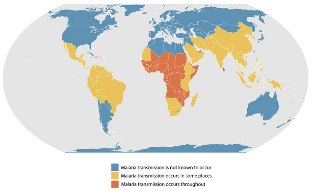
Image Source: CDC (2020). Malaria about distribution, Licensed under a creative commons licence. Available for free at: https://www.cdc.gov/malaria/about/distribution.html (Accessed:24 January 2024).
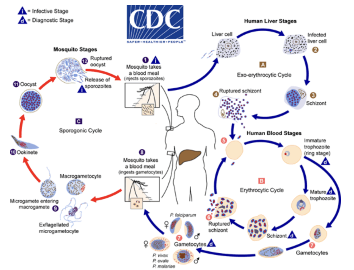
Image Source: CDC (2020). Malaria about biology, Licensed under a creative commons licence. Available for free at: https://www.cdc.gov/malaria/about/biology/index.html (Accessed: 24 January 2024).
References
- CDC Malaria species and resistance by country [Internet]. Cdc.gov [cited April 2022]. Available from: https://www.cdc.gov/malaria/travelers/country_table/a.html
- WHO Guidelines for malaria, 3 June 2022. Geneva: World Health Organization; 2022
- Lalloo DG, Shingadia D, Bell DJ, Beeching NJ, Whitty CJM, Chiodini PL; PHE Advisory Committee on Malaria Prevention in UK Travellers. UK malaria treatment guidelines 2016. J Infect. 2016 Jun;72(6):635-649. doi: 10.1016/j.jinf.2016.02.001. Epub 2016 Feb 12. PMID: 26880088; PMCID: PMC7132403
- Lebrun D, Floch T, Brunet A, Julien G, Romaru J, N’Guyen Y, Cousson J, Giltat A, Toubas D, Bani-Sadr F. Severe post-artesunate delayed onset anaemia responding to corticotherapy: a case report. J Travel Med. 2018 Jan 1;25(1). doi: 10.1093/jtm/tax091. PMID: 29394387
- Bell D, Lalloo D. Malaria. In: Beeching N, Gill G, ed. by. Tropical Medicine Lecture Notes. 7th ed. West Sussex: Wiley Blackwell; 2014. p. 53-69
Other protozoal infections
Leishmaniasis
Leishmaniasis is a protozoal infection endemic within many tropical and subtropical countries, with an annual incidence of up to 1,000,000 cases worldwide. Transmitted by the phlebotomine sand-fly, various forms of the disease exist, including visceral (kala-azar), cutaneous, mucocutaneous, and post-kala-azar. Visceral leishmaniasis presents with symptoms that include fever, weight loss, lymphadenopathy, and hepatosplenomegaly. Thrombocytopenia, leukopenia, and anaemia are often found on a full blood count. The untreated mortality is close to 100%. There are a variety of ways to make the diagnosis, including antigen-based point of care tests, serology, polymerase chain reaction (PCR), and direct detection of amastigotes through splenic, lymph node, or bone marrow aspiration. It is important to test patients for HIV as this often co-exists with visceral leishmaniasis, with the latter occurring as an opportunistic infection. When HIV co-infection is present, leishmaniasis serology is less sensitive. Treatment of visceral leishmaniasis normally requires intensive parenteral therapy over several weeks. Agents of choice include sodium stibogluconate, amphotericin B, paromomycin and pentamidine. Miltefosine is the only oral agent currently in use. It is beneficial in that it can be administered in the community; however, this may lead to poor compliance, especially due to gastrointestinal side-effects. Teratogenicity is another potential issue with this medication. Post-kala-azar leishmaniasis presents with diffuse maculopapular or nodular lesions, which start at the mouth before potentially spreading to the rest of the body. Again, this is associated with HIV co-infection. Lesions can be biopsied for diagnosis. The natural history of post-kala-azar leishmaniasis varies between regions. It often resolves spontaneously in Sudan, whereas it normally requires treatment in India, using the same medications as for visceral leishmaniasis.
Cutaneous leishmaniasis presents with one or more skin lesions, which can vary in appearance. They are classically said to resemble volcanoes, with a raised outer edge and depressed ulcerated centre. Cutaneous leishmaniasis is generally self-limiting and resolves over several months. The lesions can be biopsied for diagnosis, with PCR used to identify the subgenus. This can be important as the subgenus Viannia can cause mucocutaneous leishmaniasis, which involves disfiguring spread of lesions to the mucosa of the mouth and nose, often months to years after apparent healing of the primary lesions. Whether to treat cutaneous leishmaniasis with anything more than topical therapy (or at all) generally depends on whether it is classified as simple or complex. Cutaneous leishmaniasis may be deemed complex if there are several lesions involved, if the lesions are large, if the lesions are having a significant cosmetic impact, or if they involve the lymphatics. Simple lesions can be left to heal over time or treated with topical or oral therapy. Complex lesions require parenteral treatment, such as intravenous or intramuscular sodium stibogluconate. If there is suspicion that the causative subgenus is Viannia, either by local endemicity or by formal PCR analysis, then parenteral therapy would be used to reduce the risk of subsequent mucosal spread.
African Trypanosomiasis
African trypanosomiasis, or African sleeping sickness, is caused by the protozoal species Trypanosoma brucei. West African trypanosomiasis is caused by the subspecies Trypanosoma brucei gambiense, with the rarer East African trypanosomiasis being caused by the subspecies Trypanosoma brucei rhodesiense. Spread by the tsetse fly, these diseases are fatal unless treated. T. gambiense infection presents with an early stage consisting of low-grade fever, joint pain, and lymphadenopathy. Posterior cervical lymphadenopathy may be present. This is known as Winterbottom’s sign, after the physician and abolitionist who noted its prognostic use by slave traders. The latter stage involves central nervous system (CNS) infection, with personality change, disturbance of circadian rhythm, and death within months if left untreated. T. rhodesiense infection follows a more acute course. The initial stage is more severe and more likely to result in death, with pericarditis, myocarditis, and severe anaemia common. The CNS stage also appears more quickly and follows a rapid course to fatality if left untreated. Blood films, bone marrow biopsies and lymph node biopsies can be useful in diagnosis. Suramin can be used to treat both early-stage variants, though pentamidine is often used for T. gambiense disease. Neither drug penetrates the CNS and therefore, a lumbar puncture is necessary to diagnose late-stage disease. Melarsoprol, which can be used for the late stage of both diseases, carries a significant risk of encephalopathy and death. Eflornithine, which is safer, can only be used for late-stage T. gambiense disease.
Chagas’ disease
South American trypanosomiasis, or Chagas’ disease, is caused by the protozoa Trypanosoma cruzi. It is excreted in the faeces of the triatomine bugs and gains entry to the individual via the site of the triatomine bite. An iatrogenic spread has also been reported via organ and blood transfusions. As with African trypanosomiasis, there is an acute phase and a late phase. However, the acute early phase is often asymptomatic and there is a latency period of years between acute infection and chronic manifestations. Acute infection may cause fever, lymphadenopathy, and splenomegaly. A chagoma is a swelling of the skin related to parasite entry. Romaña’s sign involves periorbital swelling and conjunctivitis after ophthalmic parasite entry. Rarely, the acute phase can cause pericarditis and meningoencephalitis. This is more common in young children and immunosuppressed patients. Chronic disease leads to gastrointestinal tract pathology – megaoeseophagus and megacolon – and cardiac pathology – cardiomegaly and associated conduction abnormalities. The acute phase can generally be diagnosed using a blood film or serology. Early treatment with nifurtimox and benznidazole can prevent acute complications and reduce the risk of chronic infection. Chronic infection is more difficult to diagnose due to low level parasitaemia. The mainstay of treatment for chronic chagas disease involves managing complications. For example, the insertion of a pacemaker for a complete heart block. Xenodiagnosis is a valid method of establishing the diagnosis with low-level parasitaemia. Uninfected triatomine bugs are fed blood from suspected patients and then examined for evidence of parasites. This is not commonly used due to several factors, such as the incubation time needed for low-level parasite replication and the need for a supply of uninfected triatomine bugs.
References
- CDC leishmaniasis factsheet [Internet]. Cdc.gov [cited April 2022]. Available from: https://www.cdc.gov/dpdx/leishmaniasis/
- Mukhopadhyay D, Dalton J, Kaye P, Chatterjee M. Post kala-azar dermal leishmaniasis: an unresolved mystery. Trends Parasitol. 2014;30(2):65-74. doi:10.1016/j.pt.2013.12.004
- O’Dempsey T. Visceral leishmaniasis. In: Beeching N, Gill G, ed. by. Tropical Medicine Lecture Notes. 7th ed. West Sussex: Wiley Blackwell; 2014. p. 70-76
- CDC African trypanosomiasis factsheet [Internet]. Cdc.gov [cited April 2022]. Available from: https://www.cdc.gov/parasites/sleepingsickness/
- Lalloo D. African trypanosomiasis. In: Beeching N, Gill G, ed. by. Tropical Medicine Lecture Notes. 7th ed. West Sussex: Wiley Blackwell; 2014. p. 141-147
- Büscher P, Cecchi G, Jamonneau V, Priotto G. Human African trypanosomiasis. Lancet. 2017;390(10110):2397-2409. doi: 10.1016/S0140-6736(17)31510-6. Epub 2017 Jun 30
- CDC Chagas’ factsheet [Internet]. Cdc.gov [cited April 2022]. Available from: https://www.cdc.gov/parasites/chagas’/
- Echeverria L, Morillo C. American Trypanosomiasis (Chagas’ Disease). Infect Dis Clin North Am. 2019;33(1):119-134. doi: 10.1016/j.idc.2018.10.015
- Lalloo D. South American trypanosomiasis (Chagas’ disease). In: Beeching N, Gill G, ed. by. Tropical Medicine Lecture Notes. 7ᵗʰ ed. West Sussex: Wiley Blackwell; 2014. p. 148-150
Parasitic worm infections
Trematode infections
Trematodes, also known as flatworms or flukes, are parasitic worms that can cause human disease.
Blood flukes – Schistosomiasis
Schistosomiasis is a condition caused by trematodes of the genus Schistosoma. Various species are widely distributed across the tropics. Infection is caused by the larval stage cercariae penetrating the skin in freshwater. This can cause a pruritic rash known as “swimmer’s itch”, though this is less likely to occur with repeated infection. Likewise, the acute inflammatory reaction associated with initial egg production several weeks after larval entry is more likely in immune-naïve patients. Signs and symptoms of this inflammatory reaction, known as Katayama fever, include fever, rash, wheeze, and hepatosplenomegaly. Eosinophilia can be noted on a full blood count. The release of eggs from the body causes the main chronic symptoms. Schistosoma haematobium resides in the pelvic veins and causes urogenital schistosomiasis, with haematuria often being present. Chronic S. haematobium infection can lead to reproductive tract pathology, obstructive uropathy, and squamous cell bladder cancer. Other species of Schistosoma, including Schistosoma mansoni and Schistosoma japonicum, reside in the mesenteric veins. Egg passage through the bowel can cause small amounts of blood loss and anaemia over time. Portal hypertension can result from eggs and associated inflammatory responses within the portal veins. This, in turn, can lead to hepatosplenomegaly and oesophageal varices. Diagnosis may involve microscopic visualisation of eggs in urine or stool, though this can lack sensitivity. Stool and urine PCR can improve sensitivity, and rapid antigen detection point of care tests for urine samples are also available. Serology can be useful for returned travellers but is less useful for patients in endemic areas. Praziquantel is used to treat all species causing schistosomiasis.
Lung Flukes
Ingestion of freshwater crustaceans can lead to infection with the lung fluke genus Paragonimus. Paragonimus westermani is the main species to know about, being largely responsible for paragonimiasis in Asia. The disease is more rarely documented in Central Africa and South America. The immature flukes excyst through the duodenum and reach the lung via the peritoneum. This may cause a pneumothorax. The mature flukes then release eggs in the lung, which are coughed up and either released through the mouth or swallowed and released after passing through the digestive tract. Symptoms include initial abdominal pain followed by fever, wheeze, cough, and haemoptysis. Diagnosis is made either serologically or by identification of eggs in sputum or stool. Treatment is with praziquantel.
Liver Flukes
The main causative fluke for fascioliasis is Fasciola hepatica, which is found on every populated continent. Infection is via ingestion of contaminated freshwater plants. In a similar fashion to Paragonimus, immature Fasciola flukes excyst through the duodenum, though eventually invade the biliary ducts via the peritoneum. Similar symptoms are present during this stage, such as fever and abdominal pain. The infection may then be asymptomatic, aside from occasional pain from intermittent biliary obstruction. Pancreatitis and cholecystitis can also occur. Serology and stool culture can be useful in diagnosis. CT imaging can show hypodense liver lesions. Similarly, ultrasound can highlight hypoechoic liver lesions. Praziquantel – usually a safe bet for trematode treatment – is not effective for fascioliasis; triclabendazole is used instead.
Trematodes of the genus Clonorchis and Opisthorchis cause a similar clinical presentation to fascioliasis. Encysted larvae are introduced through the ingestion of freshwater fish. Infection occurs in Asia only, except in the case of rare international imports. Larvae enter the biliary tract through the ampulla of vater rather than through the peritoneum and inhabit smaller bile ducts than Fasciola. Chronic infection can lead to the development of cholangiocarcinoma. Praziquantel is the treatment of choice.
Nematode infections – Filarial disease
There are several important types of filarial disease. All are caused by nematode worms and are transmitted by diverse insect vectors.
Onchocerciasis
Onchocerca volvulus is the causative nematode in onchocerciasis, or “river blindness”. It is a major cause of preventable blindness across Central Africa. There is also some endemicity across Central and South America. Simulium blackflies allow passage of the larval stage into the subcutaneous tissue, where adult female worms mature over a period of around one year, before producing microfilariae for up to 15 years if left untreated. Dying microfilariae cause the signs and symptoms of onchocerciasis, which include depigmented skin lesions and gradual loss of sight as microfilariae die within various parts of the eye. Diagnosis can be made by microscopy and direct visualisation of microfilariae, with PCR being used on the sample if visualisation is not possible. Microfilariae can also be observed on slit lamp examination of the eye. Diethylcarbamazine (DEC) causes rapid death of microfilariae and can lead to the Mazzotti reaction, a life-threatening immunological reaction characterised by fever, hypotension, urticaria, and lymphadenopathy. This phenomenon can be exploited for diagnostic purposes, using small patches to invoke a localised immune response. Ivermectin is microfilaricidal, however is less likely to cause the Mazzotti reaction as its mechanism of action is slower. Wolbachia is an endosymbiotic bacterium found within all the filarial species we will discuss, except for Loa loa. It is, therefore, a useful target in treating adult worms. Doxycycline causes Wolbachia death and is therefore useful as an agent against adult worms (macrofilaricidal).
Loiasis
Loiasis, caused by Loa loa, is transmitted by the Chrysops fly, which resides in the rainforests of West and Central Africa. As in onchocerciasis, adult female worms develop in the subcutaneous tissue and begin to produce microfilariae after several months. Again, the adult worms may be viable for over a decade. Microfilariae are found in various bodily fluids. As the Chrysops fly is a day biter, evolution has dictated that the microfilariae are most likely to be found in the blood during this time in order to complete the lifecycle. Migration of the adult worm causes most signs and symptoms. It can be visible in the patient’s eye at times, usually causing transient local inflammation without permanent damage to sight. Calabar swellings are skin swellings associated with adult worm migration through the subcutaneous tissue. Diagnosis can be clinical, or microfilariae can be found on blood films taken from the patient during the daytime. DEC kills both adult worms and microfilariae but must be used with caution in areas where onchocerciasis is endemic. Ivermectin treatment prior to DEC administration will reduce the chance of the Mazzotti reaction on patients with both diseases. Unfortunately, treatment with both agents can precipitate encephalopathy due to rapid microfilarial death. If microfilarial load is high in blood samples, then prior treatment with albendazole (which limits the production of viable microfilaria) and concurrent use of steroids may reduce the likelihood of treatment-related encephalopathy. As there is no Wolbachia endosymbiosis, doxycycline is not effective.
Lymphatic filariasis
Lymphatic filariasis, often caused by Wuchereria bancrofti, is prevalent across much of the tropics and subtropics. The infective process is similar to other filarial disease, with mosquitos acting as the vectors. However, the adult worms reside within the lymphatics rather than subcutaneous tissue. Microfilarial death within the pulmonary capillaries may cause a syndrome called tropical pulmonary eosinophilia, characterised by an asthma-like clinical picture, with low-grade fever and eosinophilia. Chest X rays may be normal or demonstrate a reticulonodular or miliary appearance. Chronic lymphatic filariasis is associated with lymphoedema due to lymphatic obstruction, causing disfiguring leg and scrotal swelling. A blood film obtained during the night has the best chance of identifying microfilaria. Ultrasound may show adult worms. Serological tests are also available. Ideally, lymphatic filariasis is treated with DEC. However, this may precipitate the Mazzotti reaction or encephalopathy if there is concurrent infection with Onchocerca volvulus or Loa loa, respectively. Albendazole and ivermectin combination therapy may also be used. Doxycycline can be useful as there is Wolbachia endosymbiosis. Mass drug administration in areas of high helminth endemicity is a viable public health intervention, though the complex overlap of filarial diseases in certain regions and reactions to specific treatments makes this challenging.
Other nematode infections
Strongyloidiasis
Strongyloides stercoralis is a nematode parasite of the small intestine. Although more common in warmer climates, it has a worldwide distribution. Infection is caused by penetration of the larvae through open skin. They then migrate via the bloodstream and lymphatics to the lungs before subsequently being coughed up and swallowed. The mature female produces eggs from the intestine, with the resultant larvae capable of causing autoinfection by penetrating the intestine or perianal skin. The autoinfection stage of strongyloidiasis can cause an itchy, rapidly spreading but transient skin eruption called larva currens. Symptoms can also include cough and abdominal pain, however strongyloidiasis can often be asymptomatic over a period of years. The main worry is the potential for life-threatening strongyloidiasis hyperinfection if a patient becomes immunocompromised. Diagnosis can be made by stool microscopy or serology. Treatment is with standard anti-helminth drugs such as ivermectin or albendazole.
Cystic helminth disease and key differentials
Hydatid disease
Cystic echinococcosis, or hydatid disease, is caused by ingestion of the eggs of Echinococcus granulosus, a cestode (tapeworm). This can lead to cystic disease of a variety of organs, mainly the liver and lungs. Hydatid cysts can be asymptomatic for years and detected on incidental imaging. Right upper quadrant pain may occur due to cyst growth. Rupture of lung cysts may cause pulmonary manifestations such as a pneumothorax. A variety of stages can be observed using different imaging modalities, with a WHO classification system existing for ultrasound diagnosis. The water lily sign is caused by the detachment of the endocyst membrane, which then floats within the cystic fluid. Serology can also be used to help with the diagnosis. Due to the slow growth of cysts caused by E. granulosus, a watchful waiting approach can be taken to see if the cyst growth has ceased. Active treatment strategies may involve chemotherapy with albendazole, with or without more invasive management. PAIR – Puncture, Aspiration, Injection (of a scolicidal agent such as hypertonic saline), Reaspiration – is a radiologically-guided technique that can be used to avoid formal surgery. Surgery can then be reserved for more complicated cases or calcified cysts. Alveolar echinococcosis is a rarer disease caused by E. multilocularis, which involves rapid destruction and invasion of tissue, requiring more complex management.
It is important to be able to differentiate hydatid liver cysts from amoebic liver abscesses caused by the protozoa Entamoeba histolytica. Occurring worldwide, there is a strong association between poor sanitation and hygiene and amoebiasis. Amoebiasis is generally a self-limiting gastrointestinal infection, causing abdominal pain and loose stools with blood or mucus. The most common extra-intestinal manifestation is amoebic liver abscess. Abscesses are usually isolated in nature, though multiple can be present. Right upper quadrant pain and fever are the predominant symptoms. In terms of diagnosis, stool microscopy is not brilliant as it is difficult to differentiate E. histolytica from E. dispar, a non-pathogenic species. Stool antigen tests may be of use. Serology is often preferred, though it can give a false negative in early disease and a false positive if the patient has been previously infected. Amoebic liver cysts are similar in appearance to pyogenic liver cysts on imaging. Medical therapy with metronidazole is usually first line for amoebiasis and amoebic liver cysts. Aspiration and drainage are reserved for special circumstances. These include diagnostic uncertainty, failure of medical therapy, clinical or radiological evidence of impending rupture, and left-sided liver cysts (which may rupture into the pericardium). Fluid obtained from amoebic liver cysts is normally thick and brown, as opposed to the thin, clear fluid found in hydatid cysts.
Neurocysticercosis
Another tapeworm cyst to be aware of is that of Taenia solium. Once again acquired via the faecal-oral route, cysticercosis is a result of larval migration to tissues and organs including muscles and the brain, with the latter termed neurocysticercosis. Consumption of infected meat simply leads to intestinal tapeworm infection. Neurocysticercosis is a leading cause of seizures in the tropics and East Asia. As this is a pig tapeworm, cases are rarer in areas where pigs are not reared for religious reasons. Diagnosis of neurocysticercosis is usually based on imaging and clinical history. Treatment includes anti-epileptics and, if anti-helminth medication is to be used, steroids can prove useful in preventing morbidity and mortality from the ensuing inflammatory response.
References
- WHO Schisotosomiasis factsheet [Internet]. WHO.int [cited April 2022]. Available from: https://www.who.int/news-room/fact-sheets/detail/schistosomiasis
- Squire B, Stothard R. Schistosomiasis. In: Beeching N, Gill G, ed. by. Tropical Medicine Lecture Notes. 7th ed. West Sussex: Wiley Blackwell; 2014. p. 151-162
- Blair D. Paragonomiasis. In: Toledo R, Fried B, ed. by. Digenetic Trematodes. Advances in Experimental Medicine and Biology. Vol 766. New York: Springer; 2014. https://doi.org/10.1007/978-1-4939-0915-5_5
- CDC paragonimiasis factsheet [Internet]. Cdc.gov [cited April 2022]. Available from: https://www.cdc.gov/parasites/paragonimus/index.html
- CDC fascioliasis factsheet [Internet]. Cdc.gov [cited April 2022]. Available from: https://www.cdc.gov/parasites/fasciola/index.html
- Dusak A, Onur M, Cicek M et al. Radiological Imaging Features of Fasciola hepatica Infection – A Pictorial Review. J Clin Imaging Sci. 2012;2:2. doi:10.4103/2156-7514.92372
- CDC Clonorchis factsheet [Internet]. Cdc.gov [cited April 2022]. Available from: https://www.cdc.gov/parasites/clonorchis/index.html
- CDC Opisthorchis factsheet [Internet]. Cdc.gov [cited April 2022]. Available from: https://www.cdc.gov/parasites/opisthorchis/index.html
- O’Dempsey T. Onchocerciasis, filariasis and loiasis. In: Beeching N, Gill G, ed. by. Tropical Medicine Lecture Notes. 7th ed. West Sussex: Wiley Blackwell; 2014. p. 126-140
- Brattig N, Cheke R, Garms R. Onchocerciasis (river blindness) – more than a century of research and control. Acta Trop. 2021;218:105677. doi: 10.1016/j.actatropica.2020.105677. Epub 2020 Aug 26
- Lourens G, Ferrell D. Lymphatic Filariasis. Nurs Clin North Am. 2019;54(2):181-192. doi: 10.1016/j.cnur.2019.02.007. Epub 2019 Mar 27
- Mullerpattan J, Udwadia Z, Udwadia F. Tropical pulmonary eosinophilia–a review. Indian J Med Res. 2013;138(3):295-302
- Krolewiecki A, Nutman T. Strongyloidiasis: A Neglected Tropical Disease. Infect Dis Clin North Am. 2019;33(1):135-151. doi: 10.1016/j.idc.2018.10.006
- Peters L, Burkert S, Grüner B. Parasites of the liver – epidemiology, diagnosis and clinical management in the European context. J Hepatol. 2021;75(1):202-218. doi: 10.1016/j.jhep.2021.02.015. Epub 2021 Feb 24
- Takayanagui OM, Haes TM. Update on the diagnosis and management of neurocysticercosis. Arq Neuropsiquiatr. 2022;80(5 Suppl 1):296-306. doi: 10.1590/0004-282X-ANP-2022-S115
Rabies & Leprosy
Rabies
Rabies, caused by a virus of the Lyssavirus genus, is a fatal disease of the central nervous system if given time to propagate. It is transmitted through direct contact with infected mammals, often a bite. These mammals include dogs, bats, and raccoons. The first symptoms of rabies infection are a flu-like illness and irritation at the site of the bite. There is generally a long incubation period between exposure and CNS symptoms – in the order of weeks – as it takes time for the virus to travel backwards through peripheral nerves. This is shortened in children or in bites to the face as there is a smaller distance to travel. CNS symptoms include fluctuating periods of confusion, severe agitation and aggression. Other symptoms include hypersalivation and hydrophobia. The disease is almost always fatal once CNS symptoms are present.
Rabies is a vaccine-preventable disease. Post-exposure prophylaxis, involving a combination of vaccinations and immunoglobulins, is also available. As rabies is generally a disease of resource-poor areas, a patient may choose to observe a dog after a potential transmission event instead of immediately seeking medical attention. If the dog remains alive and well after around 10-15 days, then it did not have transmissible rabies infection at the time of the direct contact. As alluded to, this method may be risky if the incubation period is expected to be shorter than usual. The vaccination schedule and use of immunoglobulin therapy depends on whether it is pre-or post-exposure prophylaxis, as well as previous vaccinations, local endemicity of rabies, and severity of exposure. Please see the Green Book reference for further details and note that these are always subject to change.
Leprosy
Leprosy is caused by Mycobacterium leprae and has an almost worldwide distribution, with an incidence of over 100,000 cases per year. It is primarily a disease that affects the skin and peripheral nerves and is contracted through prolonged contact with an untreated individual. Blindness is a potential consequence of the disease.
Diagnosis of leprosy can be made clinically, with findings of stereotypical skin lesions and thickened peripheral nerves. A clinical spectrum exists between paucibacillary leprosy (tuberculoid) and multibacillary leprosy (lepromatous). Multibacillary leprosy is a failure of the cell-mediated immune response to the mycobacteria. This results in more lesions, more affected nerves, and more mycobacteria on skin snippings. Individuals with paucibacillary leprosy will have full anaesthesia over a fewer number of skin lesions, which will be asymmetrically distributed. Bacterial load can be calculated by microscopy of skin scrapings. Treatment regimens are based on whether the clinical picture is one of multibacillary or paucibacillary leprosy. Rifampicin, dapsone and clofazimine are used in combination for multibacillary leprosy, whereas a shorter course of rifampicin and dapsone alone is sufficient to treat paucibacillary leprosy. Steroids are used if new nerve damage is present on routine follow-up or if there is evidence of hypersensitivity or immune complex-mediated reactions at any point before, during, or after antimicrobial treatment.
References
- Rupprecht CE, Mani RS, Mshelbwala PP, Recuenco SE, Ward MP. Rabies in the Tropics. Curr Trop Med Rep. 2022;9(1):28-39. doi: 10.1007/s40475-022-00257-6
- Solomon T, Lalloo D. Rabies. In: Beeching N, Gill G, ed. by. Tropical Medicine Lecture Notes. 7th ed. West Sussex: Wiley Blackwell; 2014. p. 254-258
- UK Health Security Agency. Immunisation against infectious disease. Ramsay M ed by. 2021, Jan
- WHO rabies factsheet [Internet]. WHO.int [cited September 2022]. Available from: https://www.who.int/newsroom/fact-sheets/detail/rabies
- Maymone M, Laughter M, Venkatesh S et al. Leprosy: Clinical aspects and diagnostic techniques. J Am Acad Dermatol. 2020;83(1):1-14. doi: 10.1016/j.jaad.2019.12.080. Epub 2020 Mar 27
- Lockwood D, Beeching N. Leprosy. In: Beeching N, Gill G, ed. by. Tropical Medicine Lecture Notes. 7th ed. West Sussex: Wiley Blackwell; 2014. p. 163-174
- Maymone MBC, Laughter M, Venkatesh S, Dacso MM, Rao PN, Stryjewska BM, Hugh J, Dellavalle RP, Dunnick CA. Leprosy: Clinical aspects and diagnostic techniques. J Am Acad Dermatol. 2020 (1):1-14. doi: 10.1016/j.jaad.2019.12.080
- WHO leprosy factsheet [Internet]. WHO.int [cited September 2022]. Available from: https://www.who.int/newsroom/fact-sheets/detail/leprosy
Arboviruses & viral haemorrhagic fevers
Arboviruses
Arboviruses are simply arthropod-borne viruses. These include zika virus, chikungunya virus, and dengue virus. This section will focus on dengue, due to its prevalence and potential for life-threatening consequences.
Dengue, a flavivirus, is transmitted by the Aedes mosquito in the tropics and subtropics. The clinical manifestations of the virus are fever, arthralgia, and a distinctive maculopapular rash. The illness is usually self-limiting, though a small proportion of patients may develop severe, life-threatening dengue. Interestingly, past infection with one of the four serotypes of dengue increases the risk of subsequent severe dengue, likely by a process called Antibody Dependent Enhancement. This makes vaccine development difficult. The term “dengue haemorrhagic fever” was previously used to denote severe dengue, however this has been superseded by the most recent WHO classifications of dengue, with or without warning signs, and severe dengue. Serology can be used for formal diagnosis, though diagnosis in endemic regions can normally be made on clinical and laboratory findings. Leukopenia and thrombocytopenia are common. Treatment of dengue is purely supportive.

Image Source: WHO/IRIS (2009). 10665, Licensed under a creative commons licence. Available for free at: https://apps.who.int/iris/handle/10665/44188 (Accessed: 24 January 2024).
Viral haemorrhagic fevers
It is worth having a general appreciation of other viral haemorrhagic fevers. If a viral haemorrhagic fever is suspected in a returned traveller, an algorithm should be followed (see references). This considers the history of the febrile illness, the area travelled to, and any ill contacts or contacts with potential reservoirs of infection. A thorough history is necessary, as evidenced by Barr et al. (2013), where the source of infection was identified using wedding photographs.
The antiviral ribavirin may be of benefit in some viral haemorrhagic fevers, however the mainstay of treatment for viral haemorrhagic fevers is generally supportive care. It is important to note that Lassa, Ebola, and Marburg, and Crimean-Congo haemorrhagic fevers are all capable of person to person spread. Rift Valley haemorrhagic fever is generally spread via virulent livestock. Yellow fever (which is vaccine-preventable) and dengue are both arboviruses.
References
- Shastri PS, Taneja S. Dengue and Other Viral Hemorrhagic Fevers. Indian J Crit Care Med. 2021 (Suppl 2):S130-S133. doi: 10.5005/jp-journals-10071-23814
- Solomon T. Dengue and yellow fever. In: Beeching N, Gill G, ed. by. Tropical Medicine Lecture Notes. 7th ed. West Sussex: Wiley Blackwell; 2014. p. 283-288
- UK health security agency. UK viral haemorrhagic fever algorithm – https://www.gov.uk/government/publications/viral-haemorrhagic-fever-algorithm-and-guidance-on-management-of-patients (accessed April 2022)
- Barr D, Aitken C, Bell D et al. First confirmed case of Crimean-Congo haemorrhagic fever in the UK. The Lancet. 2019;382(9902), p.1458
Other tropical infections
The tropical bowel
Tropical sprue
Tropical sprue is a disease of unknown aetiology occurring in tropical and subtropical countries, though it is relatively less common on the African continent. It is characterised by malabsorption in the small bowel. It is often precipitated by an acute diarrhoeal illness. Villous blunting is observed on endoscopy. A prolonged course of antibiotics is given as treatment for the associated bacterial overgrowth, as well as nutrition supplementation, and there is usually a good resolution of pathology.
Giardiasis
The protozoan infection Giardiasis, caused by Giardia duodenalis, is spread via the faecal-oral route and has a worldwide distribution. The incubation period is generally several days but can extend into weeks and months. Signs and symptoms include foul-smelling diarrhoea and flatulence, abdominal pain, and weight loss if there is any chronicity. Stool microscopy is generally used to make a diagnosis and the treatment is a course of metronidazole.
Rickettsial infection
Rickettsial disease refers to infection caused by intracellular bacteria of the family Rickettsiaceae. All these diseases present with a typical triad of fever, arthralgia, and rash. Louse-borne – epidemic – typhus occurs when Rickettsia prowazekii is transmitted through the infected faeces of the human body louse via the skin. It is associated with areas of poor hygiene, such as refugee camps, and can cause multi-organ inflammation and failure, with a high mortality rate if left untreated. Tick typhus, including Rocky Mountain spotted fever and African tick typhus, have varying clinical courses. There is usually an eschar present with African tick typhus, and it is generally less severe than Rocky Mountain spotted fever. Finally, scrub typhus, caused by Orientia tsutsugamushi, is transmitted via biting mites from Eastern Asia and Oceania. A small eschar can be found at the site of the bite, with hepatosplenomegaly and multi-organ dysfunction possible sequalae. Rickettsial disease is generally susceptible to tetracycline antibiotics.
Anthrax
Anthrax, caused by the endospore-forming Bacillus anthracis, is classified by route of inoculation. Cutaneous and inhalational anthrax generally occurs after occupational exposure to spores from animal products. Bioterrorism may also be alluded to in single best answer questions. Consumption of contaminated meat leads to gastrointestinal anthrax. Injectable anthrax can result from injecting contaminated illicit drugs. Cutaneous anthrax has a short incubation period and generally resolves over a few weeks with appropriate treatment, though it can lead to fatal sequalae in 20% of untreated patients. The lesion typically contains a central dark eschar with significant surrounding oedema. Inhalational anthrax presents with upper respiratory tract symptoms followed by swift respiratory failure and death. Gastrointestinal anthrax presents with severe abdominal pain and bloody diarrhoea, followed often by overwhelming sepsis. All forms may rarely be complicated by meningeal spread. Diagnosis of cutaneous anthrax is made by microscopy of fluid from the lesion, with PCR if necessary. Blood cultures are also a vital investigation. Cutaneous anthrax may be treated with ciprofloxacin, doxycycline, or, when sensitivities are known, amoxicillin. Therapy for other forms is more complicated, with benzylpenicillin often part of the antimicrobial cocktail. Inhalational and gastrointestinal forms of anthrax are likely fatal, even with appropriate treatment.
Plague
Plague, caused by the bacteria Yersinia pestis, is generally transmitted by fleas and can present in three forms. Bubonic and septicaemic plague is what is classically depicted in images from the 14th century pandemic. Bubonic plague is characterised by fever, malaise, and the appearance of buboes; painful, swollen lymph nodes. Septicaemic plague is characterised by shock and tissue necrosis – “black death”. Pneumonic plague, which can be transmitted from person to person via respiratory droplets or develop from untreated bubonic or septicaemic plague, is associated with a rapidly progressive multilobar consolidative process. Buboes can be sampled for diagnosis, or samples of sputum or blood can be analysed. A variety of classes of antibiotic are used as treatment. Post-exposure antibiotic prophylaxis is recommended after contact with pneumonic plague. A vaccine exists for individuals expected to be at high risk of the disease, such as exposed healthcare workers and clinical scientists.
Other tropical infections
There are several other tropical infections which can come up in part 2 of MRCP, including bacterial gastroenteritis, leptospirosis, Q fever, and melioidosis. It is worth having some familiarity with the pertinent features of each of these.
References
- Stein C, Parry C, Frean J et al. Multi-system diseases and infections. In: Davidson R, Brent A, Seale A, ed. by. Oxford Handbook of Tropical Medicine. 4ᵗʰ ed. Oxford: Oxford University Press; 2014. p. 729-822
- Price E, Seymour M, Sarovich D et al. Molecular epidemiologic investigation of an anthrax outbreak among heroin users, Europe. Emerg Infect Dis. 2012;18(8):1307-13. doi: 10.3201/eid1808.111343
- Blanton L. Rickettsial infections in the tropics and in the traveller. Curr Opin Infect Dis. 2013;26(5):435-40. doi: 10.1097/QCO.0b013e328363811b
- Penny M, Rivera F. Diarrhoeal diseases. In: Davidson R, Brent A, Seale A, ed. by. Oxford Handbook of Tropical Medicine. 4ᵗʰ ed. Oxford: Oxford University Press; 2014. p. 233-292
Complete Guide to Clinical Genetics ST3 Application and Recruitment (2027)
Written by Medibuddy. Last updated: October 2026. Clinical Genetics is

Complete Guide to Clinical Radiology ST3 Application and Recruitment (2027)
Recruitment paused: NHS England is not running Clinical Radiology ST3+ recru

Complete Guide to Paediatric Surgery ST3 Application and Recruitment in United Kingdom (2026)
Written by Medibuddy. Last updated: October 2026. If you’re planning


Take your subscriptions with you
Our mobile app allows you to access your interview and exam question banks wherever you are.

