


 Close
Close
 16th February 2025
Pulmonary function tests (PFTs)
Spirometry
During a spirometry test, the patient inhales maximally, then exhales into the spirometer as hard as possible until no breath remains. The main values of interest are as follows, with predicted values calculated from the patient’s height and demographics.
Peak expiratory flow rate (PEFR)
Also measured using a peak flow meter, the PEFR is the maximum rate of expiratory flow the patient can achieve, in L/min, typically attained at the beginning of expiration. A reduction in PEFR is suggestive of airway obstruction, such as COPD or asthma.
In asthma, variability in PEFR during the day is typically observed, with the lowest values usually being recorded early in the morning or after exercise. PEFR is usually reduced but stable in COPD. There is also a rare phenotype of asthma where fixed PEFR is observed, as in COPD.
PEFR is frequently used for monitoring acute asthma in the inpatient setting. BTS guidelines recommend the patient is safe for discharge when PEFR is >75% of best or predicted, with a diurnal variation of less than 25%.
FVC, FEV1 and FEV1/FVC ratio
The forced vital capacity (FVC) is the total volume of air expelled from full inspiration to full expiration. Above 80% of predicted is considered normal.
The forced expiratory volume in one second (FEV1) is the volume of air expired in the first second of forced expiration. Typically, the FEV1 is interpreted using the FEV1/FVC ratio, which is more useful as it is independent of other factors. A ratio of >70% is considered normal, with a ratio of <70% suggesting airway obstruction.
Obstructive pattern
An obstructive pattern of PFTs is where the FEV1/FVC ratio is less than 70%, with normal or low FVC.
This usually indicates COPD or asthma, which can be differentiated by a reversibility test, where the test is repeated following administration of a bronchodilator such as salbutamol. If the FEV1 or FVC increases by more than 12%, the obstruction is said to be reversible or bronchodilator responsive, which is suggestive of asthma, although a small degree of reversibility can also be seen in COPD.
Alternatively, diurnal variability in PEFR can be used to differentiate the two, as mentioned above.
Restrictive pattern
A restrictive pattern of PFTs is where the FVC is reduced, with a preserved FEV1/FVC ratio (>70%). This occurs in three main aetiologies:
Reduced lung compliance/volume, such as lung fibrosis or pneumonectomy.
Muscle weakness, such as myasthenia gravis or myopathy.
Reduced chest wall compliance – see following section.
FVC monitoring
Monitoring of FVC is also used for assessing decline in respiratory function in acute neuromuscular disease, such as in Guillain-Barré syndrome and myasthenic crisis. An FVC of less than 15-20 ml/kg body weight suggests endotracheal intubation may be necessary.
Diseases of the chest wall
Pathology of the chest wall may decrease its compliance and, therefore, cause a restrictive defect. It is classified as follows:
- Congenital: for example, pectus excavatum, pectus carinatum and Poland syndrome. The latter involves a constellation of unilateral features, including incomplete development of the hands, respiratory muscles and costal cartilages
- Age related: it is normal for chest wall compliance to decrease with age, and this can be further exacerbated by kyphosis and scoliosis
- Traumatic and iatrogenic: flail chest and thoracoplasty
- Abdominal processes: obesity and ascites cause a restrictive defect via splinting of the diaphragm
- Ankylosing spondylitis: may lead to fixation of the chest wall in advanced disease
Measures of gas exchange – TLᴄᴏ and Kᴄᴏ
To quantify alveolar gas exchange, the patient breathes a mix of an inert gas (helium) and carbon monoxide (CO), breath holding for 10 seconds, then the CO in exhaled gas is measured. Only the CO will be absorbed into the blood, so the drop in CO concentration is indicative of gas exchange, as recorded in two measures:
- The transfer factor of the lung for carbon monoxide (TLᴄᴏ), also called the diffusing capacity (DLᴄᴏ). This measures the total amount of CO transferred to the blood and therefore the total gas exchange capacity of the lungs
- The carbon monoxide transfer coefficient (Kᴄᴏ) – this is the TLᴄᴏ divided by the total lung volume, giving a measure of the average gas exchange per unit volume of the lung. It is low where there is a problem with gas exchange at the level of the alveolar membrane, for example due to thickening
A low Kᴄᴏ helps differentiate between causes of respiratory disease:
- In a restrictive spirometry pattern, a low Kᴄᴏ suggests an intrapulmonary cause such as fibrosis. It will be normal in extrapulmonary causes such as kyphoscoliosis
- In an obstructive spirometry pattern, a low Kᴄᴏ suggests COPD rather than asthma
A high Kᴄᴏ is seen in two scenarios:
- In the few days after alveolar haemorrhage, due to red blood cells taking up CO. For example in SLE, Wegener’s and Goodpasture’s
- In poor lung expansion, such as due to neuromuscular disease or chest wall restrictive disease
Flow-volume loops
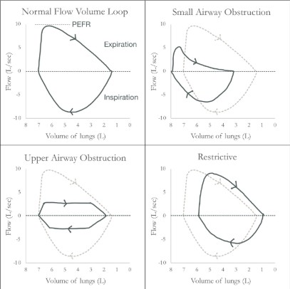
A flow-volume loop is constructed from spirometry involving forced maximal inspiration and expiration. The cycle is clockwise, with an upward deflection indicating expiration and a downward indicating inspiration. The start point of maximal inspiration (total lung capacity, TLC) is the leftmost point of the loop, with the rightmost point representing the minimal lung volume (residual volume, RV).
References
- Chapman SJ, Robinson GV, Shrimanker R, Turnbull CD, Wrightson JM. Oxford handbook of respiratory medicine. 4th ed. London, England: Oxford University Press; 2021
- Bourke SJ. Respiratory medicine: Lecture notes, 10th edition: Lecture Notes. 10th ed. Hoboken, NJ: Wiley-Blackwell; 2022
- Bessant R et al. The pocketbook for PACES. London, England: Oxford University Press; 2012
- British Thoracic Society, Scottish Intercollegiate Guidelines Network. British guideline on the management of asthma. Thorax. 2014;69 Suppl 1:1–192
- Kaminsky DA. Overview of pulmonary function testing in adults [Internet]. McCormack MC, editor. UpToDate. 2022 [cited 2022 Dec 9]. Available from: https://www.uptodate.com/contents/overview-of-pulmonary-function-testing-in-adults
- McCormack MC. Diffusing capacity for carbon monoxide [Internet]. Stoller JK, editor. UpToDate. 2022 [cited 2022 Dec 9]. Available from: https://www.uptodate.com/contents/diffusing-capacity-for-carbon-monoxide
Complete Guide to Clinical Genetics ST3 Application and Recruitment (2027)
Written by Medibuddy. Last updated: October 2026. Clinical Genetics is

Complete Guide to Clinical Radiology ST3 Application and Recruitment (2027)
Recruitment paused: NHS England is not running Clinical Radiology ST3+ recru

Complete Guide to Paediatric Surgery ST3 Application and Recruitment in United Kingdom (2026)
Written by Medibuddy. Last updated: October 2026. If you’re planning


Take your subscriptions with you
Our mobile app allows you to access your interview and exam question banks wherever you are.

